Review
. 2020 May 8;295(19):6312-6329.
doi: 10.1074/jbc.REV119.009453. Epub 2020 Mar 18.

Brain manganese and the balance between essential roles and neurotoxicity

Affiliations
Review

Brain manganese and the balance between essential roles and neurotoxicity

Rekha C Balachandran et al. J Biol Chem. .

Abstract

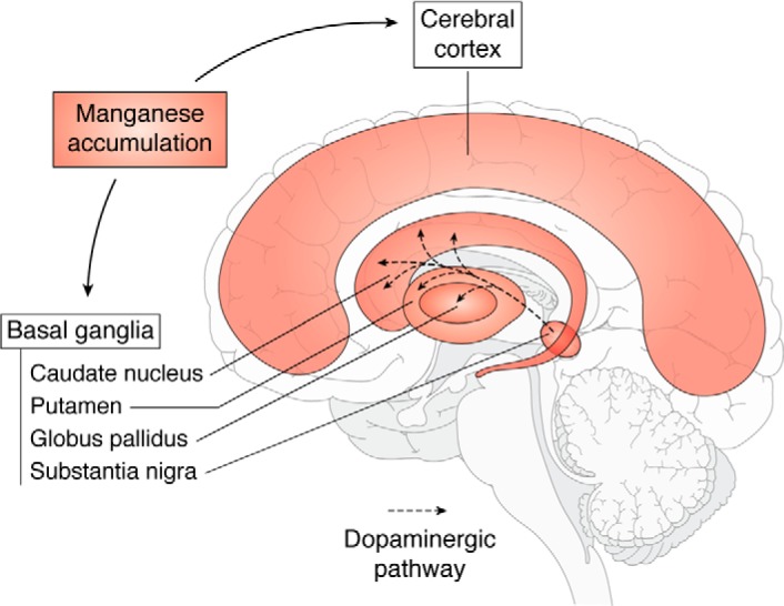
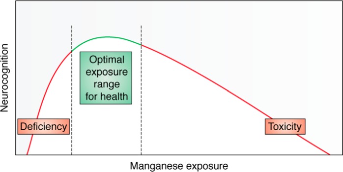
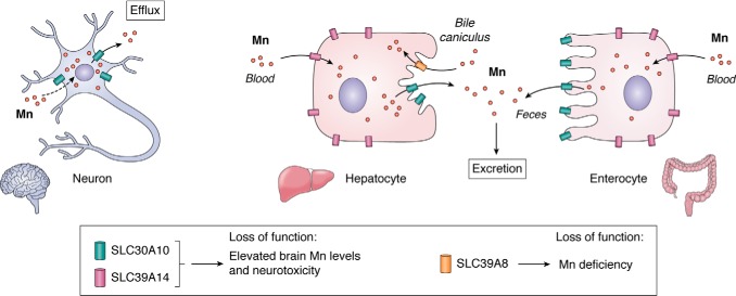
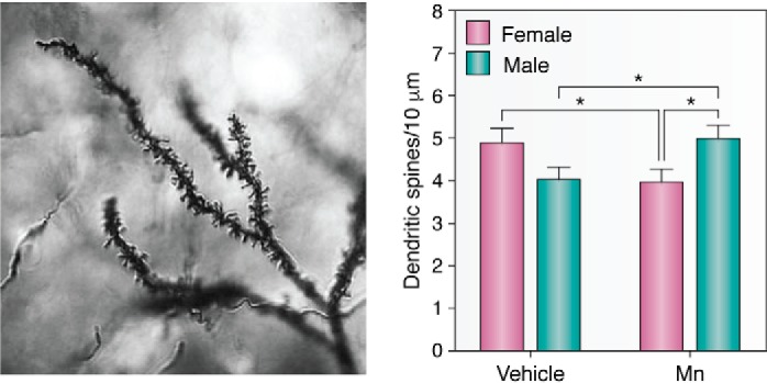
Manganese (Mn) is an essential micronutrient required for the normal development of many organs, including the brain. Although its roles as a cofactor in several enzymes and in maintaining optimal physiology are well-known, the overall biological functions of Mn are rather poorly understood. Alterations in body Mn status are associated with altered neuronal physiology and cognition in humans, and either overexposure or (more rarely) insufficiency can cause neurological dysfunction. The resultant balancing act can be viewed as a hormetic U-shaped relationship for biological Mn status and optimal brain health, with changes in the brain leading to physiological effects throughout the body and vice versa. This review discusses Mn homeostasis, biomarkers, molecular mechanisms of cellular transport, and neuropathological changes associated with disruptions of Mn homeostasis, especially in its excess, and identifies gaps in our understanding of the molecular and biochemical mechanisms underlying Mn homeostasis and neurotoxicity.

Keywords: brain; homeostasis; manganese; metal; metal homeostasis; neurodegeneration; neurodegenerative disease; neurodevelopment; neurotoxin; neurotransmitter; toxicology.


Conflict of interest statement

The authors declare that they have no conflicts of interest with the contents of this article

Figures

Figure 1.
Figure 1.
Schematic showing the sagittal section of human brain showing the brain regions where Mn predominantly accumulates (48, 200). Dopamine is a key neurotransmitter that is produced in the substantia nigra, and the dopaminergic neurotransmitters project to the basal ganglia region.
Figure 2.
Figure 2.
Mn exhibits hormetic dose response, which means an inverted “U-shaped” curve. Deficits in neurocognition are seen at both lower and higher doses, with maximum function being at the top of the inverted U-shaped curve. Adapted from Vollet et al. (6). This research was originally published in Current Environmental Health Reports. Vollet, K., Haynes, E. N., and Dietrich, K. N. Manganese exposure and cognition across the lifespan: contemporary review and argument for biphasic dose-response health effects. Curr. Environ. Health Rep. 2016; 3:392–404. © Springer.
Figure 3.
Figure 3.
Function of SLC30A10, SLC39A14, and SLC39A8. SLC30A10 and SLC39A14 synergistically mediate Mn excretion. SLC39A14 transports Mn from blood into hepatocytes and enterocytes for subsequent excretion by SLC30A10 into bile and feces. SLC30A10 also mediates Mn efflux from neuronal cells. In contrast, SLC39A8 reclaims Mn lost in bile. Elevated brain Mn levels and neurotoxicity evident on loss-of-function of SLC30A10 or SLC39A14 is a consequence of an inhibition of Mn excretion and, for SLC30A10, a block in Mn efflux from neurons. Loss-of-function of SLC39A8, in contrast, produces Mn deficiency.
Figure 4.
Figure 4.
Mn exposure alters synaptic spine density in striatal MSNs. Left, representative mouse MSN dendritic segment impregnated by the rapid Golgi method, reproduced here with permission (83). Dendritic branching is clearly visualized as well as dendritic spines. Image obtained in 2010 by Jennifer Madison, Ph.D. in the Aaron Bowman laboratory at Vanderbilt University. Right, total spine density was the only measure of neuron morphology to have a significant gender difference 3 weeks post-exposure. Mn-exposed (Mn) male mice had a higher total spine density than Mn-exposed female mice, whereas Mn exposure decreased spine density in female mice. More studies need to be done to establish the spine density levels between sexes under control conditions to consistently better-establish the differences caused by exposure to contaminants. Data are plotted as mean ± S.E. (error bars); *, p < 0.05, post hoc t test. Data were originally published in Ref. . This research was originally published in Neurotoxicology. Madison, J. L., Wegrzynowicz, M., Aschner, M., and Bowman, A. B. Gender and manganese exposure interactions on mouse striatal neuron morphology. Neurotoxicology. 2011; 32:896–906.

References

    1. Aschner J. L., and Aschner M. (2005) Nutritional aspects of manganese homeostasis. Mol. Aspects Med. 26, 353–362 10.1016/j.mam.2005.07.003 - DOI - PMC - PubMed
    1. Underwood M. J., More R. S., Thompson M. M., and Gershlick A. H. (1993) Reduction of vein graft intimal hyperplasia by ex vivo treatment with desferrioxamine manganese. J. Vasc. Res. 30, 239–240 10.1159/000158999 - DOI - PubMed
    1. Takeda A. (2003) Manganese action in brain function. Brain Res. Rev. 41, 79–87 10.1016/S0165-0173(02)00234-5 - DOI - PubMed
    1. Wedler F. C. (1993) Biological significance of manganese in mammalian systems. Prog. Med. Chem. 30, 89–133 10.1016/S0079-6468(08)70376-X - DOI - PubMed
    1. Keen C. L., Ensunsa J. L., and Clegg M. S. (2000) Manganese metabolism in animals and humans including the toxicity of manganese. Met. Ions Biol. Syst. 37, 89–121 - PubMed

Publication types

LinkOut - more resources