. 2020 Mar 18;12(535):eaav8075.
doi: 10.1126/scitranslmed.aav8075.

Design of anti-inflammatory heparan sulfate to protect against acetaminophen-induced acute liver failure

Affiliations

Design of anti-inflammatory heparan sulfate to protect against acetaminophen-induced acute liver failure

Katelyn Arnold et al. Sci Transl Med. .

Abstract

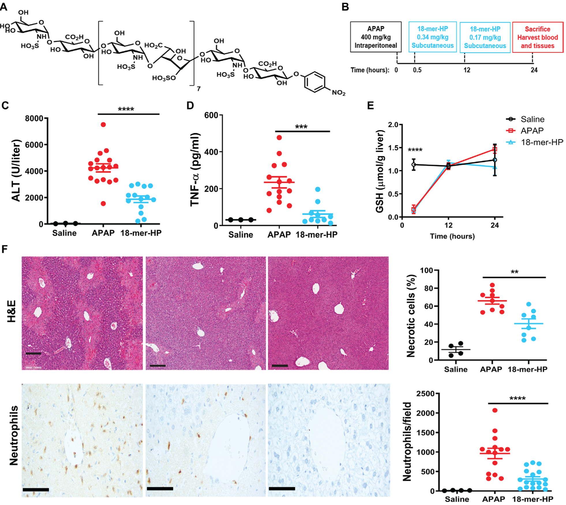
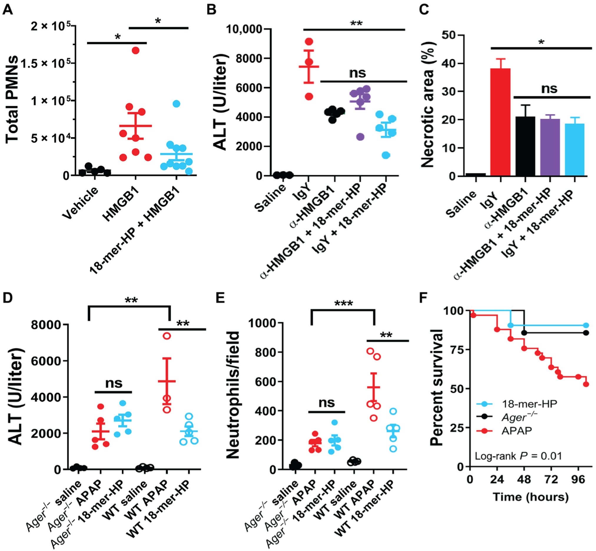
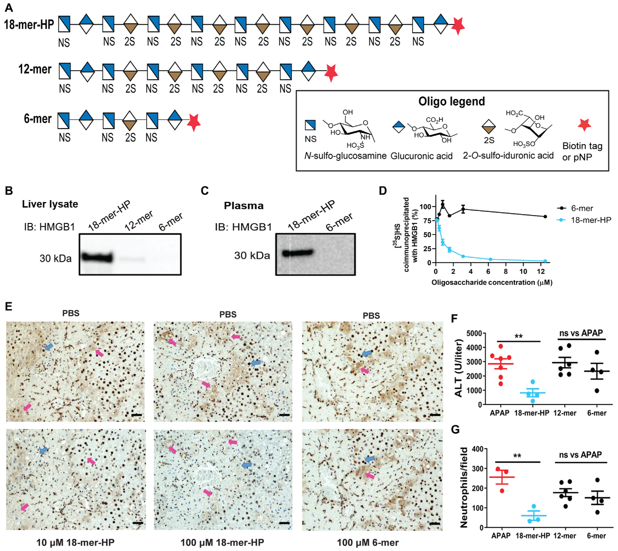
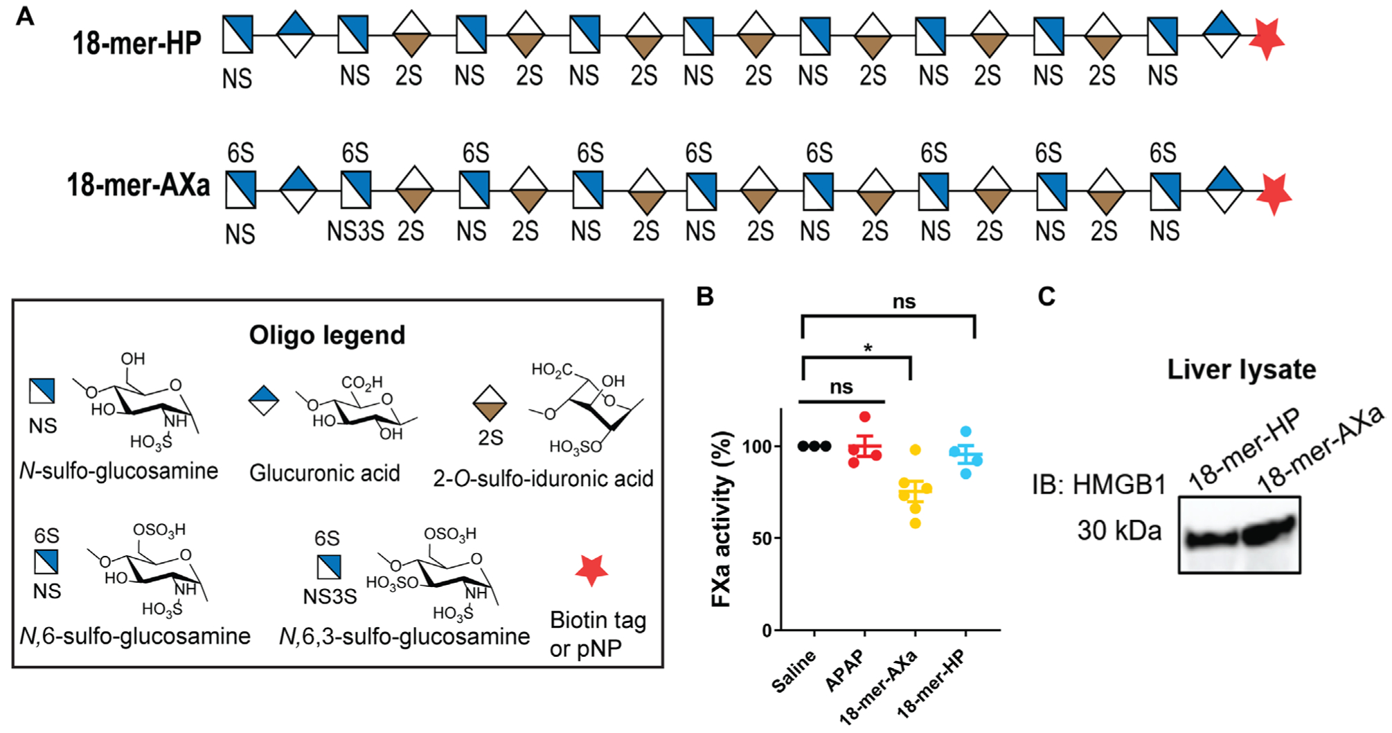
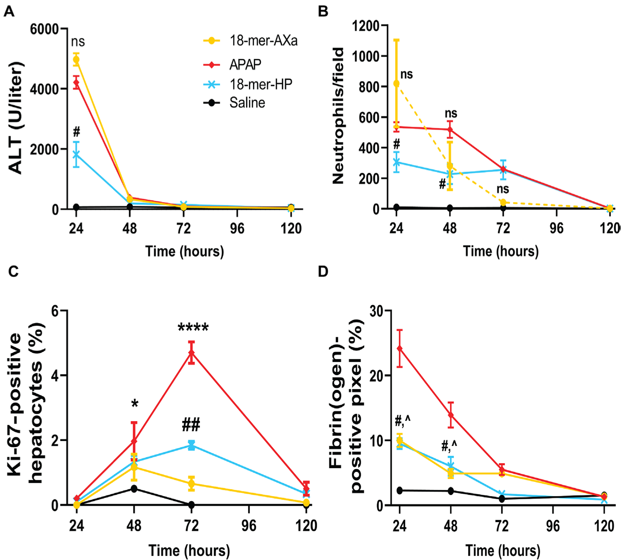
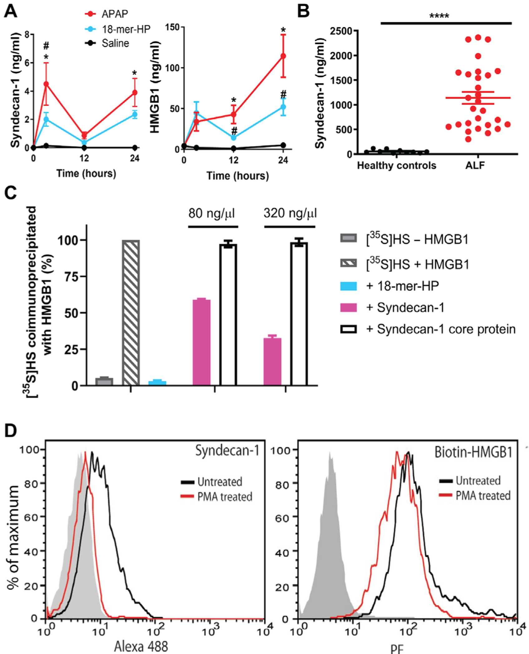
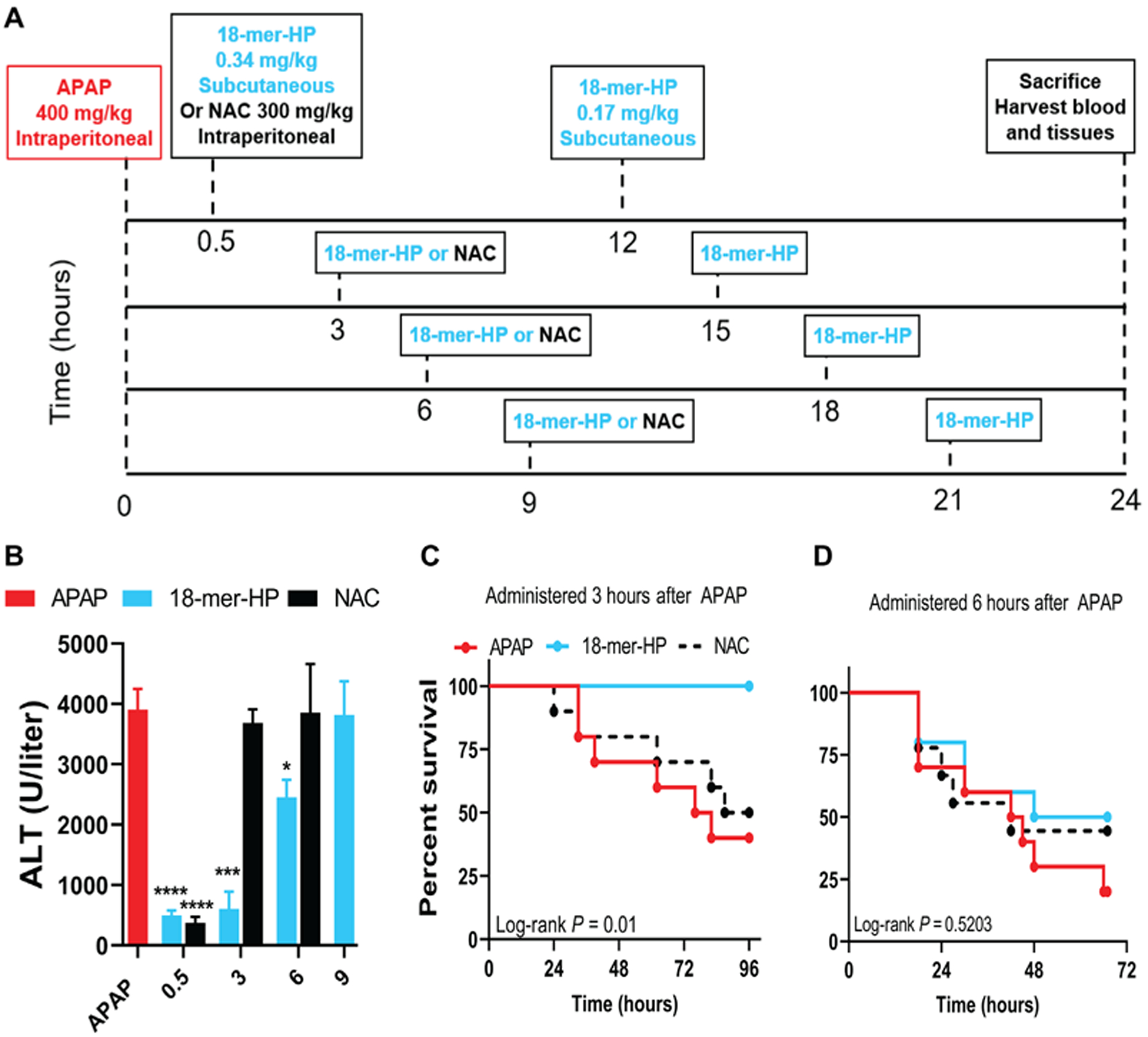
Acetaminophen/paracetamol (APAP) overdose is the leading cause of drug-induced acute liver failure (ALF) in the United States and Europe. The progression of the disease is attributed to sterile inflammation induced by the release of high mobility group box 1 (HMGB1) and the interaction with receptor for advanced glycation end products (RAGE). A specific, effective, and safe approach to neutralize the proinflammatory activity of HMGB1 is highly desirable. Here, we found that a heparan sulfate (HS) octadecasaccharide (18-mer-HP or hepatoprotective 18-mer) displays potent hepatoprotection by targeting the HMGB1/RAGE axis. Endogenous HS proteoglycan, syndecan-1, is shed in response to APAP overdose in mice and humans. Furthermore, purified syndecan-1, but not syndecan-1 core protein, binds to HMGB1, suggesting that HMGB1 binds to HS polysaccharide side chains of syndecan-1. Last, we compared the protection effect between 18-mer-HP and N-acetyl cysteine, which is the standard of care to treat APAP overdose. We demonstrated that 18-mer-HP administered 3 hours after a lethal dose of APAP is fully protective; however, the treatment of N-acetyl cysteine loses protection. Therefore, 18-mer-HP may offer a potential therapeutic advantage over N-acetyl cysteine for late-presenting patients. Synthetic HS provides a potential approach for the treatment of APAP-induced ALF.


Conflict of interest statement

Competing interests: J.L. and Y.X. are founders of Glycan Therapeutics, LLC. V.P. is an employee of Glycan Therapeutics, LLC and has the equity. K.A., Y.X., J.L., D.X., and R.P. are inventors for a U.S. patent (US 62/581,443). All other authors declare that they have no competing interests.

Figures

Fig. 1.
Fig. 1.. 18-mer-HP protects from liver injury after APAP overdose.
(A) Chemical structure of 18-mer-HP. (B) Murine model experimental design schematic including time, dose, and administration route of APAP and 18-mer-HP. (C) Plasma ALT concentrations from mice treated with saline, APAP alone, and APAP + 18-mer-HP. (D) Plas- ma TNF-α concentrations are decreased in the 18-mer-HP—treated group compared to APAP alone. (E) 18-mer-HP— and APAP-treated mice both have decreased GSH concentrations immediately after APAP overdose compared to the saline control mice. 18-mer-HP does not affect APAP’s metabolism to NAPQI. Saline n= 3, APAP n = 6, 18-mer-HP n = 6. (F) Representative images of hematoxylin and eosin (H&E) staining of formalin-fixed paraffin-embedded liver tissues and neutrophil immunohistochemistry (IHC). Quantitation of H&E-and IHC neutrophil—stained liver tissues from 100× fields are shown on the right. Scale bars, 200 μm. Data represent means ± SEM (C, D, E, and F). **P < 0.01, ***P < 0.001, ****P < 0.0001 by one-way ANOVA followed by Tukey’s post hoc test.
Fig. 2.
Fig. 2.. 18-mer-HP targets HMGB1/RAGE axis to decrease inflammation and liver injury.
(A) 18-mer-HP prevents neutrophil infiltration induced by HMGB1 in the air pouch model. Number of polymorphonuclear neutrophils (PMNs) migrating to the air pouch was determined by flow cytometry. (B) HMGB1-neutralizing antibody (α-HMGB1; 4 mg/kg) decreases ALT at 24 hours after APAP. 18-mer-HP (0.34 mg/kg) in combination with α-HMGB1 (4 mg/kg) shows no further protection compared to α-HMGB1 or 18-mer-HP alone. (C) α-HMGB1 decreases liver necrotic area at 24 hours after APAP as determined by H&E staining. 18-mer-HP in the presence of α-HMGB1 shows no further protection compared to α-HMGB1 or 18-mer-HP alone. (D and E) 18-mer-HP lacks anti-inflammatory effects in Ager−/− mice as demonstrated by ALT (D) and neutrophil infiltration (E). (F) 18-mer-HP improves survival after a lethal overdose of APAP (600 mg/kg) comparable to the protection seen in Ager−/− mice (APAP, n = 33; 18-mer, n = 21; Ager−/− mice, n = 7). *P < 0.05, **P < 0.01, and ***P < 0.001 by one-way ANOVA followed by Dunnett’s post hoc test (A) and by one-way ANOVA followed by Tukey’s post hoc test (B to E). ns, not significant.
Fig. 3.
Fig. 3.. Size of HS oligosaccharide is important for HS/HMGB1 interaction and hepatoprotection.
(A) Symbolic structures of HS oligosaccharides. (B) Western analysis of HMGB1 pulldown from mouse liver lysate by 18-mer-HP, 12-mer, and 6-mer biotinylated oligosaccharides using avidin-Sepharose. Full image is presented in fig. S11A. IB, immunoblotting. (C) Western analysis of plasma circulating HMGB1 pulldown from APAP overdose mouse plasma by 18-mer-HP and 6-mer biotinylated oligosaccharides. A higher—molecular weight protein band was also detected, possibly because of the cross-link with other plasma proteins from the action of transglutaminase-2 (fig. S11B) (41). We detected the presence of trasglutaminase-2, which is known to bind to heparin (42), in the plasma sample (fig. S11D). (D) HMGB1 binding competition assay using 35S-labeled HS with oligosaccharides as competitor ligands. (E) Liver sections from mice treated with APAP (400 mg/kg) and euthanized at 24 hours, stained with anti-HMGB1 antibody (top). Blue arrows indicate HMGB1 staining in the nucleus, and pink arrows indicate diffuse HMGB1 staining outside the cell. Incubation with 18-mer-HP before anti-HMGB1 results in decreased staining (bottom) compared to incubation with PBS (top). 6-mer has no effect on anti-HMGB1 staining. (F and G) 18-mer-HP decreases ALT (F) and neutrophil infiltration (G) after APAP overdose, whereas 12-and 6-mer do not. Data represent means ± SEM. **P < 0.01 by one-way ANOVA followed by Dunnett’s posttest.
Fig. 4.
Fig. 4.. 18-mer-AXa binds to HMGB1 and displays anti-FXa activity.
(A) Symbolic structure of 18-mer-HP and 18-mer-AXa. (B) Factor Xa (FXa) activity measured in plasma from saline-treated mice and APAP overdose mice treated with saline (APAP control group), 18-mer-AXa, or 18-mer-HP. Plasma was collected at the time of sacrifice, which was 12 hours after the second oligosaccharide injections and 24 hours after APAP overdose. (C) Western analysis of HMGB1 pulldown from liver lysate by 18-mer-HP and 18-mer-AXa biotinylated oligosaccharides using avidin-Sepharose. Full image is presented in fig. S15. Data represent means ± SEM. *P < 0.05 by one-way ANOVA followed my Dunnett’s posttest (B).
Fig. 5.
Fig. 5.. 18-mer-AXa impairs liver regeneration after APAP overdose.
18-mer-AXa or 18-mer-HP was administered every 12 hours for 120 hours after APAP overdose. (A) ALT is increased after APAP overdose. At 24 hours, 18-mer-HP decreased ALT compared to APAP (#P = 0.0121), whereas there was no difference between 18-mer-AXa and APAP. (B) Neutrophils accumulate in the liver of APAP and 18-mer-AXa—treated mice up to 48 hours after APAP overdose. At 24 and 48 hours, 18-mer-HP decreased neutrophil infiltration (#P = 0.0355 and #P = 0.0301 at 24 and 48 hours, respectively). (C) APAP overdose mice began to increase proliferating hepatocytes at 48 hours (*P = 0.0280) and peaked at 72 hours (****P < 0.0001) before returning to baseline. 18-mer-HP treatment resulted in mild liver regeneration with an increase at 72 hours compared to saline (##P = 0.0045). In contrast, 18-mer-AXa treatment did not result in significant changes in Ki-672014positive hepatocytes at any time point after APAP overdose. (D) At 24 and 48 hours, APAP overdose resulted in elevated fibrin(ogen) deposits in the liver. 18-mer-HP and 18-mer-AXa have decreased the concentration of firbrin(ogen) at 24 hours (#P = 0.01 and ^P = 0.02, respectively) and 48 hours (#P = 0.05 and ^P = 0.02). n = 3 for all groups at all time points. Data represent means ± SEM. One-way ANOVA was followed by Dunnett’s posttest.
Fig. 6.
Fig. 6.. Endogenous syndecan-1 is shed in response to liver injury after APAP overdose and binds to HMGB1.
(A) Mouse plasma syndecan-1 and HMGB1 concentrations after APAP overdose with or without 18-mer-HP treatment or only saline as a control. At 3 hours after APAP overdose, plasma syndecan-1 concentrations were elevated in APAP mice compared to saline (*P = 0.0152) and 18-mer-—treated mice (#P = 0.0444). Plasma syndecan-1 concentrations decreased at 12 hours and then rose again at 24 hours in the APAP group compared to saline (*P = 0.0215). Plasma HMGB1 concentrations were elevated in the APAP group at 12 (*P = 0.0354 versus saline, #P = 0.0354 versus 18-mer-HP) and 24 hours (*P = 0.0169 versus saline, #P = 0.0306 versus 18-mer); n = 2 to 3 for saline, n = 5 to 6 for APAP, and n = 5 to 7 for 18-mer-HP at all time points. (B) Plasma syndecan-1 in healthy individuals and in patients with APAP-ALF. (C) HMGB1 binding competition assay using [35S]HS with competitor ligands 18-mer-HP, syndecan-1, and syndecan-1 core protein. (D) Syndecan-1 shedding reduces the binding of HMGB1 to Hep3B cells. Left: PMA treatment induces syndecan-1 shedding. Right: Binding of HMGB1 to Hep3B cells before and after syndecan-1 shedding. The shaded histogram is from cells stained with isotype control rat IgG and secondary antibody (left) or with streptavidin—R-phycoerythrin (PE) only (right). Data represent means ± SEM. One-way ANOVA was followed by Sidak’s post hoc test (A) and by unpaired Student’s t test (B).
Fig. 7.
Fig. 7.. Delayed treatment with 18-mer-HP after APAP overdose remains effective.
(A) Murine model experimental design schematic including time, dose, and route of administration of APAP, NAC, and 18-mer-HP. All groups received APAP at time 0 and were euthanized at 24 hours. The dosing remained the same in all 18-mer-HP—treated groups in that the first injection was 18-mer-HP (0.34 mg/kg), and the second injection was 18-mer-HP (0.17 mg/kg). NAC (300 mg/kg) was injected once in the indicated groups because of the known mechanism of action and was not used in the 9-hour delay group because no protection was seen at 3 and 6 hours. (B)18-mer-HP decreased ALT when given at 0.5, 3, and 6 hours after APAP (APAP, n = 24; 18-mer, n = 5 to 7; NAC, n = 6 to 9). Data represent means ± SEM. *P < 0.05, ***P < 0.001, and ****P < 0.0001 by two-way ANOVA followed by Tukey’s post hoc test. (C) 18-mer-HP administered 3 hours after APAP (600 mg/kg) increased the survival rate compared to both APAP and NAC given at 3 hours after APAP. (n = 10 for all groups). (D) 18-mer-HP administered 6 hours after APAP (600 mg/kg) had no protective effect (18-mer-HP, n = 10; NAC, n = 9; APAP, n = 10).

References

    1. Zitvogel L, Kepp O, Kroemer G, Decoding cell death signals in inflammation and immunity. Cell 140, 798–804 (2010). - PubMed
    1. Chen GY, Nuñez G, Sterile inflammation: Sensing and reacting to damage. Nat. Immunol 10, 826–837 (2010). - PMC - PubMed
    1. Bianchi ME, Crippa MP, Manfredi AA, Mezzapelle R, Querini PR, Venereau E, High-mobility group box 1 protein orchestrates responses to tissue damage via inflammation, innate and adaptive immunity, and tissue repair. Immunol. Rev 280, 74–82 (2017). - PubMed
    1. Huebener P, Pradere J-P, Hernandez C, Gwak G-Y, Caviglia JM, Mu X, Loike JD, Schwabe RF, The HMGB1/RAGE axis triggers neutrophil-mediated injury amplification following necrosis. J. Clin. Invest 125, 539–550 (2015). - PMC - PubMed
    1. Heard KJ, Acetylcystein for acetaminophen poisoning. N. Engl. J. Med 359, 285–292 (2008). - PMC - PubMed

Publication types