. 2020 May 18;94(11):e02143-19.
doi: 10.1128/JVI.02143-19. Print 2020 May 18.

Epidermal Growth Factor Receptor and Abl2 Kinase Regulate Distinct Steps of Human Papillomavirus 16 Endocytosis

Affiliations

Epidermal Growth Factor Receptor and Abl2 Kinase Regulate Distinct Steps of Human Papillomavirus 16 Endocytosis

Carina Bannach et al. J Virol. .

Abstract

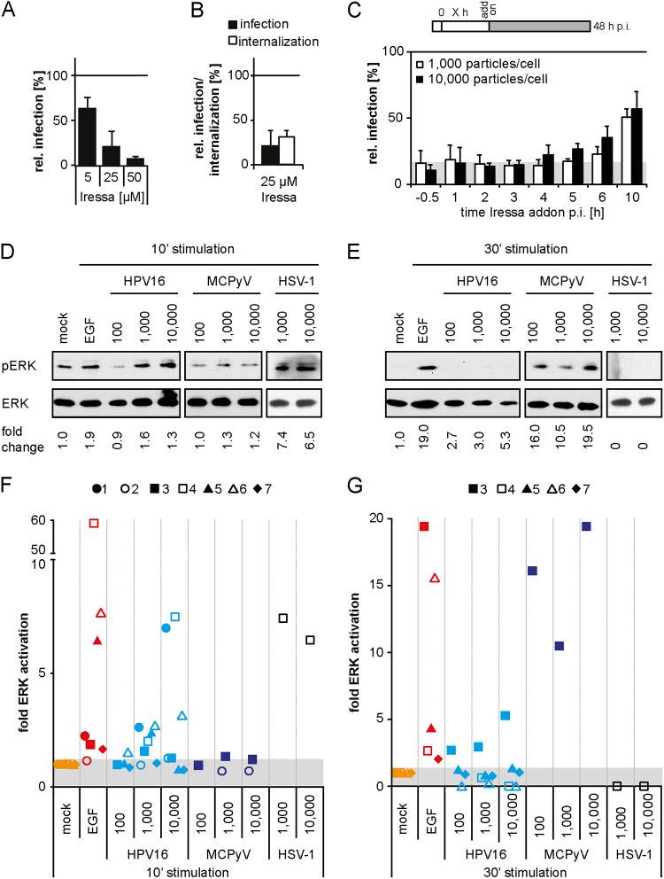
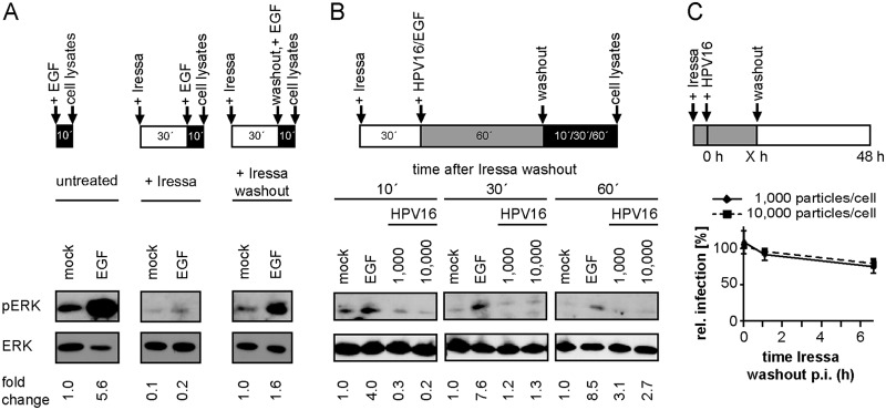
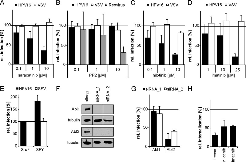
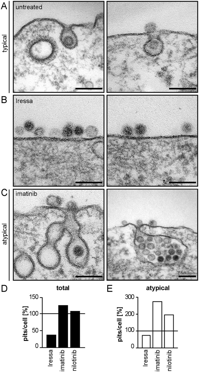
Human papillomavirus 16 (HPV16), the leading cause of cervical cancer, exploits a novel endocytic pathway during host cell entry. This mechanism shares many requirements with macropinocytosis but differs in the mode of vesicle formation. Previous work indicated a role of the epidermal growth factor receptor (EGFR) in HPV16 endocytosis. However, the functional outcome of EGFR signaling and its downstream targets during HPV16 uptake are not well characterized. Here, we analyzed the functional importance of signal transduction via EGFR and its downstream effectors for endocytosis of HPV16. Our findings indicate two phases of EGFR signaling as follows: a-likely dispensable-transient activation with or shortly after cell binding and signaling required throughout the process of asynchronous internalization of HPV16. Interestingly, EGFR inhibition interfered with virus internalization and strongly reduced the number of endocytic pits, suggesting a role for EGFR signaling in the induction of HPV16 endocytosis. Moreover, we identified the Src-related kinase Abl2 as a novel regulator of virus uptake. Inhibition of Abl2 resulted in an accumulation of misshaped endocytic pits, indicating Abl2's importance for endocytic vesicle maturation. Since Abl2 rather than Src, a regulator of membrane ruffling during macropinocytosis, mediated downstream signaling of EGFR, we propose that the selective effector targeting downstream of EGFR determines whether HPV16 endocytosis or macropinocytosis is induced.IMPORTANCE Human papillomaviruses are small, nonenveloped DNA viruses that infect skin and mucosa. The so-called high-risk HPVs (e.g., HPV16, HPV18, HPV31) have transforming potential and are associated with various anogenital and oropharyngeal tumors. These viruses enter host cells by a novel endocytic pathway with unknown cellular function. To date, it is unclear how endocytic vesicle formation occurs mechanistically. Here, we addressed the role of epidermal growth factor receptor signaling, which has previously been implicated in HPV16 endocytosis and identified the kinase Abl2 as a novel regulator of virus uptake. Since other viruses, such as influenza A virus and lymphocytic choriomeningitis virus, possibly make use of related mechanisms, our findings shed light on fundamental strategies of virus entry and may in turn help to develop new host cell-targeted antiviral strategies.

Keywords: HPV; endocytosis; papillomavirus; signaling; virus entry.


Figures

FIG 1
FIG 1
EGFR signaling is essential for HPV16 endocytosis. (A) HeLa cells were pretreated with the EGFR inhibitor Iressa at the indicated concentrations for 30 min before they were infected with HPV16 PsVs. To reduce cytotoxicity, Iressa was exchanged for NH4Cl at 12 h p.i. Infection was scored by flow cytometry at 48 h p.i. Infection values were normalized to solvent controls and are depicted as the mean of three independent experiments ± standard deviation (SD). (B) HeLa cells were preincubated with 25 μM Iressa for 30 min and subsequently infected with HPV16 PsVs. At 12 h p.i., cells were either treated as in panel A (infection), or extracellular viruses were inactivated by treatment with a high pH buffer (infectious internalization). Infection was scored by flow cytometry and normalized to inhibitor reversibility. Depicted is the mean ± SD (n = 3). (C) HaCaT cells were infected with HPV16 PsVs at different MOIs (in particles/cell). Iressa (25 μM) was added at the indicated time points p.i., and infection was continued in the presence of the inhibitor. Infection was analyzed by flow cytometry at 48 h p.i., and infection values were normalized to solvent-treated controls. The area in gray indicates baseline infection levels upon Iressa treatment. Depicted is the mean of three independent experiments ± SD. (D, E) Serum-starved HaCaT cells were either incubated with PBS (mock), EGF, or different MOIs of HPV16, MCPyV, or HSV-1 for 10 (D) or 30 min (E). Cell lysates were first blotted against pERK and thereafter against ERK. (F, G) pERK levels were quantified and normalized to the respective mock samples and ERK (loading control). Depicted are the values from each experiment as fold activation of the background of n = 7 independent experiments (for HPV16). The individual experiments are indicated by individual geometric data points, whereas the data points are grouped by color according to the stimulus.
FIG 2
FIG 2
Early ERK activation is dispensable for HPV16 infection. (A) Serum-starved HaCaT cells were either directly incubated with PBS (mock) or EGF for 10 min (left) or pretreated with 25 μM Iressa for 30 min and subsequently stimulated with EGF for 10 min (middle). To test for the reversibility of Iressa treatment, cells were preincubated with 25 μM Iressa for 30 min and stimulated with EGF after washout of the inhibitor for 10 min (right). Cell lysates were first blotted against pERK and thereafter against ERK. pERK levels were quantified and normalized to the mock of the untreated condition and ERK (loading control). (B) Serum-starved HaCaT cells were incubated with 25 μM Iressa for 30 min prior to binding of HPV16 PsVs or EGF for 1 h (mock = PBS). Subsequently, Iressa was washed out, and incubation was continued in the presence of HPV16 or EGF for the indicated time points. Lysates were processed as in panel A. pERK levels were quantified and normalized to the respective mock samples and ERK (loading control). (C) HaCaT cells were preincubated with 25 μM Iressa for 30 min and infected with HPV16 PsVs at different MOIs. At the indicated time points p.i., Iressa was washed out and infection was continued in the absence of the inhibitor. Cells were scored for infection 48 h p.i. by flow cytometry, and infection was normalized to the untreated control. Depicted is the mean of three experiments ± SD.
FIG 3
FIG 3
HPV16 internalization depends on the tyrosine kinase Abl2. (A to D) HeLa cells were incubated with the small-compound inhibitors saracatinib (Src, Abl) (A), PP2 (Src) (B), nilotinib (Abl) (C), or imatinib (Abl) (D) at the indicated concentrations for 30 min. The cells were then either infected with HPV16 PsVs (black bars) for 48 h, with VSV-GFP for 5 h (white bars), or with reovirus for 16 h (gray bars). In the case of HPV16 infection, the inhibitor was exchanged by NH4Cl at 12 h p.i. to reduce cytotoxicity. Infection was scored by flow cytometry and normalized to solvent-treated control cells. (E) Mouse embryonic fibroblasts with a knockout of Src, Fyn, and Yes (SFY) as well as the littermate control cell line (Src+/+) were infected with HPV16 PsVs (black bars) for 48 h or SFV (white bars) for 5 h. Infection was scored by flow cytometry. (F) HeLa Kyoto cells were lysed 48 h after transfection with indicated siRNAs. Lysates were immunoblotted with Abl1- or Abl2-specific antibodies. (G) HeLa Kyoto cells were transfected with siRNAs against Abl1 and 2. At 48 h posttransfection, cells were infected with HPV16 PsVs for 48 h. Infection was analyzed by microscopy and automated cell scoring, and values were normalized to the control siRNA. (H) HeLa cells were preincubated with 25 μM Iressa, 25 μM imatinib, or 10 μM nilotinib for 30 min and subsequently infected with HPV16 PsVs. Cells were treated with a high-pH buffer at 12 h p.i. Infection was continued in the absence of the inhibitor. Infectious internalization was scored 48 h p.i. by flow cytometry and normalized to solvent-treated controls. All infection values are depicted as the mean of three independent experiments ± SD.
FIG 4
FIG 4
EGFR and Abl2 regulate pit induction and dilation/maturation, respectively. (A to C) HeLa ATCC cells were treated with 25 μM Iressa (B) or 25 μM imatinib (C) for 30 min or were left untreated (A) and subsequently infected with HPV16 PsVs. Cells were fixed at 6 h p.i., and endocytic pits were analyzed by ultrathin section TEM. (D) Virus-containing plasma membrane invaginations were counted and normalized to untreated control cells. At least 10 cells were counted per experiment in several independent experiments, excluding nilotinib where only one experiment was done. (E) The amount of virus-filled atypical pits per cell (e.g., in panel C) was determined and normalized to the amount of atypical pits in control cells.

References

    1. Zur Hausen H. 2002. Papillomaviruses and cancer: from basic studies to clinical application. Nat Rev Cancer 2:342–350. doi: 10.1038/nrc798. - DOI - PubMed
    1. Doorbar J, Quint W, Banks L, Bravo IG, Stoler M, Broker TR, Stanley MA. 2012. The biology and life-cycle of human papillomaviruses. Vaccine 30(Suppl):F55–F70. doi: 10.1016/j.vaccine.2012.06.083. - DOI - PubMed
    1. Raff AB, Woodham AW, Raff LM, Skeate JG, Yan L, Da Silva DM, Schelhaas M, Kast WM. 2013. The evolving field of human papillomavirus receptor research: a review of binding and entry. J Virol 87:6062–6072. doi: 10.1128/JVI.00330-13. - DOI - PMC - PubMed
    1. Mikuličić S, Florin L. 2019. The endocytic trafficking pathway of oncogenic papillomaviruses. Papillomavirus Res 7:135–137. doi: 10.1016/j.pvr.2019.03.004. - DOI - PMC - PubMed
    1. Schmitt A, Zeltner R, Iftner T, Rochat A, Barrandon Y, Borenstein L, Wettstein FO. 1996. The primary target cells of the high-risk cottontail rabbit papillomavirus colocalize with hair follicle stem cells. J Virol 70:1912–1922. doi: 10.1128/JVI.70.3.1912-1922.1996. - DOI - PMC - PubMed

Publication types