. 2020 Mar 19;15(1):62.
doi: 10.1186/s11671-020-3295-1.

Novel Chemo-Photothermal Therapy in Breast Cancer Using Methotrexate-Loaded Folic Acid Conjugated Au@SiO2 Nanoparticles

Affiliations

Novel Chemo-Photothermal Therapy in Breast Cancer Using Methotrexate-Loaded Folic Acid Conjugated Au@SiO2 Nanoparticles

Reza Agabeigi et al. Nanoscale Res Lett. .

Abstract

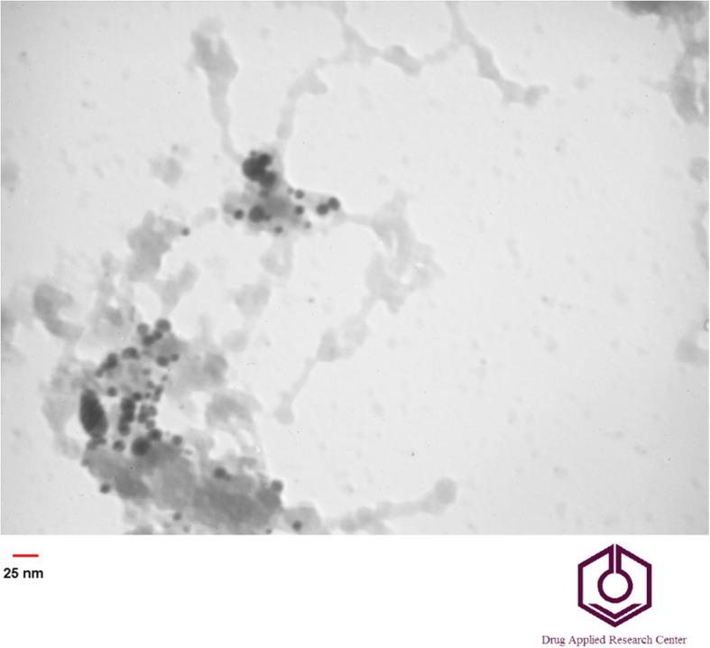
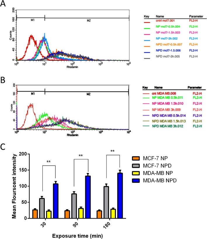
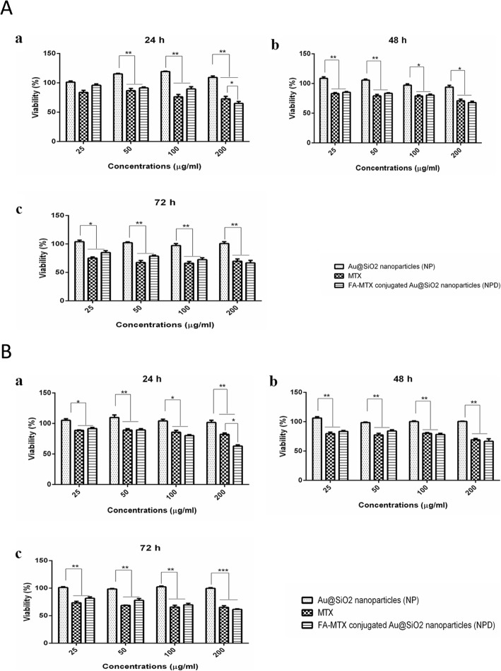
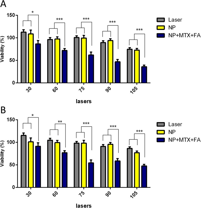
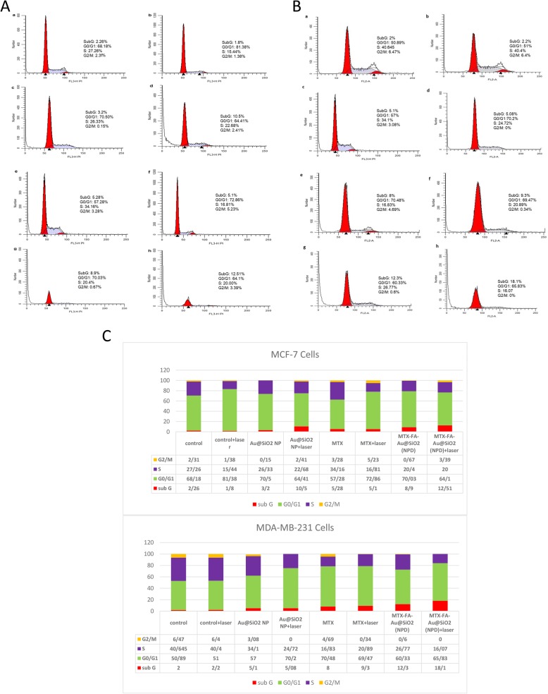
Low level laser therapy (LLLT) is known as a safe type of phototherapy to target tumor tissue/cells. Besides, using targeted nanoparticles increases the successfulness of cancer therapy. This study was designed for investigating the combined effect of folate (FA)/Methotrexate (MTX) loaded silica coated gold (Au@SiO2) nanoparticles (NPs) and LLLT on the fight against breast cancer.NPs were synthesized and characterized using FTIR, TEM and DLS-Zeta. The NPs had spherical morphology with mean diameter of around 25 nm and positive charge (+13.3 mV) while after conjugation with FA and MTX their net charge reduced to around -19.7 mV.Our findings in cell uptake studies clearly showed enhanced cellular uptake of NPs after FA and MTX loaded NPs in both breast cancer cell lines especially on MDA-MB-231 due to high expression of folate receptors. The results indicated that LLLT had a proliferative effect on both breast cancer cell lines but in the presence of engineered breast cancer targeted nanoparticle, the efficacy of combination chemo-photothermal therapy was significantly increased using MTT assay (p<0.05), DAPI staining, and cell cycle findings. The highest apoptotic effect on breast cancer cell lines was observed in the cells exposed to a combination of MTX-FA loaded Au@SiO2 NP and LLLT proved by DAPI staining and cell cycle(by increasing the cell arrest in subG0/G1). Taken together a combination of chemotherapy and LLLT improves the potential of breast cancer therapy with minimum side effects.

Keywords: Au@SiO2 nanoparticles; Breast cancer; Folic acid; Low level laser therapy; Methotrexate.


Conflict of interest statement

The authors declare that they have no competing interests.

Figures

Fig. 1
Fig. 1
The stepwise synthetic scheme for the preparation of folate and methotrexate loaded biocompatible Au@SiO2 NPs nanoparticles
Fig. 2
Fig. 2
a) FTIR spectra of Au@SiO2 nanoparticles, b) size distribution of FA-MTX conjugated Au@SiO2 NPs measured by dynamic light scattering (DLS) c) The zeta potential of Au@SiO2 and FA-MTX conjugated Au@SiO2 NPs measured by dynamic light scattering (DLS) at pH=7.4 and T=25 °C, d) Chromatogram of unloaded MTX and FA separated from FA-MTX conjugated Au@SiO2 NP measured simultaneously by HPLC method
Fig. 3
Fig. 3
TEM image of Au@SiO2 nanoparticles
Fig. 4
Fig. 4
Quantitative cell uptake assay of rhodamine B-labelled Au@SiO2 nanoparticles (NP) or rhodamine B-labelled MTX-FA loaded Au@SiO2 nanoparticles (NPD) in MCF-7 (a) and MDA-MB-231(b) cell lines for exposure durations of 0.5 h, 1.5 h and 3h obtained by flow cytometry. Non-treated cells of both cell lines were employed as negative control. c Comparison of mean fluorescent intensity of rhodamine B-labelled Au@SiO2 nanoparticles (NP) or rhodamine B-labelled MTX-FA loaded Au@SiO2 nanoparticles (NPD) for exposure durations of 0.5 h, 1.5 h and 3h obtained by flow cytometry
Fig. 5
Fig. 5
A Qualitative cell uptake assay using Rhodamine B-labelled Au@SiO2 nanoparticles (NP) in MCF7 with exposure durations of 30 (a), 90 (b) and 180 (c) min or rhodamine B-labelled MTX-FA loaded Au@SiO2 nanoparticles (NPD) with exposure durations of 30 (d), 90 (e) and 180 (f) min and (B) Qualitative cell uptake assay using Rhodamine B-labelled Au@SiO2 nanoparticles (NP) in MDA-MB-231 with exposure durations of 30 (a), 90 (b) and 180 (c) min or rhodamine B-labelled MTX-FA loaded Au@SiO2 nanoparticles (NPD) with exposure durations of 30 (d), 90 (e) and 180 (f) min captured by florescent microscopy
Fig. 6
Fig. 6
a MCF-7 and (b) MDA-MB-231 cells growth inhibition rates after treatment with different concentration of NP, MTX and FA-MTX loaded Au@SiO2 nanoparticles (NPD) after exposure time of 24, 48 and 72h
Fig. 7
Fig. 7
A comparison of cell growth inhibition rates exposed to different laser powers (30, 60, 75, 90 and 105 J/cm2) for treatment groups of laser alone, laser + Au@SiO2 nanoparticles and laser + MTX-FA loaded Au@SiO2 nanoparticles directed for two cell line MCF-7 (a) and MDA-MB-231(b) with subsequent checking after 24h
Fig. 8
Fig. 8
Apoptosis assay using DAPI staining for MCF-7 or MDA-MB-231 cells, images captured using an inverted microscope. The untreated cells as the negative control (a), cells treated with laser (75 J/cm2) as positive control (b) cells treated with Au@SiO2 nanoparticles (NP (c), cells treated with laser (75 J/cm2) and Au@SiO2 nanoparticles (NP) (d), cells treated with MTX without laser irradiation (e), cells treated with MTX and laser irradiation (f), cells treated with MTX-FA-loaded Au@SiO2 nanoparticles (NPD) without laser exposure (g), and cells treated with MTX-FA-loaded Au@SiO2 nanoparticles (NPD) with Laser exposure (h)
Fig. 9
Fig. 9
Cell cycle distributions investigated for MCF-7 (A) or MDA-MB-231 (B) cells. The untreated cells as negative control (a), cells treated with laser (75 J/cm2) as positive control (b) cells treated with Au@SiO2 nanoparticles (NP) (c), cells treated with laser (75 J/cm2) and Au@SiO2 nanoparticles (NP) (d), cells treated with MTX without laser irradiation (e), cells treated with MTX and laser irradiation (f), cells treated with MTX-FA-loaded Au@SiO2 nanoparticles (NPD) without laser exposure (g), and cells treated with MTX-FA-loaded Au@SiO2 nanoparticles (NPD) with Laser exposure (h), C) Quantitative results of cell cycle arrest and its distribution

References

    1. Ferlay J, Soerjomataram I, Dikshit R, Eser S, Mathers C, Rebelo M, et al. Cancer incidence and mortality worldwide: sources, methods and major patterns in GLOBOCAN 2012. Int J Cancer. 2015;136(5):E359–EE86. doi: 10.1002/ijc.29210. - DOI - PubMed
    1. Aghaee F, Pirayesh Islamian J, Baradaran B. Enhanced radiosensitivity and chemosensitivity of breast cancer cells by 2-deoxy-d-glucose in combination therapy. J Breast Cancer. 2012;15(2):141–147. doi: 10.4048/jbc.2012.15.2.141. - DOI - PMC - PubMed
    1. Dias MF, Sousa E, Cabrita S, Patrício J, Oliveira CF. Chemoprevention of DMBA-induced mammary tumors in rats by a combined regimen of alpha-tocopherol, selenium, and ascorbic acid. Breast J. 2000;6(1):14–19. doi: 10.1046/j.1524-4741.2000.98071.x. - DOI - PubMed
    1. Parkin DM, Bray F, Ferlay J, Pisani P. Global cancer statistics, 2002. CA Cancer J Clin. 2005;55(2):74–108. doi: 10.3322/canjclin.55.2.74. - DOI - PubMed
    1. Movahedi M, Haghighat S, Khayamzadeh M, Moradi A, Ghanbari-Motlagh A, Mirzaei H, et al. Survival rate of breast cancer based on geographical variation in Iran, a national study. Iran Red Crescent Med J. 2012;14(12):798. doi: 10.5812/ircmj.3631. - DOI - PMC - PubMed

LinkOut - more resources