. 2020 Jul;154(1):77-95.
doi: 10.1007/s00418-020-01866-w. Epub 2020 Mar 18.

Genes regulating hormone stimulus and response to protein signaling revealed differential expression pattern during porcine oocyte in vitro maturation, confirmed by lipid concentration

Affiliations

Genes regulating hormone stimulus and response to protein signaling revealed differential expression pattern during porcine oocyte in vitro maturation, confirmed by lipid concentration

Błażej Chermuła et al. Histochem Cell Biol. 2020 Jul.

Abstract

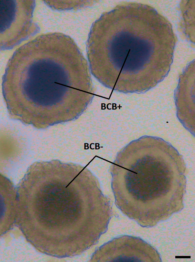
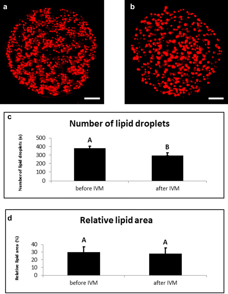
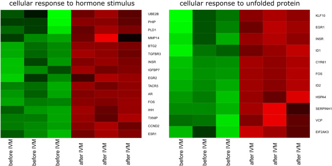
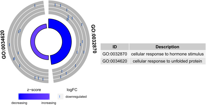
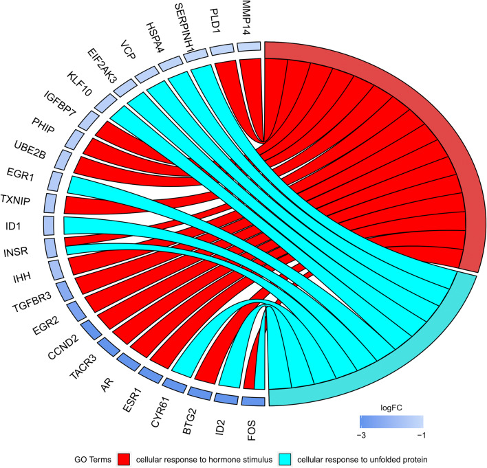
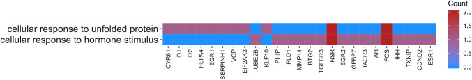
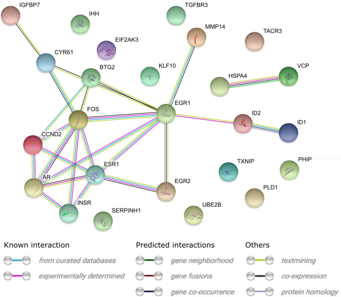
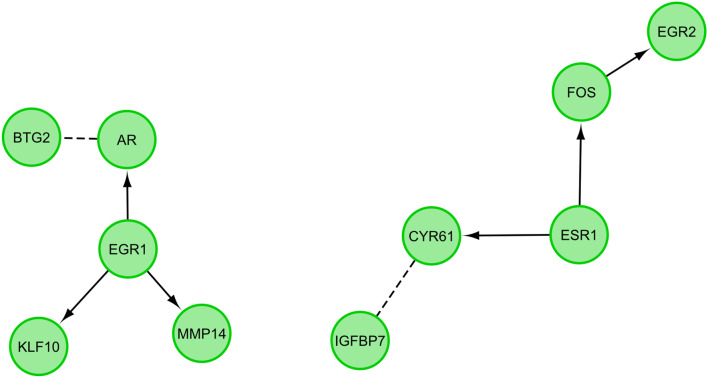
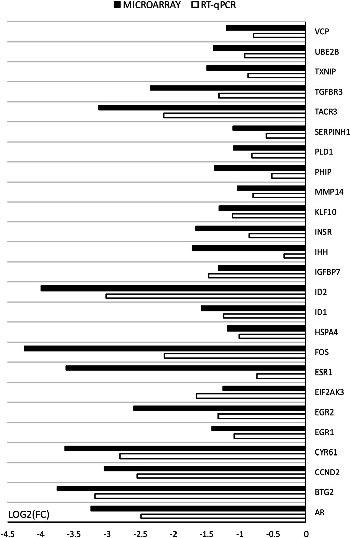
Genes influencing oocyte maturation may be valuable for predicting their developmental potential, as well as discerning the mechanistic pathways regulating oocyte development. In the presented research microarray gene expression analysis of immature and in vitro matured porcine oocytes was performed. Two groups of oocytes were compared in the study: before (3 × n = 50) and after in vitro maturation (3 × n = 50). The selection of viable oocytes was performed using the brilliant cresyl blue (BCB) test. Furthermore, microarrays and RT-qPCR was used to analyze the transcriptome of the oocytes before and after IVM. The study focused on the genes undergoing differential expression in two gene-ontology groups: "Cellular response to hormone stimulus" and "Cellular response to unfolded protein", which contain genes that may directly or indirectly be involved in signal transduction during oocyte maturation. Examination of all the genes of interest showed a lower level of their expression after IVM. From the total number of genes in these gene ontologies ten of the highest change in expression were identified: FOS, ID2, BTG2, CYR61, ESR1, AR, TACR3, CCND2, EGR2 and TGFBR3. The successful maturation of the oocytes was additionally confirmed with the use of lipid droplet assay. The genes were briefly described and related to the literature sources, to investigate their potential roles in the process of oocyte maturation. The results of the study may serve as a basic molecular reference for further research aimed at improving the methods of oocyte in vitro maturation, which plays an important role in the procedures of assisted reproduction.

Keywords: Microarray; Mitochondrial activity; Oocyte maturation; Pig.


Conflict of interest statement

The authors declare no conflict of interest.

Figures

Fig. 1
Fig. 1
Briliant cresyl blue (BCB) staining of porcine oocytes. Representative picture of BCB+ and BCB− oocytes after their staining by BCB. During the washing procedure, oocytes were examined under an inverted microscope and classified as blue (BCB+) or colorless (BCB−). Only BCB+ oocytes were used for subsequent molecular analysis (immature group) or IVM. The contrast of the Figure was globally adjusted to better visualize the difference between blue and colorless oocytes
Fig. 2
Fig. 2
Sample images taken during fluorescence microscopy confirmation of oocyte maturity. a Immature oocyte after isolation at germinal vesicle stage (GV), b mature oocyte after 44 h of cultivation at metaphase of second meiotic division (MII); N nucleolus, PB polar body, CH condensed chromosomes. Scale bar represents 30 µm
Fig. 3
Fig. 3
Microphotograph representing ovaries with follicles in different stages of development. ah ovaries and follicles. Arrows: 1, primordial follicle; 2, oocyte; 3, follicular cells; 4, tunica albuginea; 5, germinal epithelium; 6, unilaminar primary follicle; 7, granulosa cells; 8, multilaminar primary follicle; 9, secondary follicle; 10, zona pellucida; 11, antrum; 12, corona radiate; 13, theca interna and theca externa; 14, Graafian follicle; 15, cumulus oophorus. Scale bar sizes: a, f 5000 µm; g 1000 µm; h 200 µm; ce 100 µm; b 50 µm
Fig. 4
Fig. 4
Contents of lipid droplets during IVM. Representative images of porcine oocytes before (a) and after (b) in vitro maturation. Oocytes were stained by Nile red (red colour—lipid droplets). Scale bar represents 20 µm. (c) Number of lipid droplets in porcine oocytes before (0 h) and after maturation (44 h). The oocytes were examined by confocal microscopy after either collection or maturation in standard conditions. (d) Relative lipid area in an optical section of scanned porcine oocytes before (0 h) and after maturation (44 h). The oocytes were examined by confocal microscopy after either collection or maturation in standard conditions. Values with different superscripts are significantly different (a, b, p < 0.05)
Fig. 5
Fig. 5
Heat map representations of differentially expressed genes belonging to the “cellular response to hormone stimulus” and “cellular response to unfolded protein” GO BP terms. Arbitrary signal intensity acquired from microarray analysis is represented by colours (green, higher; red, lower expression). Log2 signal intensity values for any single gene were resized to Row Z-Score scale (from − 2, the lowest expression to + 2, the highest expression for a single gene)
Fig. 6
Fig. 6
The circle plot showing the differently expressed genes and z-score “cellular response to hormone stimulus” and “cellular response to unfolded protein”. The outer circle shows a scatter plot for each term of the fold change of the assigned genes. Purple circles display up-regulation and blue ones down-regulation. The inner circle shows the z-score of each GO BP term. The width of each bar corresponds to the number of genes within GO BP term and the color corresponds to the z-score
Fig. 7
Fig. 7
The representation of the mutual relationship between differently expressed genes that belong to the “cellular response to hormone stimulus” and “cellular response to unfolded protein”. The ribbons indicate which gene belongs to which categories. The middle circle represents logarithm from fold change (LogFC) after IVM. The genes were sorted by logFC from most to least changed gene
Fig. 8
Fig. 8
Heatmap showing the gene occurrence between differently expressed genes that belongs to the “cellular response to hormone stimulus” and “cellular response to unfolded protein”. The red color is associated with gene occurrence in the GO Term. The intensity of the color is corresponding to the amount of GO BP terms that each gene belongs to
Fig. 9
Fig. 9
STRING-generated interaction network between genes that belongs to the “cellular response to hormone stimulus” and “cellular response to unfolded protein”. The intensity of the edges reflects the strength of the interaction score
Fig. 10
Fig. 10
Functional interaction (FI) between genes that belongs to the “cellular response to hormone stimulus” and “cellular response to unfolded protein”. In the following figure “→” stands for activating/catalyzing inputs, and “---” for predicted FIs
Fig. 11
Fig. 11
Comparison of gene expression analysis of oocytes before IVM (3xn = 50) and after IVM (3xn = 50) using microarray assay and RT-qPCR. RT-qPCR analysis was normalized to the expression of three housekeeping genes (PBGD, β-actin, 18S rRNA). All of the differences in expression were statistically significant at: p < 0.05

Similar articles

Cited by

References

    1. Al-Edani T, Assou S, Ferrières A, Bringer Deutsch S, Gala A, Lecellier C-H, Hamamah S, et al. Female Aging alters expression of human cumulus cells genes that are essential for oocyte quality. Biomed Res Int. 2014 doi: 10.1155/2014/964614. - DOI - PMC - PubMed
    1. Artini PG, Tatone C, Sperduti S, D’Aurora M, Franchi S, Di Emidio G, Gatta V, et al. Cumulus cells surrounding oocytes with high developmental competence exhibit down-regulation of phosphoinositol 1, 3 kinase/protein kinase B (PI3K/AKT) signalling genes involved in proliferation and survival. Hum Reprod. 2017;32(12):2474–2484. doi: 10.1093/humrep/dex320. - DOI - PMC - PubMed
    1. Blaha M, Nemcova L, Kepkova KV, Vodicka P, Prochazka R. Gene expression analysis of pig cumulus-oocyte complexes stimulated in vitro with follicle stimulating hormone or epidermal growth factor-like peptides. Reprod Biol Endocrinol. 2015;13(1):113. doi: 10.1186/s12958-015-0112-2. - DOI - PMC - PubMed
    1. Borys-Wójcik S, Kocherova I, Celichowski P, Popis M, Jeseta M, Bukowska D, Kempisty B, et al. Protein oligomerization is the biochemical process highly up-regulated in porcine oocytes before in vitro maturation (IVM) Med J Cell Biol. 2018;6(4):155–162. doi: 10.2478/acb-2018-0025. - DOI
    1. Borys S, Brązert M, Jankowski M, Kocherova I, Ożegowska K, Celichowski P, Kempisty B, et al. Enzyme linked receptor protein signaling pathway is one of the ontology groups that are highly up-regulated in porcine oocytes before in vitro maturation. J Biol Regul Homeost Agents. 2018;32(5):21–35. - PubMed