

Decreased density of cholinergic interneurons in striatal territories in Williams syndrome
- PMID: 32189114
- PMCID: PMC10254093
- DOI: 10.1007/s00429-020-02055-0
Decreased density of cholinergic interneurons in striatal territories in Williams syndrome
Abstract
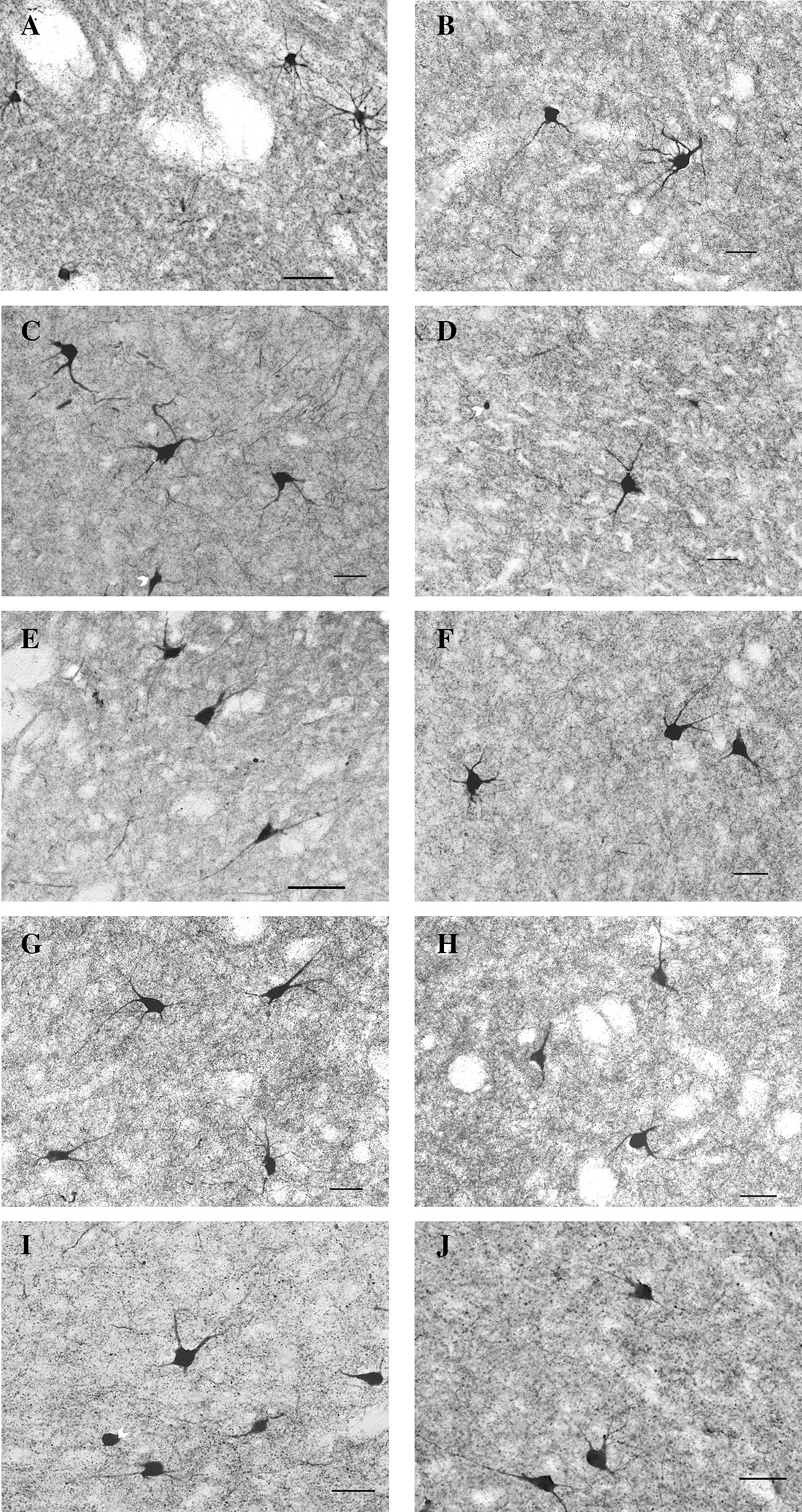
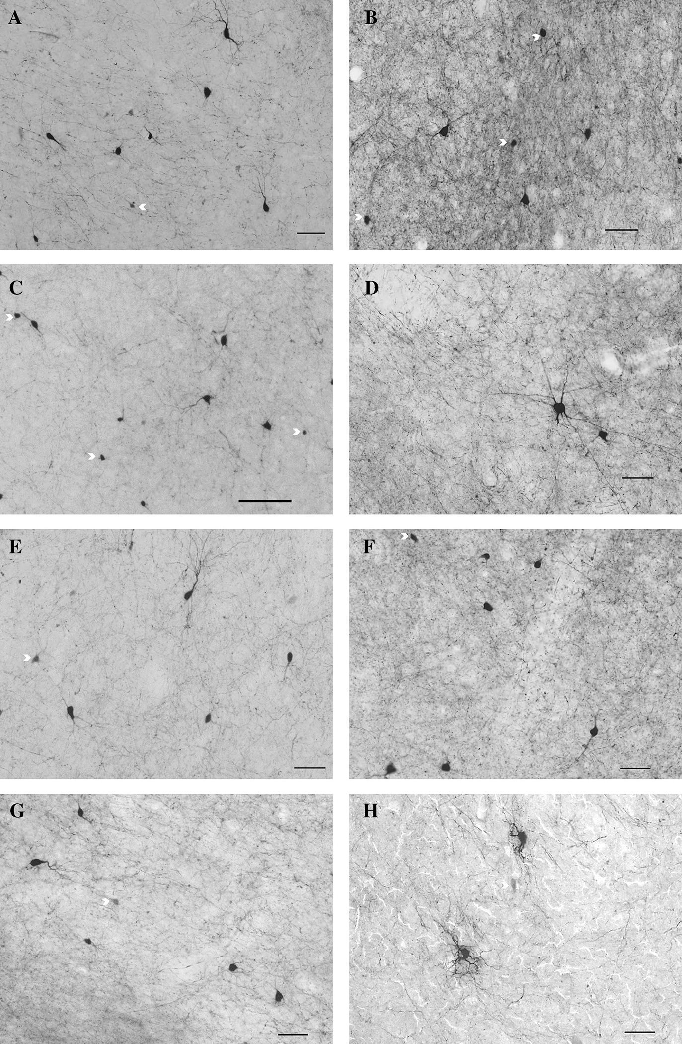
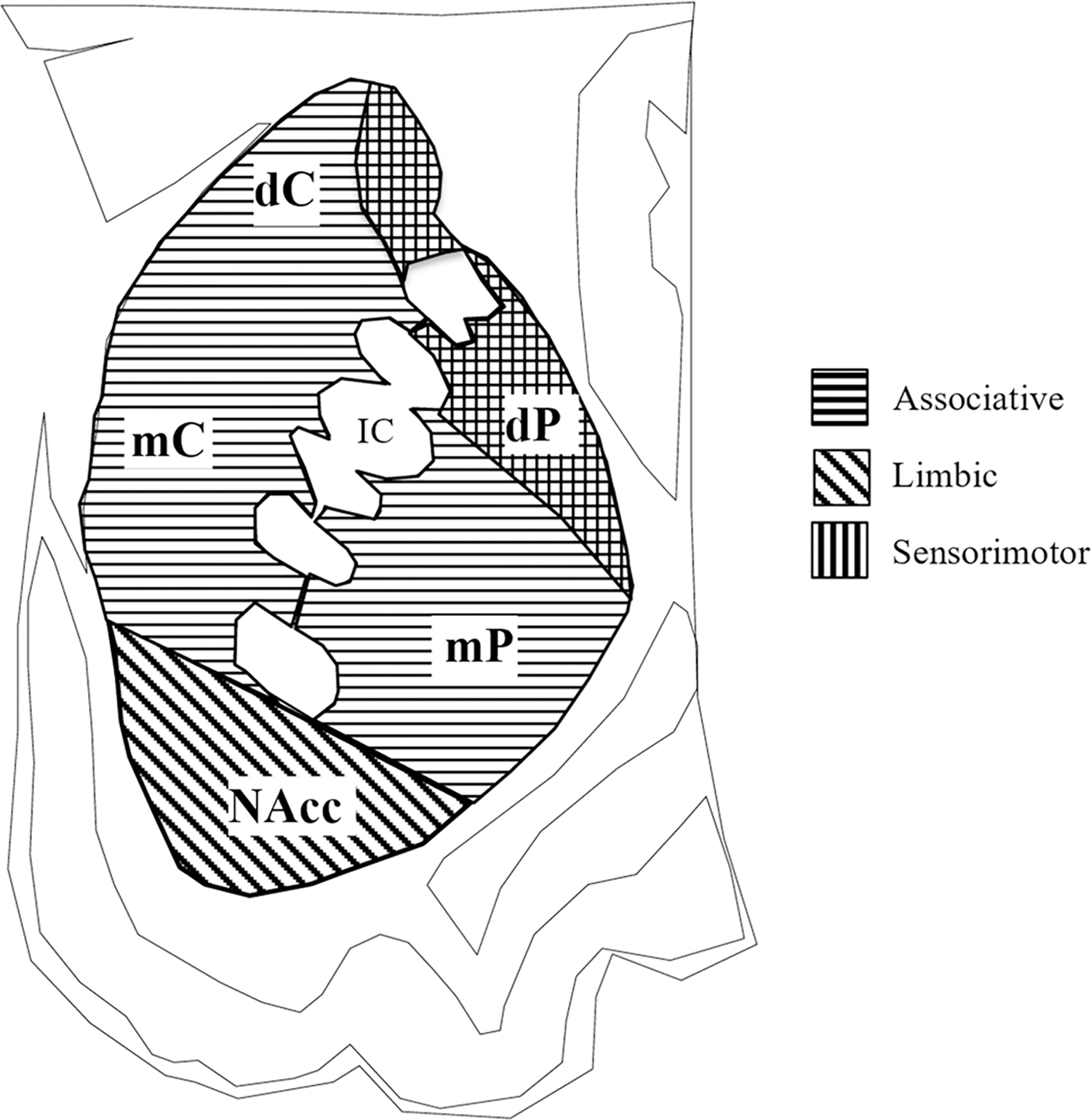
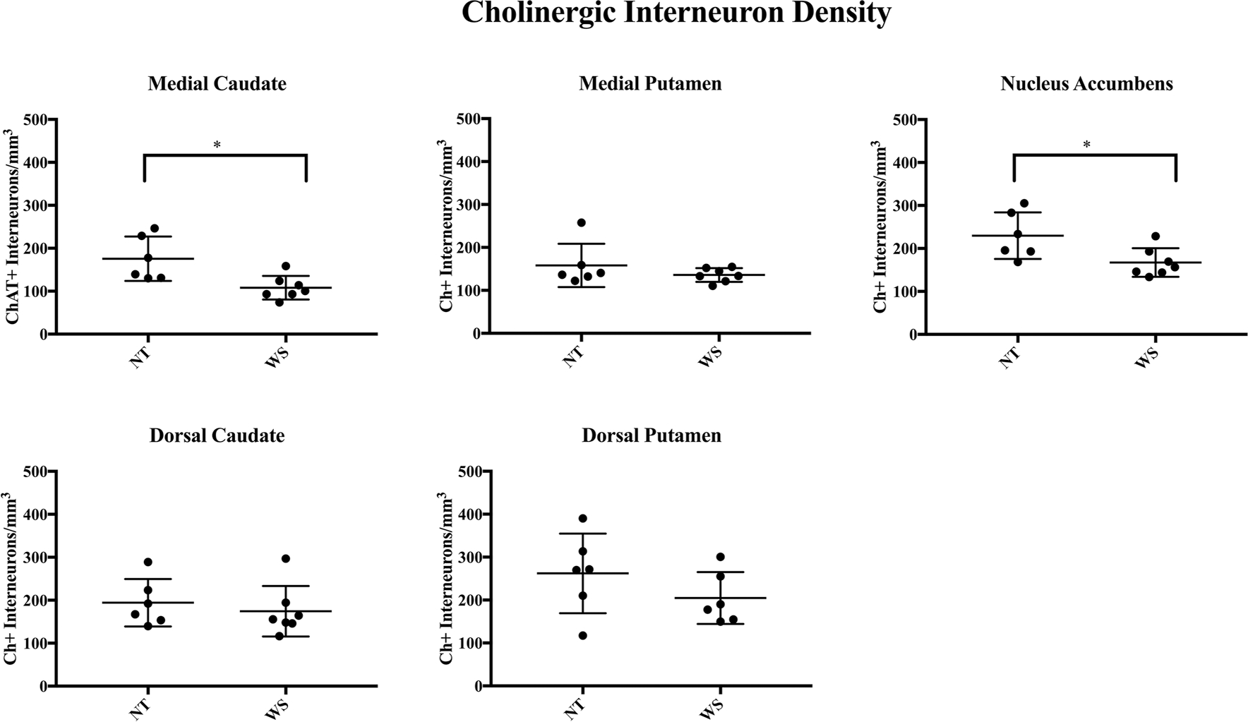
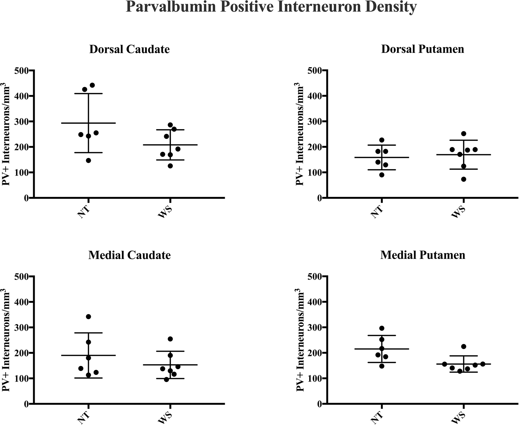
Williams syndrome (WS) is a rare neurodevelopmental disorder caused by the hemideletion of approximately 25-28 genes at 7q11.23. Its unusual social and cognitive phenotype is most strikingly characterized by the disinhibition of social behavior, in addition to reduced global IQ, with a relative sparing of language ability. Hypersociality and increased social approach behavior in WS may represent a unique inability to inhibit responses to specific social stimuli, which is likely associated with abnormalities of frontostriatal circuitry. The striatum is characterized by a diversity of interneuron subtypes, including inhibitory parvalbumin-positive interneurons (PV+) and excitatory cholinergic interneurons (Ch+). Animal model research has identified an important role for these specialized cells in regulating social approach behavior. Previous research in humans identified a depletion of interneuron subtypes associated with neuropsychiatric disorders. Here, we examined the density of PV+ and Ch+ interneurons in the striatum of 13 WS and neurotypical (NT) subjects. We found a significant reduction in the density of Ch+ interneurons in the medial caudate nucleus and nucleus accumbens, important regions receiving cortical afferents from the orbitofrontal and ventromedial prefrontal cortex, and circuitry involved in language and reward systems. No significant difference in the distribution of PV+ interneurons was found. The pattern of decreased Ch+ interneuron densities in WS differs from patterns of interneuron depletion found in other disorders.
Keywords: Basal ganglia; Interneurons; Striatum; Williams syndrome.
Conflict of interest statement
Compliance with ethical standards
Figures
Similar articles
-
Increased glia density in the caudate nucleus in williams syndrome: Implications for frontostriatal dysfunction in autism.Dev Neurobiol. 2018 May;78(5):531-545. doi: 10.1002/dneu.22554. Epub 2017 Nov 13. Dev Neurobiol. 2018. PMID: 29090517 Free PMC article.
-
Decreased number of parvalbumin and cholinergic interneurons in the striatum of individuals with Tourette syndrome.J Comp Neurol. 2010 Feb 1;518(3):277-91. doi: 10.1002/cne.22206. J Comp Neurol. 2010. PMID: 19941350 Free PMC article.
-
Evidence for a deficit in cholinergic interneurons in the striatum in schizophrenia.Neuroscience. 1999;94(1):21-31. doi: 10.1016/s0306-4522(99)00279-1. Neuroscience. 1999. PMID: 10613493
-
Cholinergic interneurons in the dorsal and ventral striatum: anatomical and functional considerations in normal and diseased conditions.Ann N Y Acad Sci. 2015 Sep;1349(1):1-45. doi: 10.1111/nyas.12762. Epub 2015 Apr 15. Ann N Y Acad Sci. 2015. PMID: 25876458 Free PMC article. Review.
-
Recurrent Implication of Striatal Cholinergic Interneurons in a Range of Neurodevelopmental, Neurodegenerative, and Neuropsychiatric Disorders.Cells. 2021 Apr 15;10(4):907. doi: 10.3390/cells10040907. Cells. 2021. PMID: 33920757 Free PMC article. Review.
Cited by
-
Targeted Tshz3 deletion in corticostriatal circuit components segregates core autistic behaviors.Transl Psychiatry. 2022 Mar 15;12(1):106. doi: 10.1038/s41398-022-01865-6. Transl Psychiatry. 2022. PMID: 35292625 Free PMC article.
-
Levodopa-responsive dystonia, parkinsonism, and treatment-resistant schizoaffective disorder in Williams syndrome.Neurol Sci. 2025 Jan;46(1):463-468. doi: 10.1007/s10072-024-07705-3. Epub 2024 Jul 18. Neurol Sci. 2025. PMID: 39023712 Free PMC article.
-
iPSC toolbox for understanding and repairing disrupted brain circuits in autism.Mol Psychiatry. 2022 Jan;27(1):249-258. doi: 10.1038/s41380-021-01288-7. Epub 2021 Sep 8. Mol Psychiatry. 2022. PMID: 34497379 Free PMC article. Review.
-
Inhibitory Systems in Brain Evolution: Pathways of Vulnerability in Neurodevelopmental Disorders.Brain Behav Evol. 2025;100(1):29-48. doi: 10.1159/000540865. Epub 2024 Aug 13. Brain Behav Evol. 2025. PMID: 39137740 Free PMC article. Review.
References
MeSH terms
Substances
Grants and funding
LinkOut - more resources
Full Text Sources
Research Materials
