

Review
doi: 10.1007/s12105-020-01152-0.
Epub 2020 Mar 18.
Tonsillar p16-Positive Follicular Dendritic Cell Sarcoma Mimicking HPV-Related Oropharyngeal Squamous Cell Carcinoma: A Case Report and Review of Reported Cases
Affiliations
- PMID: 32189159
- PMCID: PMC8010052
- DOI: 10.1007/s12105-020-01152-0
Item in Clipboard
Review
Tonsillar p16-Positive Follicular Dendritic Cell Sarcoma Mimicking HPV-Related Oropharyngeal Squamous Cell Carcinoma: A Case Report and Review of Reported Cases
Head Neck Pathol.
2021 Mar.
Abstract
Follicular dendritic cell sarcoma (FDCS) is a rare entity which can share morphologic features with non-keratinizing squamous cell carcinoma. Recent reports suggest that up to half of FDCSs show immunohistochemical positivity for p16 (Zhang et al., in Hum Pathol 66:40-47, 2017), a stain that is conventionally used in the risk stratification of oropharyngeal squamous cell carcinoma (OPSCC). Herein, we report a case of p16-positive FDCS with clinical and histomorphologic overlap with human papilloma virus (HPV)-related OPSCC.
Conflict of interest statement
All authors declare that they have no conflict of interest.
Figures

A CT scan showed non-specific enlargement of the right palatine tonsil up to
2.8 cm in diameter. The oropharynx and neck were otherwise unremarkable

The well-circumscribed mass consisted of sheets of epithelioid cells with
pseudonuclear inclusions. Patchy lymphocytic infiltrates and giant cells were present
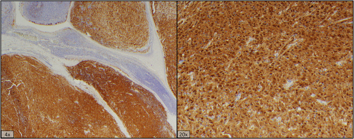
p16 immunostaining shows cytoplasmic and nuclear positivity in > 70% of
lesional cells
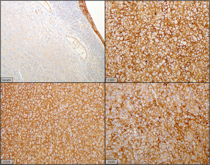
The lesional cells were negative for cytokeratin and were strongly positive for
dendritic cell markers CD21, CD23, and CD35
References
-
- Massoth LR, Hung YP, Ferry JA, Hasserjian RP, Louissaint A, Montesion M, Sokol ES, Pavlick DC, et al. Comprehensive genomic profiling of 104 rare histiocytic and dendritic cell neoplasms reveals shared and distinct targetable genomic alterations. Blood. 2019;134(Supplement 1):2541. doi: 10.1182/blood-2019-123729. - DOI
Publication types
MeSH terms
Substances
Grants and funding
LinkOut - more resources
Full Text Sources
Miscellaneous
