Review
. 2020 Apr;12(2):519-539.
doi: 10.1007/s12551-020-00680-x. Epub 2020 Mar 18.

Biological phase separation: cell biology meets biophysics

Affiliations
Review

Biological phase separation: cell biology meets biophysics

Takuya Yoshizawa et al. Biophys Rev. 2020 Apr.

Abstract

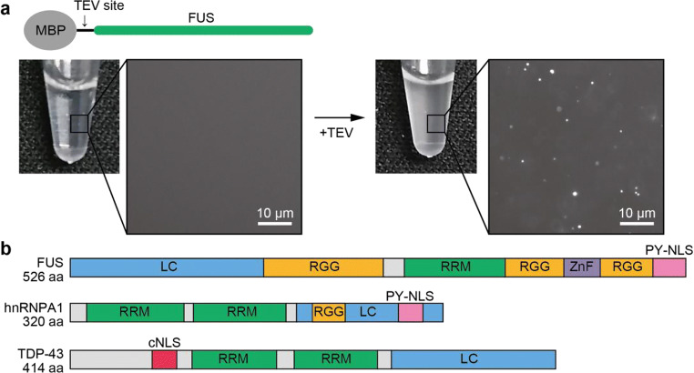
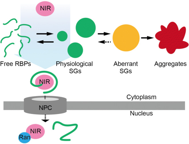
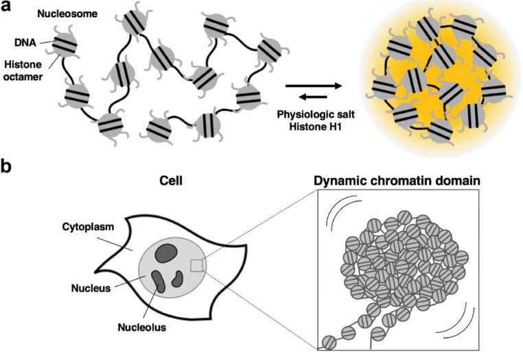
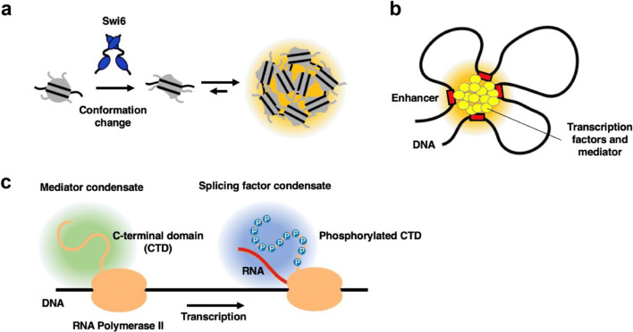
Progress in development of biophysical analytic approaches has recently crossed paths with macromolecule condensates in cells. These cell condensates, typically termed liquid-like droplets, are formed by liquid-liquid phase separation (LLPS). More and more cell biologists now recognize that many of the membrane-less organelles observed in cells are formed by LLPS caused by interactions between proteins and nucleic acids. However, the detailed biophysical processes within the cell that lead to these assemblies remain largely unexplored. In this review, we evaluate recent discoveries related to biological phase separation including stress granule formation, chromatin regulation, and processes in the origin and evolution of life. We also discuss the potential issues and technical advancements required to properly study biological phase separation.

Keywords: Intrinsically disordered region/protein (IDR/IDP); Liquid-liquid phase separation (LLPS); Low-complexity (LC) domain; Membrane-less organelle.


Conflict of interest statement

The authors declare that they have no conflict of interest.

Figures

Fig. 1
Fig. 1
a In vitro LLPS initiation of fused in sarcoma (FUS) using maltose-binding protein (MBP)-FUS. A TEV cleavage site was introduced between MBP and FUS (top). MBP-FUS is monodisperse and clear in the test tube. The solution became cloudy after TEV treatment (bottom). Close-up panels show microscope images of the solutions. b Domain map of RNA-binding proteins (RBPs) FUS, hnRNPA1, and TDP-43 imported by nuclear import receptors (NIRs). LC, low-complexity domain (blue); RGG, Arginine-Glycine-Glycine repeat (orange); RRM, RNA recognition motif (green); ZnF, zinc finger (purple); PY-NLS, proline-tyrosine nuclear localization signal (pink); cNLS, canonical-NLS (red)
Fig. 2
Fig. 2
Model of chaperone function of NIRs for RBPs in cells. NIRs (pink) actively carry nascently translated proteins (green lines) or proteins in physiological SGs (green circles) into the nucleus, preventing LLPS from occurring in the cytoplasm. Small GTPase Ran (blue) displaces the proteins and binds NIR in the nucleus. In the absence of this regulatory system, physiological SGs could then subsequently transform into aberrant SGs (yellow) and irreversible aggregates (red) in the cytoplasm
Fig. 3
Fig. 3
A new view of chromatin behavior. a A nucleosome array forms phase-separated condensate in physiological salt concentrations facilitated by histone H1 (Gibson et al. 2019). b Visualizing individual nucleosomes using live-cell superresolution imaging technique suggests chromatin forms a large irregular structure behaving like “liquid drops” in the cell (Nozaki et al. 2017)
Fig. 4
Fig. 4
The distinct microenvironment on chromatin driven by LLPS. aSchizosaccharomyces pombe HP1 protein Swi6 induces a conformation change of nucleosome, resulting in the formation of condensed, highly concentrated nucleosomes (Sanulli et al. 2019). b Super-enhancers are a cluster of enhancers bound by transcription factors and a mediator that concentrate to create a highly transcriptionally active genomic region (Hnisz et al. 2017). c Phosphorylation of the C-terminal domain (CTD) of RNA polymerase II switches its affinity from the mediator to splicing factors (Guo et al. 2019)
Fig. 5
Fig. 5
Poly (ethylene) glycol (PEG)/dextran phase-separated droplets encapsulated within oleic acid vesicles. The top row shows images acquired by an epifluorescence microscope, while the bottom row shows images acquired by a confocal microscope. a, b Merged images of (c) and (e) or (d) and (f), respectively, which show separated fluorescence channels. Cyanine 5 (Cy5)-labeled RNA (red in (a) and (b)) partitioned into the outer dextran phase as shown in (c) and (d), while HTPS (8-hydroxypyrene-1,3,6-trisulfonate) (green in (a) and (b)) partitioned into the PEG phase as shown in (e) and (f). g, h Phase contrast and bright field images. Image reprinted with permission from Jia, Tony Z., Hentrich, Christian, Szostak, Jack W.: “Rapid RNA Exchange in Aqueous Two-Phase System and Coacervate Droplets.” Origins of Life and Evolution of Biospheres, 44, 1–12 (2014) (Jia et al. 2014) under a Creative Commons License
Fig. 6
Fig. 6
Example of a coacervate droplet system composed of a nucleic acid (polyU RNA) and a simple polyamine (a). The negative charges on the RNA backbone bind strongly to the positive charges on the polyamine, resulting in a condensed coacervate phase consisting of both components (blue), and a dilute aqueous phase. b A micrograph of coacervate droplets produced from polyU and spermine. Image reprinted with permission from Aumiller, William M., Pir Cakmak, Fatma, Davis, Bradley W., Keating, Christine D.: “RNA-Based Coacervates as a Model for Membraneless Organelles: Formation, Properties, and Interfacial Liposome Assembly” Langmuir, 32(39), 10,042–10,053 (2016), (10.1021/acs.langmuir.6b02499) (Aumiller et al. 2016). Copyright American Chemical Society; further permissions related to the material excerpted should be directed to the American Chemical Society
Fig. 7
Fig. 7
Membrane-less microdroplets assembled from the rehydration of polyesters synthesized by the drying of mixtures of simple, prebiotically abundant alpha hydroxy acids. Scale bars are 100 μm. LA, lactic acid; GA, glycolic acid; PA, phenyllactic acid; SA, 2-hydroxy-4-(methylsulfanyl) butanoic acid; MA, 2-hydroxy-4-methylpentanoic acid. The labels above each image represent the mixture present in that sample. Reprinted with permission from Jia, Tony Z., Chandru, Kuhan, Hongo, Yayoi, Afrin, Rehana, Usui, Tomohiro, Myojo, Kunihiro, and Cleaves, H. James: “Membraneless polyester microdroplets as primordial compartments at the origins of life.” Proceedings of the National Academy of Science of the USA, 116, 15,830–15,835 (2019) (Jia et al. 2019). Copyright Jia, Tony Z., Chandru, Kuhan, et al.
Fig. 8
Fig. 8
1H-15N Heteronuclear single quantum coherence (HSQC) spectrum of a disordered protein. In principle, one amide group gives one resonance in the spectrum. If the protein exists in multiple conformational states, each of the conformational states provides distinct sets of resonances. The spectrum of LC domain proteins/IDPs typically shows a narrow chemical shift dispersion
Fig. 9
Fig. 9
Paramagnetic effects for NMR structural studies on LC proteins. a Schematic representation of the paramagnetic effects frequently used in protein structural studies by NMR. Pseudocontact shifts (PCSs) are generated by anisotropic paramagnetic ions such as lanthanide ions, while paramagnetic relaxation enhancements (PREs) are generated by nitroxide spin labels as well as lanthanide ions. b Observation of intermolecular and intramolecular paramagnetic effects. When the paramagnetic center is attached to an isotopically labeled protein, observed paramagnetic effects represent intramolecular information (left). When the paramagnetic center is attached to an unlabeled protein, observed paramagnetic effects represent intermolecular information (right)

Similar articles

Cited by

References

    1. Adamala K, Szostak JW. Competition between model protocells driven by an encapsulated catalyst. Nat Chem. 2013;5:495–501. doi: 10.1038/nchem.1650. - DOI - PMC - PubMed
    1. Adamala K, Szostak JW. Nonenzymatic template-directed RNA synthesis inside model protocells. Science. 2013;342(80):1098–1100. doi: 10.1126/science.1241888. - DOI - PMC - PubMed
    1. Alberti S. The wisdom of crowds: regulating cell function through condensed states of living matter. J Cell Sci. 2017;130:2789–2796. doi: 10.1242/jcs.200295. - DOI - PubMed
    1. Alberti S. Phase separation in biology. Curr Biol. 2017;27:R1097–R1102. doi: 10.1016/j.cub.2017.08.069. - DOI - PubMed
    1. Alberti S, Dormann D. Liquid–liquid phase separation in disease. Annu Rev Genet. 2019;53:1–24. doi: 10.1146/annurev-genet-112618-043527. - DOI - PubMed

LinkOut - more resources