

CT image of novel coronavirus pneumonia: a case report
- PMID: 32189175
- PMCID: PMC7088615
- DOI: 10.1007/s11604-020-00945-1
CT image of novel coronavirus pneumonia: a case report
Abstract
Objective: Knowledge of CT characteristics of COVID-19 pneumonia might be helpful to the early diagnosis and treatment of patients, and to control the spread of infection.
Methods: The chest CT images of the patient were collected to describe the CT manifestations and characteristics, and they were compared with the previous studies.
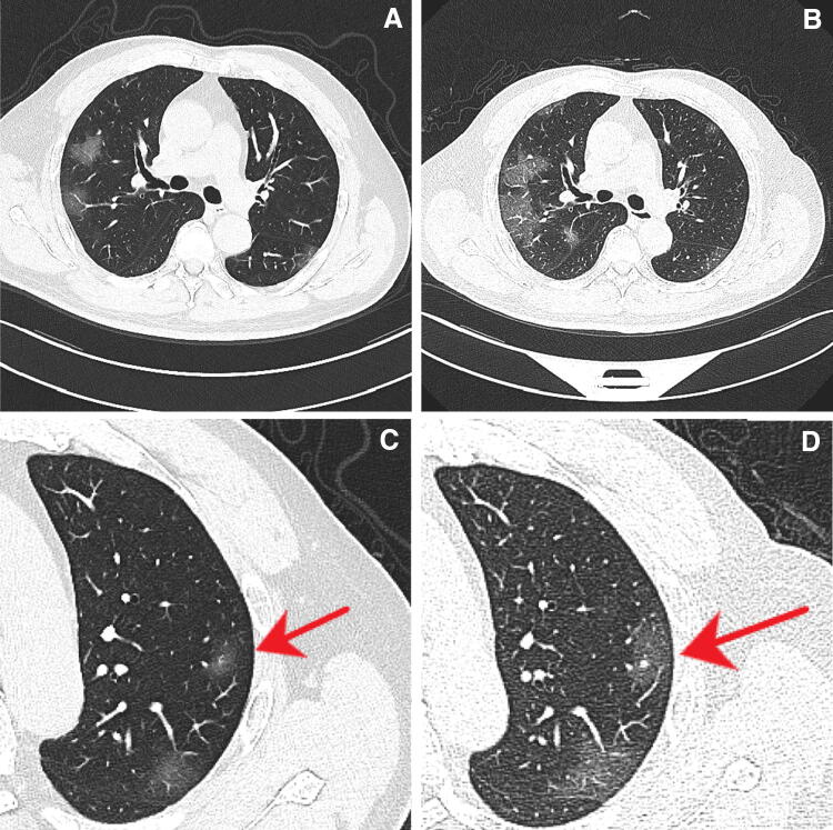
Results: Multiple patchy ground-glass opacities (GGOs) were seen in bilateral lung, mostly in subpleural areas. They progressed within 3 days, and nodular GGOs were also seen together with subpleural patchy GGOs.
Conclusion: Our case of COVID-19 pneumonia showed multiple subpleural GGOs in bilateral lung, rapid progression, and it also accompanied nodular GGOs on chest CT. These findings were consistent with the previous reports, and they might be useful for early detection and evaluation of severity of COVID-19 pneumonia.
Keywords: COVID-19 pneumonia; CT; Ground-glass opacities.
Conflict of interest statement
The authors declare that they have no conflict of interest.
Figures
Similar articles
-
CT in coronavirus disease 2019 (COVID-19): a systematic review of chest CT findings in 4410 adult patients.Eur Radiol. 2020 Nov;30(11):6129-6138. doi: 10.1007/s00330-020-06975-7. Epub 2020 May 30. Eur Radiol. 2020. PMID: 32474632 Free PMC article.
-
Imaging and clinical features of patients with 2019 novel coronavirus SARS-CoV-2.Eur J Nucl Med Mol Imaging. 2020 May;47(5):1275-1280. doi: 10.1007/s00259-020-04735-9. Epub 2020 Feb 28. Eur J Nucl Med Mol Imaging. 2020. PMID: 32107577 Free PMC article.
-
CT findings of COVID-19 in follow-up: comparison between progression and recovery.Diagn Interv Radiol. 2020 Jul;26(4):301-307. doi: 10.5152/dir.2019.20176. Diagn Interv Radiol. 2020. PMID: 32436847 Free PMC article.
-
A Comparison of Clinical and Chest CT Findings in Patients With Influenza A (H1N1) Virus Infection and Coronavirus Disease (COVID-19).AJR Am J Roentgenol. 2020 Nov;215(5):1065-1071. doi: 10.2214/AJR.20.23214. Epub 2020 May 26. AJR Am J Roentgenol. 2020. PMID: 32452731
-
Clinical and radiological features of novel coronavirus pneumonia.J Xray Sci Technol. 2020;28(3):391-404. doi: 10.3233/XST-200687. J Xray Sci Technol. 2020. PMID: 32538893 Free PMC article. Review.
Cited by
-
A review on the use of artificial intelligence for medical imaging of the lungs of patients with coronavirus disease 2019.Diagn Interv Radiol. 2020 Sep;26(5):443-448. doi: 10.5152/dir.2019.20294. Diagn Interv Radiol. 2020. PMID: 32436845 Free PMC article. Review.
-
Clinical Features, Diagnosis, and Treatment of COVID-19 in Hospitalized Patients: A Systematic Review of Case Reports and Case Series.Front Med (Lausanne). 2020 May 15;7:231. doi: 10.3389/fmed.2020.00231. eCollection 2020. Front Med (Lausanne). 2020. PMID: 32574328 Free PMC article.
-
CARE-radiology statement explanation and elaboration: reporting guideline for radiological case reports.BMJ Evid Based Med. 2024 Nov 22;29(6):399-408. doi: 10.1136/bmjebm-2023-112695. BMJ Evid Based Med. 2024. PMID: 38458654 Free PMC article.
-
Medical imaging and computational image analysis in COVID-19 diagnosis: A review.Comput Biol Med. 2021 Aug;135:104605. doi: 10.1016/j.compbiomed.2021.104605. Epub 2021 Jun 23. Comput Biol Med. 2021. PMID: 34175533 Free PMC article. Review.
-
Chest Computed Tomography Findings in Asymptomatic Patients with COVID-19.Respiration. 2020;99(9):748-754. doi: 10.1159/000509334. Epub 2020 Sep 7. Respiration. 2020. PMID: 32894853 Free PMC article.
References
-
- National Health Commission of the People’s Republic of China. Diagnosis and Treatment of Pneumonia Infected by 2019-nCoV. (trial implementation 5th Edition). Chin J Integr Tradit West Med. 2020. https://kns.cnki.net/kcms/detail/11.2787.R.20200208.1034.002.html.
Publication types
MeSH terms
LinkOut - more resources
Full Text Sources
