Review
. 2020:94:345-382.
doi: 10.1007/978-3-030-41769-7_14.

Hemoglobin: Structure, Function and Allostery

Affiliations
Review

Hemoglobin: Structure, Function and Allostery

Mostafa H Ahmed et al. Subcell Biochem. 2020.

Abstract

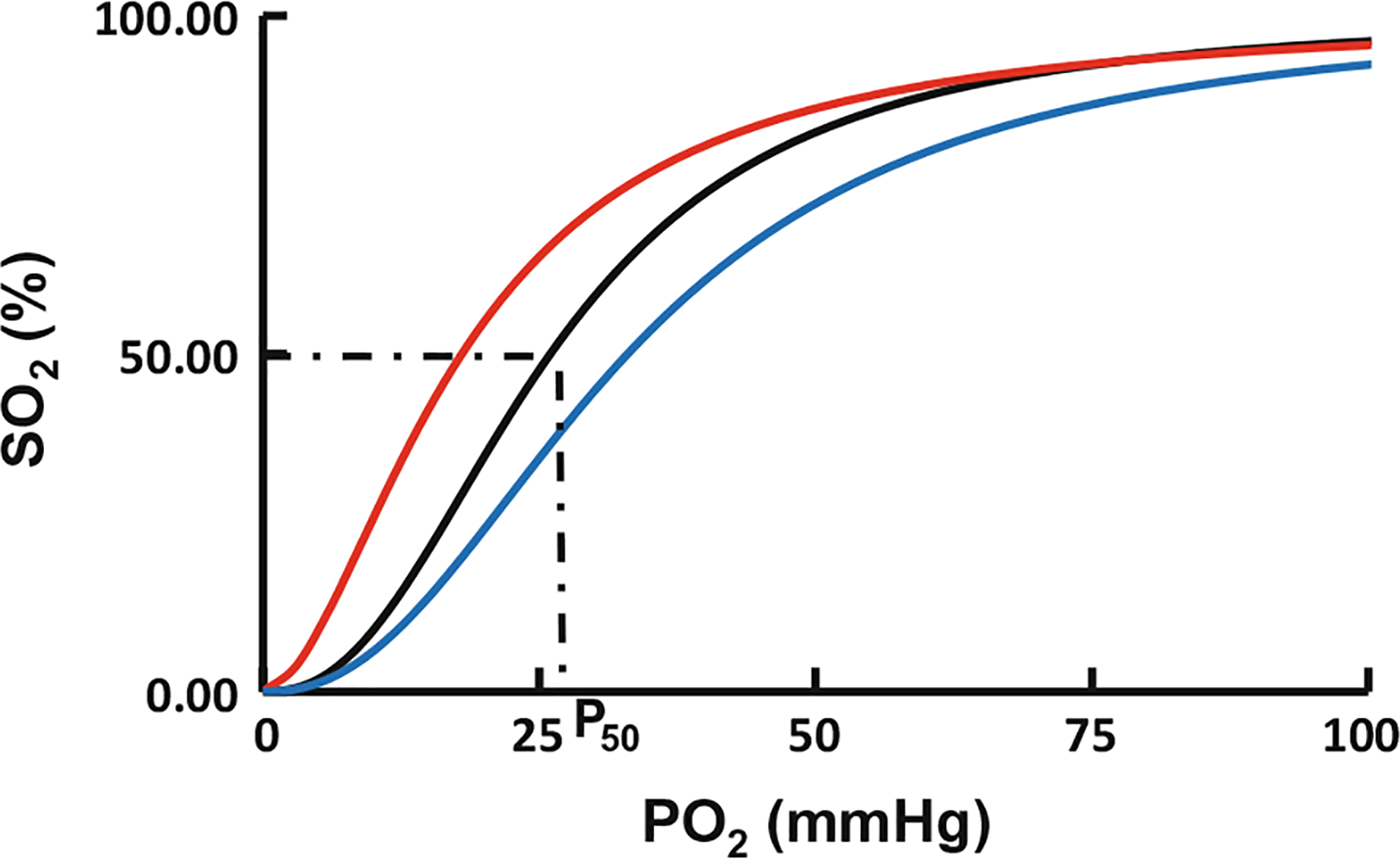
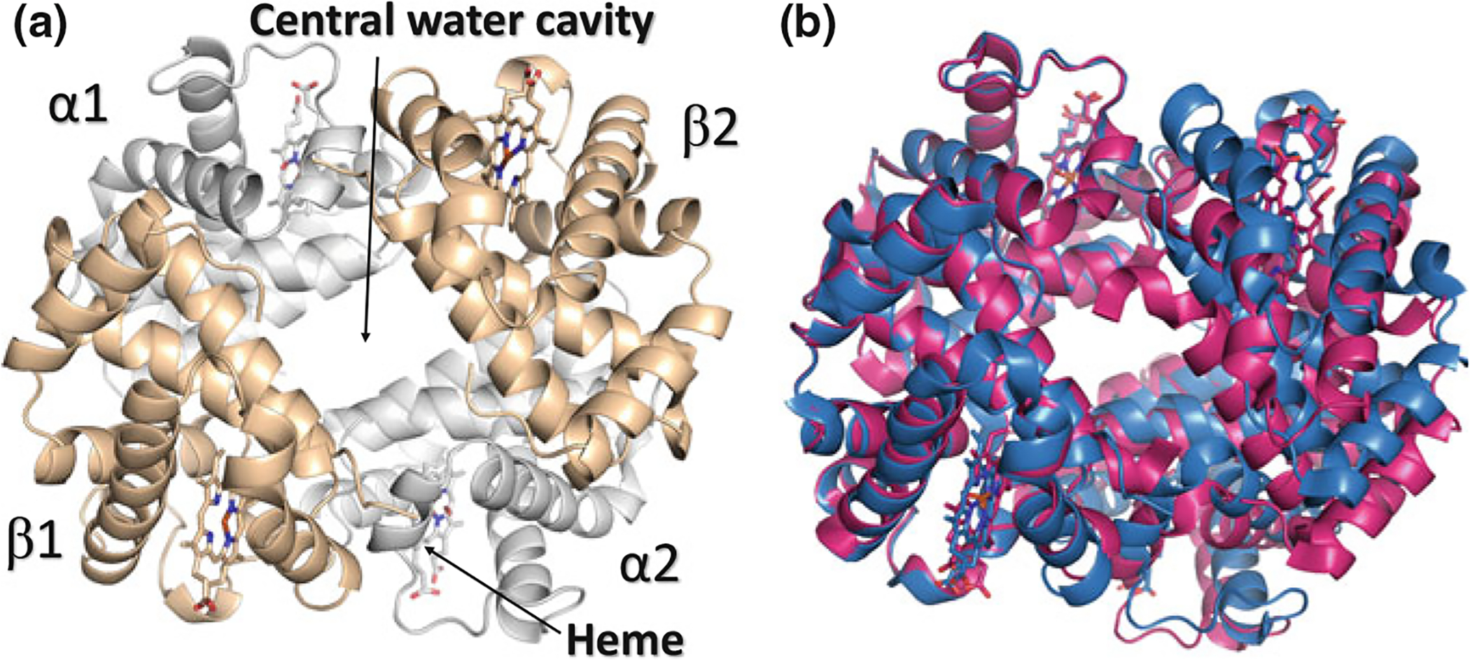
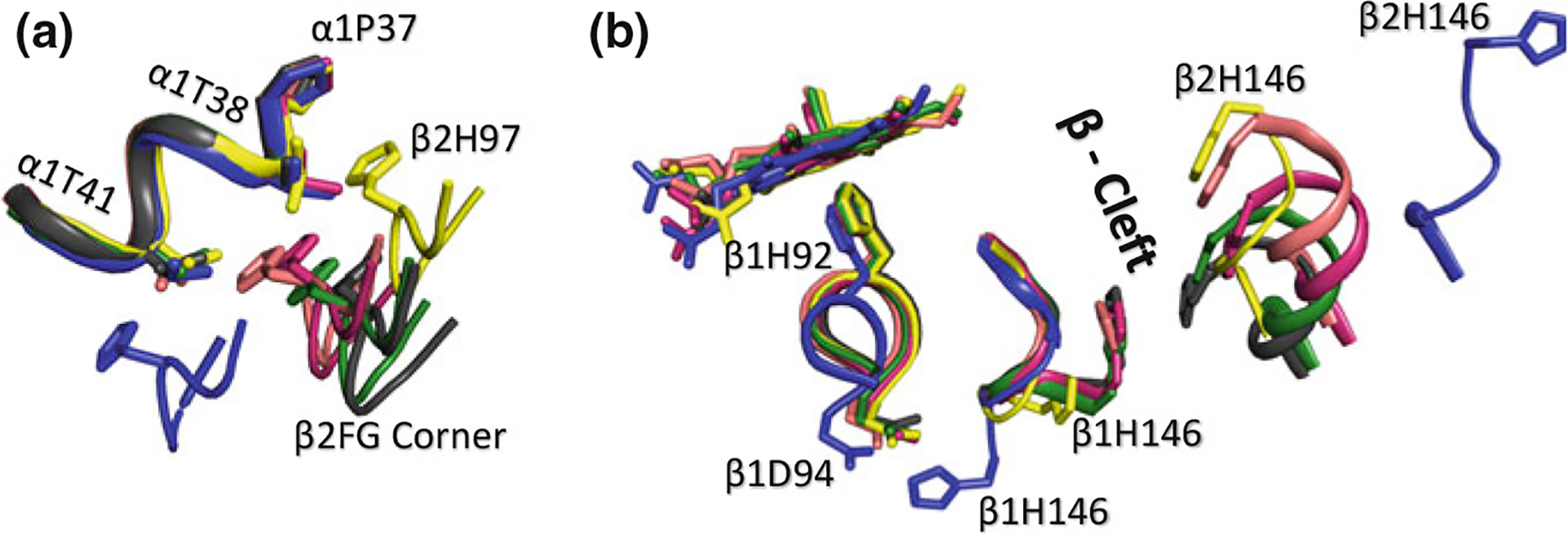
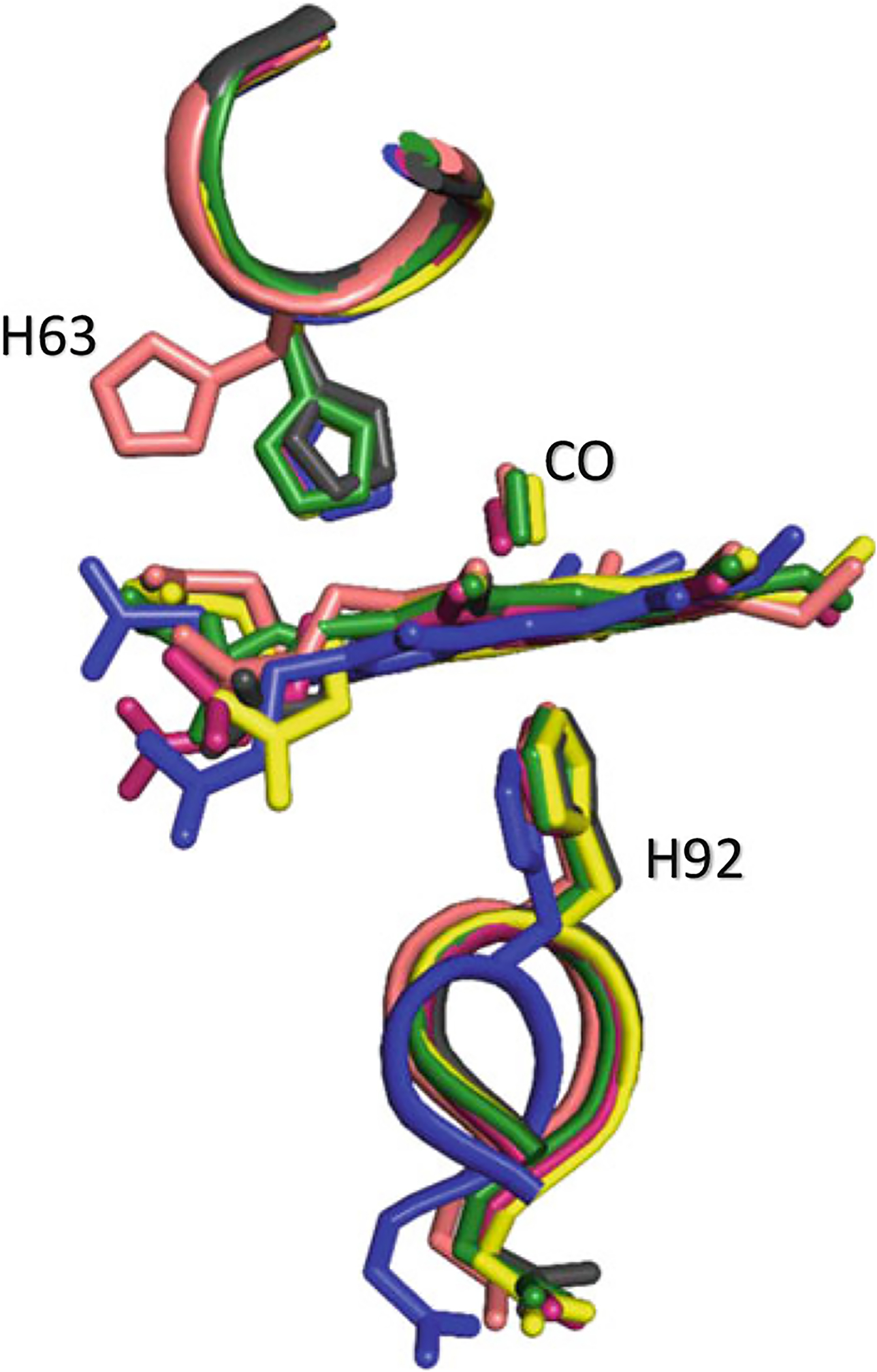
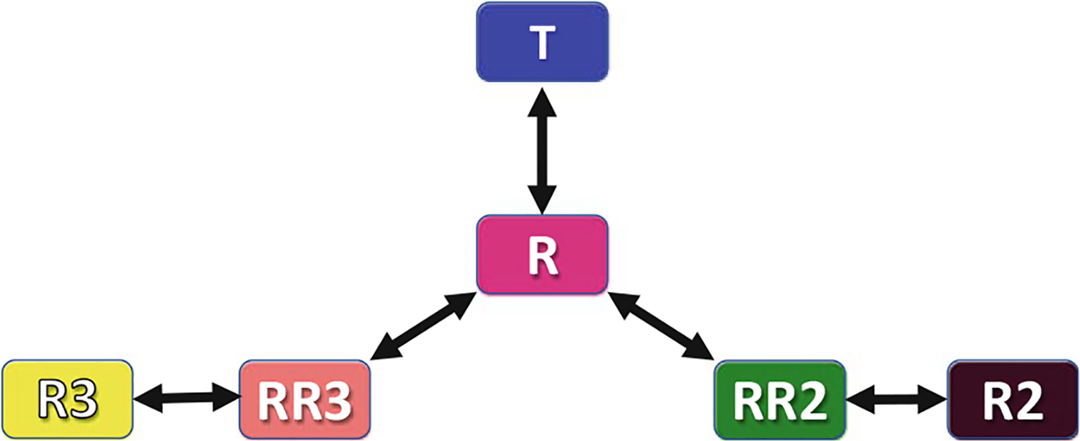
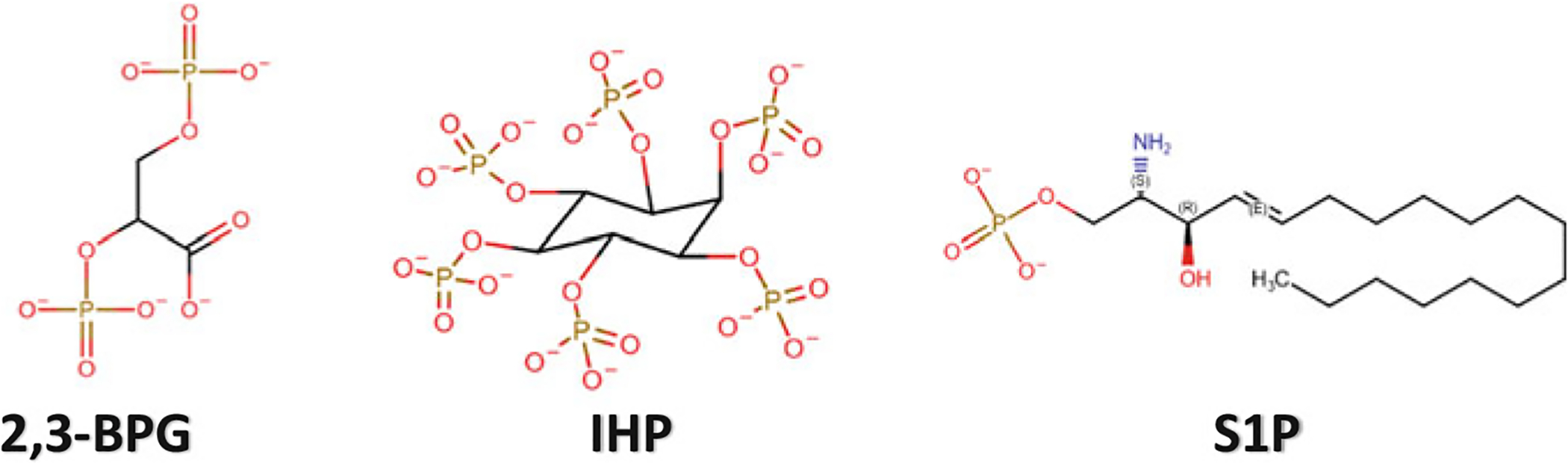
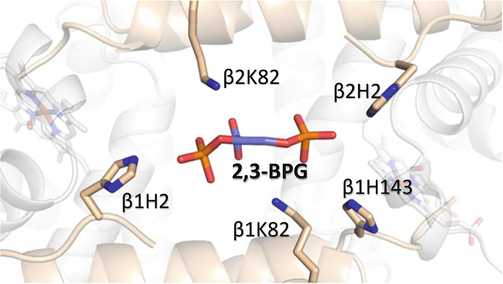
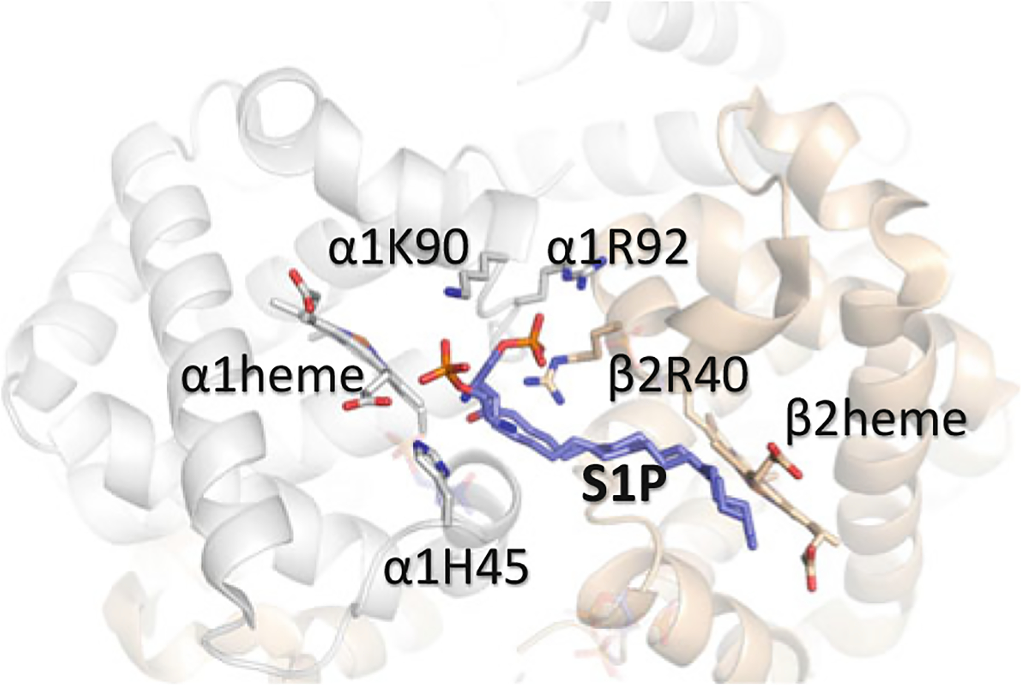
This chapter reviews how allosteric (heterotrophic) effectors and natural mutations impact hemoglobin (Hb) primary physiological function of oxygen binding and transport. First, an introduction about the structure of Hb is provided, including the ensemble of tense and relaxed Hb states and the dynamic equilibrium of Hb multistate. This is followed by a brief review of Hb variants with altered Hb structure and oxygen binding properties. Finally, a review of different endogenous and exogenous allosteric effectors of Hb is presented with particular emphasis on the atomic interactions of synthetic ligands with altered allosteric function of Hb that could potentially be harnessed for the treatment of diseases.

Keywords: Allosteric effectors; Allostery; Hemoglobin; Hemoglobin variants; Oxygen affinty; Relaxed state; T state; X-ray crystallography.


Conflict of interest statement

Conflict-of-Interest Disclosure Virginia Commonwealth University and Martin K. Safo have patents related to several aromatic aldehydes mentioned in the chapter.

Figures

Fig. 14.1
Fig. 14.1
Oxygen equilibrium curve of Hb. The normal P50 value is indicated by dashed lines. The left-shift and right-shift in the curves are colored red and blue respectively
Fig. 14.2
Fig. 14.2
Crystal structure of hemoglobin. a Overall quaternary structure of Hb with the two α chains and β chains colored grey and tan, respectively. b Structure of oxygenated (R state) Hb (magenta) superimposed on the structure of deoxygenated (T state) Hb (blue). Note the larger central water cavity in the T structure
Fig. 14.3
Fig. 14.3
Superposed structures of T (blue), R (magenta), R3 (yellow), RR2 (green), R2 (black), and RR3 (salmon) on their α1β1 dimers. a Transitions between the different states lead to significant changes (sliding motion) at the α1β2 dimer interface switch regions. b Transitions from the T state to the relaxed states breaks a T state stabilizing salt-bridge interaction between βAsp94 and βHis146. In the R2 and RR2 structures β1His146 makes close contact with β2His146, while in the other relaxed structures, βHis146 becomes highly disordered. There is also a significant size decrease in the β-cleft of the relaxed structures compared to the T structure
Fig. 14.4
Fig. 14.4
Superposed β heme structures of R (magenta), R3 (yellow), RR2 (green), R2 (black), and RR3 (salmon) showing the positions of βHis63. Note the rotation of βHis63 out of the distal pocket in the RR3 structure, creating a ligand channel to the bulk solvent, while in R, RR2, and R2 structures, βHis63 is still located in the pocket making hydrogen-bond interaction with the ligand. The R3 structure shows a partially opened ligand channel
Fig. 14.5
Fig. 14.5
Schematic representation of the proposed allosteric pathway between the different Hb states
Fig. 14.6
Fig. 14.6
Chemical structures of IHP, 2,3-BPG and S1P
Fig. 14.7
Fig. 14.7
Binding of 2,3-BPG (purple) at the β-cleft of Hb. The α-subunits are colored in gray and the β-subunits are colored in tan
Fig. 14.8
Fig. 14.8
Binding of S1P (purple) on the surface of deoxygenated Hb. The α-subunits are colored in gray and the β-subunits are colored in tan. Note that S1P only binds to the surface of the protein when 2,3-BPG binds to the β-cleft
Fig. 14.9
Fig. 14.9
Superposed structures of T (blue), R (magenta), R3 (yellow), RR2 (green), R2 (black), and RR3 (salmon) on their α1β1 dimers. αArg141 participates in inter-subunit salt-bridge interaction with αAsp126 (as well as αLys127—not shown) in deoxygenated Hb stabilizing the low-affinity T state and facilitating O2 release. At higher pH, this interaction is broken increasing the mobility of αArg141, which facilitates the T → R transition, and increase Hb oxygen affinity
Fig. 14.10
Fig. 14.10
Chemical structures of BZF (and derivative L35) and RSR-13 and derivatives (RSR-4 and TB-27)
Fig. 14.11
Fig. 14.11
a Binding of a pair of RSR-13 (purple) at the central water cavity of deoxygenated (T state) Hb. b Detailed interactions between one of the RSR-13 molecules and the protein. The other molecule makes similar symmetry-related interactions. The two α-subunits are colored grey, while the β-subunit is colored tan
Fig. 14.12
Fig. 14.12
a Chemical structure of IRL-2500. b Binding of IRL-2500 (purple) at the β-cleft of Hb. The α-subunits are colored in gray and the β-subunits are colored in tan
Fig. 14.13
Fig. 14.13
Chemical structures of high-O2 affinity antisickling aromatic aldehydes
Fig. 14.14
Fig. 14.14
Binding of 5-HMF (purple) in a symmetry-related fashion at the α-cleft Hb. The α-subunits are colored in gray and the β-subunits are colored in tan. Water molecules are red spheres
Fig. 14.15
Fig. 14.15
a Binding of INN-298 (purple and cyan) in a symmetry-related fashion at the α-cleft Hb. Unlike most aromatic aldehydes, four molecules of INN-298 bind per one Hb molecule. The molecules in purple make Schiff-base interactions with αVal1 nitrogen (primary), while the molecules in cyan (secondary) bind non-covalently and significantly weaker. b Detailed interactions of two of the INN-298 molecules (purple and cyan) with Hb. The meta-positioned methoxy-pyridine group of the primary bound INN-298 disposes further down the central water-cavity
Fig. 14.16
Fig. 14.16
Binding of INN-312 (purple) in a symmetry-related fashion at the α-cleft of Hb. The α-subunits are colored in gray. The ortho-positioned methoxy-pyridine group disposes toward the surface of the Hb to make interaction with the αF-helix residue of Pro77
Fig. 14.17
Fig. 14.17
Binding of GBT-440 (purple) at the α-cleft Hb. The α-subunits are colored in gray and the β-subunits are colored in tan. Unlike other aldehyde effectors only one molecule of GBT-440 binds per one Hb molecule
Fig. 14.18
Fig. 14.18
Chemical structures of antisickling thiol-containing agents
Fig. 14.19
Fig. 14.19
Crystal structure of TD-3 in complex with T and R structures, where TD-3 forms disulfide bond with βCys93 sulfur atom. Hb α and β subunits are shown as grey and tan, respectively. a The binding of TD-3 with βCys93 in deoxygenated Hb leads to disruption of the T state stabilizing salt-bridge interaction between βAsp94 and βHis146. b The binding of TD-3 in CO-liganded Hb (R structure) prevents possible interaction between βAsp94 and βHis146 that is required to shift the allosteric transition to the T state
Fig. 14.20
Fig. 14.20
Chemical structures of right-shifting (low-O2 affinity) aromatic aldehydes
Fig. 14.21
Fig. 14.21
(A) Crystal structures of deoxygenated Hb in complex with the mono-aldehyde-acid molecule of 5-FSA

Similar articles

Cited by

References

    1. Abdulmalik O, Safo MK, Lerner NB et al. (2004) Characterization of hemoglobin bassett (alpha94Asp → Ala), a variant with very low oxygen affinity. Am J Hematol 77:268–276. 10.1002/ajh.20184 - DOI - PubMed
    1. Abdulmalik O, Safo MK, Chen Q et al. (2005) 5-hydroxymethyl-2-furfural modifies intracellular sickle haemoglobin and inhibits sickling of red blood cells. Br J Haematol 128:552–561. 10.1111/j.1365-2141.2004.05332.x - DOI - PubMed
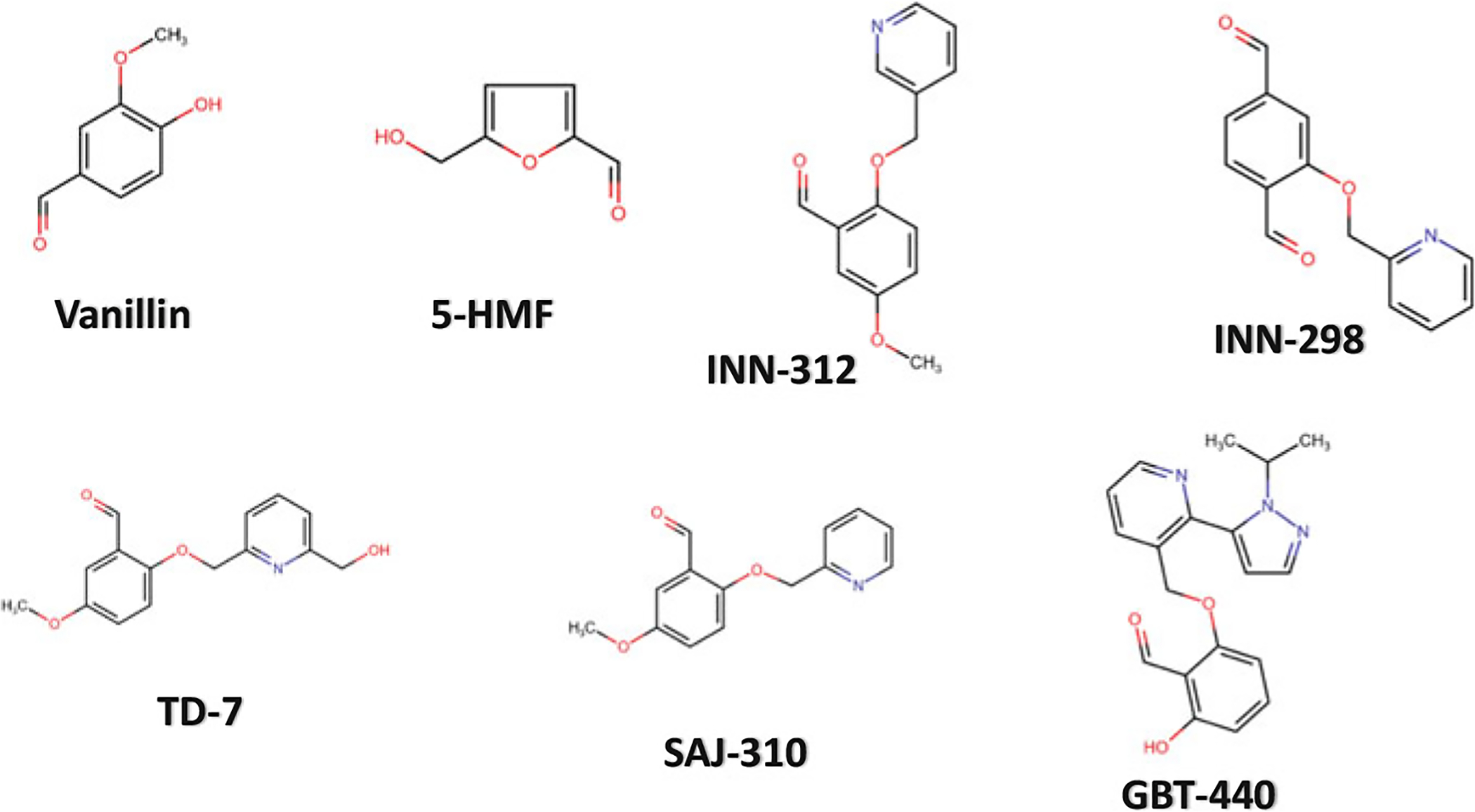
    1. Abdulmalik O, Ghatge MS, Musayev FN et al. (2011) Crystallographic analysis of human hemoglobin elucidates the structural basis of the potent and dual antisickling activity of pyridyl derivatives of vanillin Corrigendum. Acta Crystallogr D Biol Crystallogr 67:1076 10.1107/S0907444911045860 - DOI - PMC - PubMed
    1. Abraham DJ, Mehanna AS, Williams FL (1982) Design, synthesis, and testing of potential antisickling agents. 1. halogenated benzyloxy and phenoxy acids. J Med Chem 25:1015–1017. 10.1021/jm00351a002 - DOI - PubMed
    1. Abraham DJ, Perutz MF, Phillips SE (1983a) Physiological and x-ray studies of potential antisickling agents. Proc Natl Acad Sci USA 80:324–328. 10.1073/pnas.80.2.324 - DOI - PMC - PubMed

LinkOut - more resources