

Focal cemento-osseous dysplasia
- PMID: 32189898
- PMCID: PMC7069129
- DOI: 10.4103/jomfp.JOMFP_209_19
Focal cemento-osseous dysplasia
Abstract
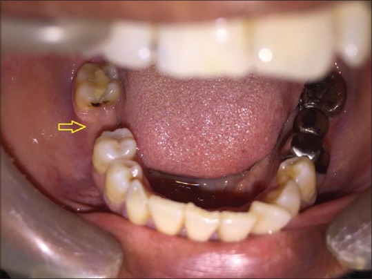
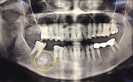
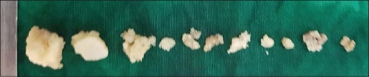
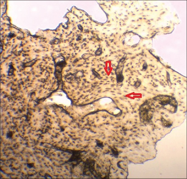
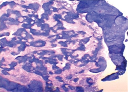
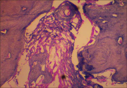
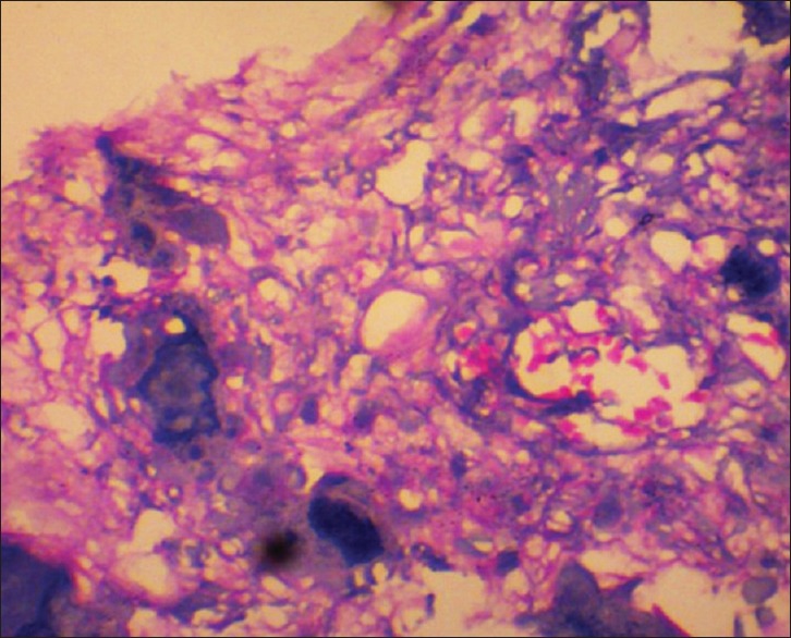
Focal cemento-osseous dysplasia (FCOD) is a benign fibro-osseous lesion of bone characterized by the replacement of normal bone by fibrous tissue and subsequently followed by its calcification with osseous and cementum-like material. It is mostly asymptomatic in nature and requires no treatment. When secondarily infected, it becomes symptomatic and intervention is required. Here, we report a case of symptomatic FCOD of mandible in a 52-year-old female patient. Histopathological evaluation of the biopsy specimen by ground sections and decalcified sections aided in the final diagnosis of the case.
Keywords: Cementum; decalcification; dysplasia; fibro-osseous; ground section.
Copyright: © 2020 Journal of Oral and Maxillofacial Pathology.
Conflict of interest statement
There are no conflicts of interest.
Figures
References
-
- Shah A, Modgill O, Patel V, Kwok J, Sproat C. Cemento-osseous dysplasia: To treat or not to treat? J Oral and Maxillofac Surg. 2016;74:e60.
-
- Salem YM, Osman YI, Norval EJ. Focal cemento-osseous dysplasia: Review and a case report. SADJ. 2010;65:422–3. - PubMed
-
- Ariji Y, Ariji E, Higuchi Y, Kubo S, Nakayama E, Kanda S. Florid cemento-osseous dysplasia. Radiographic study with special emphasis on computed tomography. Oral Surg Oral Med Oral Pathol. 1994;78:391–6. - PubMed
-
- Singh A, Gorea RK, Singla U. Few tips for making ground sections of teeth for research purpose. J Punjab Acad Forensic Med Toxicol. 2006;6:11–3.
Publication types
LinkOut - more resources
Full Text Sources