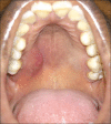
Benign lymphoepithelial lesion of the minor salivary gland - A rare presentation as a palatal swelling
- PMID: 32189901
- PMCID: PMC7069127
- DOI: 10.4103/jomfp.JOMFP_17_20
Benign lymphoepithelial lesion of the minor salivary gland - A rare presentation as a palatal swelling
Abstract
Benign lymphoepithelial lesion (BLEL) is characterized by extensive lymphocytic infiltration of the major salivary glands and may be associated with Sjogren's syndrome or HIV infection. The involvement of the palatal minor salivary glands is extremely rare. We report an isolated case of BLEL affecting the palatal minor salivary glands, presenting as a palatal swelling in a 37-year-old female patient. Serological tests ruled out potential comorbid conditions. Cone-beam computed tomography showed a palatal soft-tissue mass with thinning of the adjacent cortical plates. A histopathological examination revealed salivary gland tissue with significant acinar destruction, dense lymphocytic infiltration and focal myoepithelial islands. Therefore, BLEL may be considered as a rare differential diagnostic possibility of a palatal soft-tissue mass lesion.
Keywords: Benign lymphoepithelial lesion; minor salivary gland; palate.
Copyright: © 2020 Journal of Oral and Maxillofacial Pathology.
Conflict of interest statement
There are no conflicts of interest.
Figures


Similar articles
-
Lymphoepithelial infiltrate of palatal minor salivary glands: implications for diagnostic work-up.Int J Oral Maxillofac Surg. 2016 Dec;45(12):1626-1629. doi: 10.1016/j.ijom.2016.08.016. Epub 2016 Sep 13. Int J Oral Maxillofac Surg. 2016. PMID: 27634688
-
Minor salivary gland swelling in patient with Sjögren's syndrome.Acta Pathol Jpn. 1987 Oct;37(10):1603-9. doi: 10.1111/j.1440-1827.1987.tb02470.x. Acta Pathol Jpn. 1987. PMID: 3434282
-
MALT Lymphoma of Minor Salivary Glands in a Sjögren's Syndrome Patient: a Case Report and Review of Literature.J Oral Maxillofac Res. 2017 Mar 31;8(1):e5. doi: 10.5037/jomr.2017.8105. eCollection 2017 Jan-Mar. J Oral Maxillofac Res. 2017. PMID: 28496965 Free PMC article.
-
[Differential diagnosis of lymphoepithelial lesions of salivary glands. With particular reference to characteristic duct lesions].Pathologe. 2000 Nov;21(6):424-32. doi: 10.1007/s002920000415. Pathologe. 2000. PMID: 11148822 Review. German.
-
Papillary cystadenoma of a minor salivary gland: report of a case involving cytological analysis and review of the literature.Oral Surg Oral Med Oral Pathol Oral Radiol Endod. 2008 Jan;105(1):e28-33. doi: 10.1016/j.tripleo.2007.07.019. Oral Surg Oral Med Oral Pathol Oral Radiol Endod. 2008. PMID: 18155598 Review.
Cited by
-
Emerging patterns in nanoparticle-based therapeutic approaches for rheumatoid arthritis: A comprehensive bibliometric and visual analysis spanning two decades.Open Life Sci. 2025 Mar 21;20(1):20251071. doi: 10.1515/biol-2025-1071. eCollection 2025. Open Life Sci. 2025. PMID: 40129468 Free PMC article.
-
Chronic sclerosing sialadenitis of the bilateral submandibular glands in childhood - a diagnostic dilemma.Rom J Morphol Embryol. 2024 Jan-Mar;65(1):113-118. doi: 10.47162/RJME.65.1.14. Rom J Morphol Embryol. 2024. PMID: 38527991 Free PMC article.
References
-
- Schneider M, Rizzardi C. Lymphoepithelial carcinoma of the parotid glands and its relationship with benign lymphoepithelial lesions. Arch Pathol Lab Med. 2008;132:278–82. - PubMed
-
- Metwaly H, Cheng J, Suzuki M, Takata Y, Saitoh C, Hayashi T, et al. Benign lymphoepithelial lesion of minor salivary gland: Report of a case involving the palatal mucosa. Oral Oncol Extra. 2004;40:113–6.
-
- Yoshihara T, Morita M, Ishii T. Ultrastructure and three-dimensional imaging of epimyoepithelial islands in benign lymphoepithelial lesions. Eur Arch Otorhinolaryngol. 1995;252:106–11. - PubMed
-
- Chaudhry AP, Cutler LS, Yamane GM, Satchidanand S, Labay G, Sunderraj M. Light and ultrastructural features of lymphoepithelial lesions of the salivary glands in Mikulicz's disease. J Pathol. 1986;148:239–50. - PubMed
-
- Ihrler S, Zietz C, Sendelhofert A, Riederer A, Löhrs U. Lymphoepithelial duct lesions in Sjögren-type sialadenitis. Virchows Arch. 1999;434:315–23. - PubMed
Publication types
LinkOut - more resources
Full Text Sources
