

A miscellany of cribriform pattern, squamous metaplasia and clear cells in pleomorphic adenoma of upper lip: A diagnostic paradox
- PMID: 32189904
- PMCID: PMC7069151
- DOI: 10.4103/jomfp.JOMFP_354_19
A miscellany of cribriform pattern, squamous metaplasia and clear cells in pleomorphic adenoma of upper lip: A diagnostic paradox
Abstract
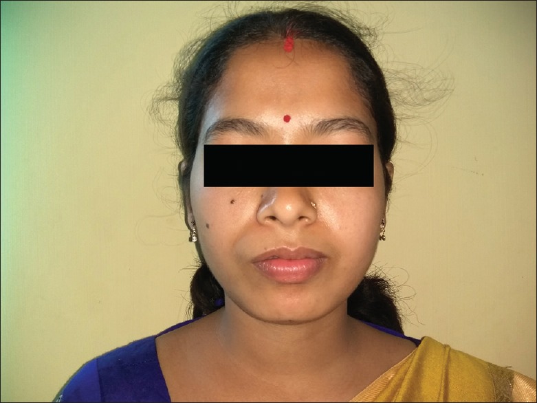
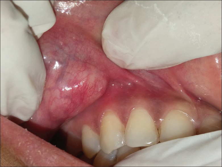
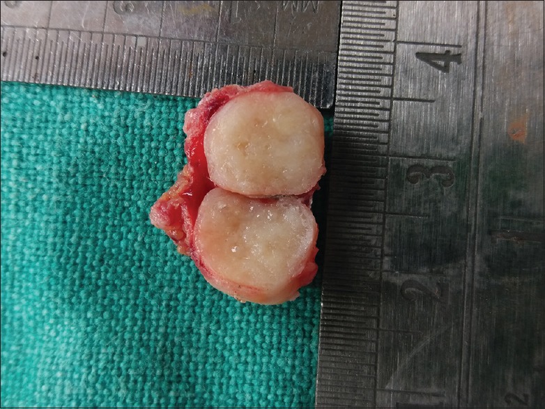
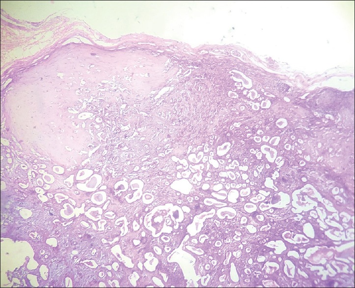
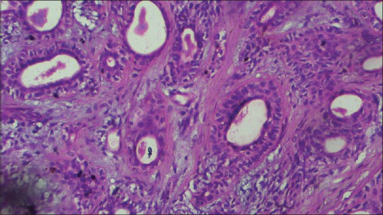
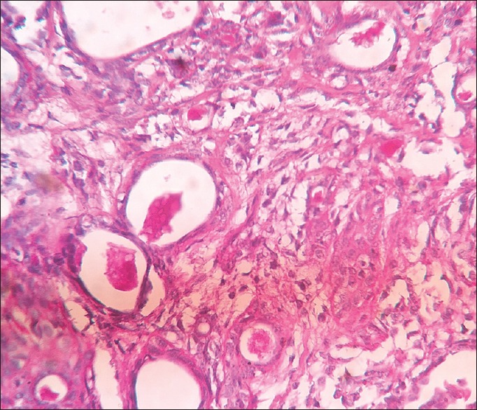
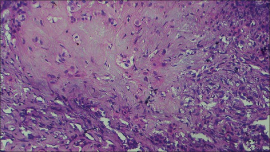
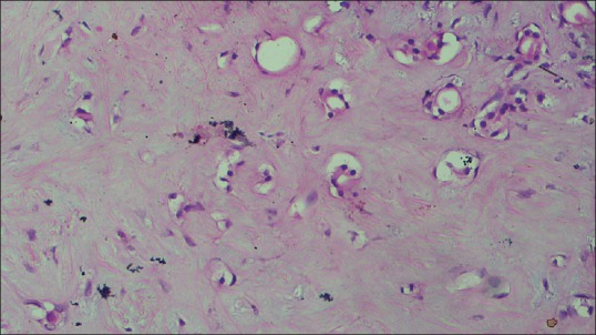
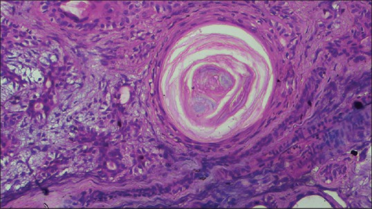
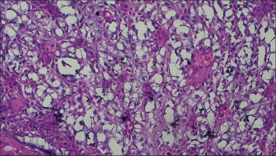
Pleomorphic Adenoma (PA), a benign neoplasm of glandular origin most commonly involves major salivary glands. It is rare in minor salivary glands such as hard palate, upper lip and buccal mucosa, frequently affecting middle aged females. PA comprises diverse histopathologic features of epithelial, myoepithelial and mesenchymal components. Aberrant histopathologic features in Pleomorhic Adenoma thus calls for judicious discrimination from alike entities which facilitates appropriate surgical management. Here we present a case report of PA in upper lip in a 25 year old female patient showing uncommon findings like clear cells, squamous metaplasia and cribriform pattern.
Keywords: Clear cell; cribriform pattern; pleomorphic adenoma; squamous metaplasia; upper lip.
Copyright: © 2020 Journal of Oral and Maxillofacial Pathology.
Conflict of interest statement
There are no conflicts of interest.
Figures
References
-
- Neville BW, Damm DD, Allen CM, Chi A. Salivary Gland Pathology Oral and Maxillofacial Pathology. First South Asia Edition. India: Elsevier; 2015. pp. 444–8.
-
- Verma P, Sachdeva SK, Verma KG, Sachdeva K. Pleomorphic adenoma of cheek: A rare case report and review of literature. Indian J Dent Res. 2014;25:122–4. - PubMed
Publication types
LinkOut - more resources
Full Text Sources