

HIPK3 Promotes Growth and Metastasis of Esophageal Squamous Cell Carcinoma via Regulation of miR-599/c-MYC Axis
- PMID: 32189968
- PMCID: PMC7064370
- DOI: 10.2147/OTT.S217087
HIPK3 Promotes Growth and Metastasis of Esophageal Squamous Cell Carcinoma via Regulation of miR-599/c-MYC Axis
Abstract
Background/aims: this experimental design was based on HIPK3 to explore the pathogenesis of ESCC.
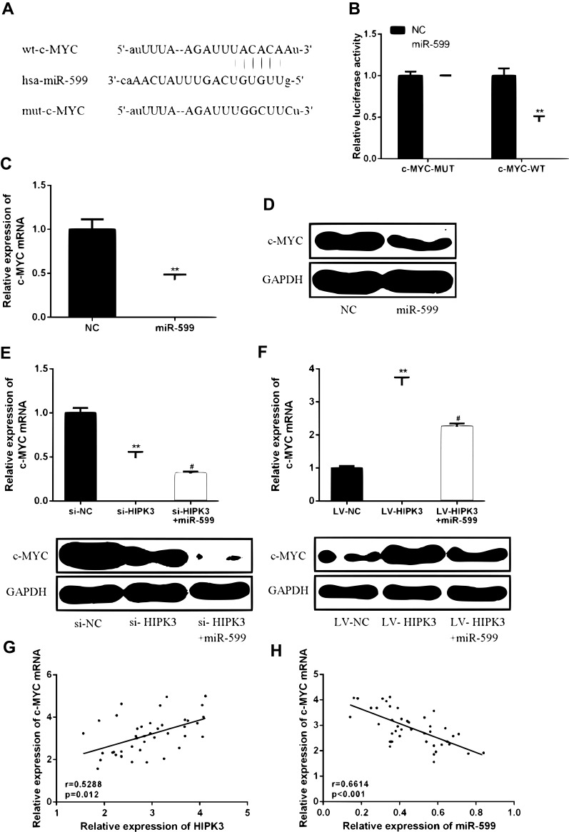
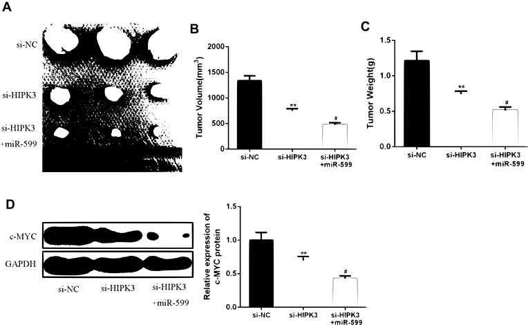
Methods: RT-qPCR was used to detect the expression of CircHIPK3 and miR-599 in ESCC tissues and cell lines.CCK-8, colony formation, flow cytometry and transwell assay were used to detect the effects of CircHIPK3 and miR-599 on tumor cell proliferation, apoptosis and migration and invasion. Target gene prediction and screening, luciferase reporter assays were used to validate downstream target genes of CircHIPK3 and miR-599.mRNA and protein expression of c-MYC were detected by RT-qPCR and Western blotting. The tumor changes in mice were detected by in vivo experiments in nude mice.
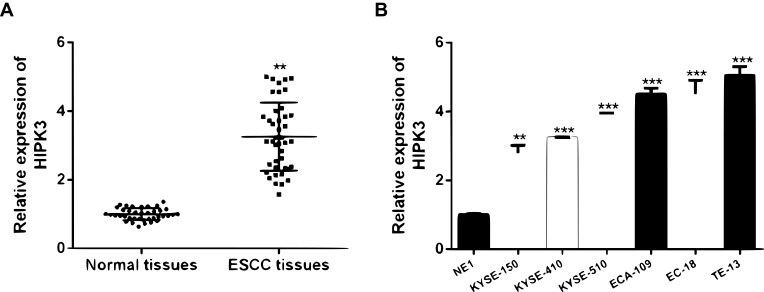
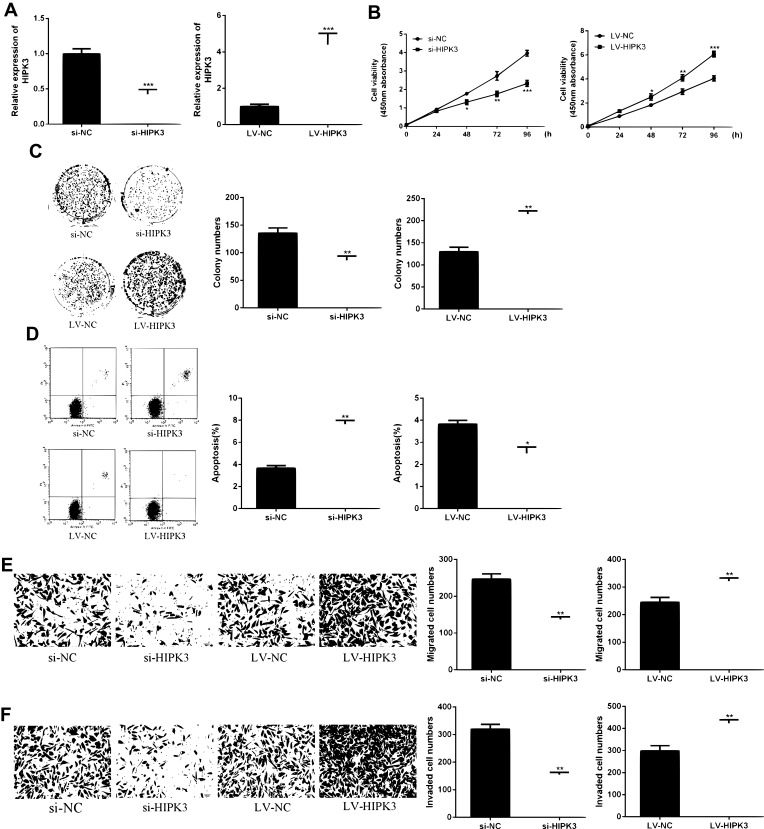
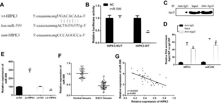
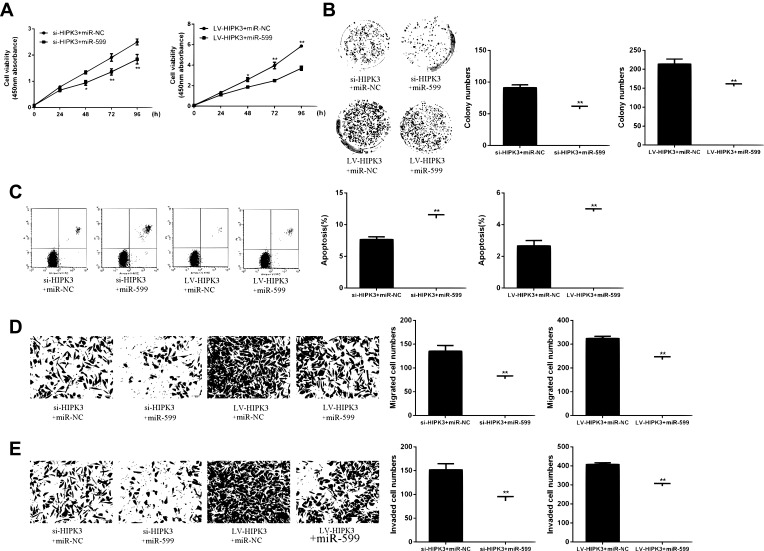
Results: HIPK3 was highly expressed in ESCC tissues and cell lines. In addition, HIPK3 expression levels were associated with advanced TNM stage, lymph node metastasis and tumor size. Moreover, HIPK3 was significantly promoted cell proliferation and migration of ESCC cells. In addition, HIPK3 was able to inhibit miRNA-599 expression and up-regulate the expression level of c-MYC. Finally, the results of in vivo animal models confirmed that HIPK3 promoted ESCC progression by modulating the miR-599/c-MYC axis.
Conclusion: HIPK3 can regulate the proliferation of esophageal squamous cell carcinoma cells by regulating miR-599/c-MYC axis, thereby inhibiting the occurrence and development of esophageal squamous cell carcinoma.
Keywords: HIPK3; c-MYC; esophageal squamous cell carcinoma; miR-599; proliferation.
© 2020 Ba et al.
Conflict of interest statement
The authors declare that they have no competing interests in this work.
Figures
Similar articles
-
Circular RNA circNTRK2 facilitates the progression of esophageal squamous cell carcinoma through up-regulating NRIP1 expression via miR-140-3p.J Exp Clin Cancer Res. 2020 Jul 11;39(1):133. doi: 10.1186/s13046-020-01640-9. J Exp Clin Cancer Res. 2020. PMID: 32653032 Free PMC article.
-
[Effects of microRNA-182-5p on cell proliferation and invasion of esophageal squamous cell carcinoma and related molecular mechanisms].Zhonghua Zhong Liu Za Zhi. 2020 Aug 23;42(8):635-643. doi: 10.3760/cma.j.cn112152-20200310-00191. Zhonghua Zhong Liu Za Zhi. 2020. PMID: 32867454 Chinese.
-
Low expression of CircRNA HIPK3 promotes osteoarthritis chondrocyte apoptosis by serving as a sponge of miR-124 to regulate SOX8.Eur Rev Med Pharmacol Sci. 2020 Aug;24(15):7937-7945. doi: 10.26355/eurrev_202008_22476. Eur Rev Med Pharmacol Sci. 2020. PMID: 32767319
-
miR-516b functions as a tumor suppressor by directly modulating CCNG1 expression in esophageal squamous cell carcinoma.Biomed Pharmacother. 2018 Oct;106:1650-1660. doi: 10.1016/j.biopha.2018.07.074. Epub 2018 Jul 29. Biomed Pharmacother. 2018. PMID: 30119241
-
[Expression of microRNA-17-5p in esophageal squamous cell carcinoma and its effects on cell proliferation and invasion].Zhonghua Zhong Liu Za Zhi. 2020 Feb 23;42(2):105-113. doi: 10.3760/cma.j.issn.0253-3766.2020.02.004. Zhonghua Zhong Liu Za Zhi. 2020. PMID: 32135643 Chinese.
Cited by
-
Understanding the Dual Roles of CircHIPK3 in Tumorigenesis and Tumor Progression.J Cancer. 2022 Nov 28;13(15):3674-3686. doi: 10.7150/jca.78090. eCollection 2022. J Cancer. 2022. PMID: 36606192 Free PMC article. Review.
-
Double matrix completion for circRNA-disease association prediction.BMC Bioinformatics. 2021 Jun 8;22(1):307. doi: 10.1186/s12859-021-04231-3. BMC Bioinformatics. 2021. PMID: 34103016 Free PMC article.
-
circHIPK3 regulates cell proliferation and migration by sponging microRNA-124 and regulating serine/threonine kinase 3 expression in esophageal squamous cell carcinoma.Bioengineered. 2022 Apr;13(4):9767-9780. doi: 10.1080/21655979.2022.2060776. Bioengineered. 2022. PMID: 35443871 Free PMC article.
-
Hsa_circ_0006916 Knockdown Represses the Development of Hepatocellular Carcinoma via Modulating miR-599/SRSF2 Axis.Onco Targets Ther. 2020 Nov 4;13:11301-11313. doi: 10.2147/OTT.S267471. eCollection 2020. Onco Targets Ther. 2020. Retraction in: Onco Targets Ther. 2021 Dec 07;14:5419-5420. doi: 10.2147/OTT.S352188. PMID: 33177838 Free PMC article. Retracted.
-
Biological functions and molecular mechanisms of circHIPK3 in digestive system tumors.Hum Cell. 2025 May 29;38(4):113. doi: 10.1007/s13577-025-01239-2. Hum Cell. 2025. PMID: 40442364 Review.
References
LinkOut - more resources
Full Text Sources
