. 2020 Mar 12:20:76.
doi: 10.1186/s12935-019-1092-7. eCollection 2020.

MicroRNA-93-5p promotes epithelial-mesenchymal transition in gastric cancer by repressing tumor suppressor AHNAK expression

Affiliations

MicroRNA-93-5p promotes epithelial-mesenchymal transition in gastric cancer by repressing tumor suppressor AHNAK expression

Erdong Shen et al. Cancer Cell Int. .

Retraction in

Abstract

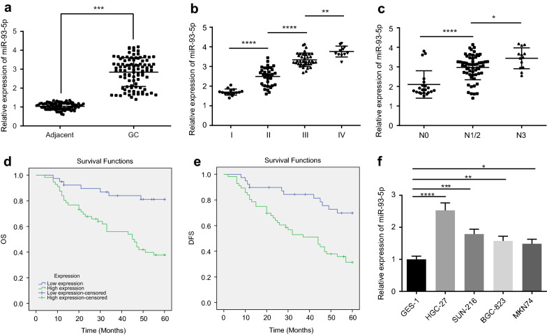
Background: Gastric cancer (GC) is a common cause of cancer-related mortality worldwide, and microRNAs (miRNAs) have been shown to play an important role in GC development. This study aims to explore the effect of microRNA-93-5p (miR-93-5p) on the epithelial-mesenchymal transition (EMT) in GC, via AHNAK and the Wnt signaling pathway.

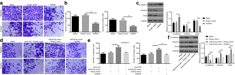
Methods: Microarray-based gene expression analysis was performed to identify GC-related differentially expressed miRNAs and genes. Then the expression of the miR-93-5p was examined in GC tissues and GC cell lines. The targeting relationship between miR-93-5p and AHNAK was verified by a dual luciferase reporter gene assay. In an attempt to ascertain the contributory role of miR-93-5p in GC, miR-93-5p mimic or inhibitor, as well as an AHNAK overexpression vector, were introduced to HGC-27 cells. HGC-27 cell migration and invasive ability, and EMT were assayed using Transwell assay and western blot analysis. Regulation of the Wnt signaling pathway was also assessed using TOP/FOP flash luciferase assay.

Results: miR-93-5p was highly expressed in GC tissue samples and cells. Notably, miR-93-5p could target and negatively regulate AHNAK. Down-regulation of miR-93-5p or overexpression of AHNAK could suppress the migration and invasion abilities, in addition to EMT in GC cells via inactivation of the Wnt signaling pathway.

Conclusion: Taken together, downregulation of miR-93-5p attenuated GC development via the Wnt signaling pathway by targeting AHNAK. These findings provide an enhanced understanding of miR-93-5p as a therapeutic target for GC treatment.

Keywords: AHNAK; Epithelial–mesenchymal transition; Gastric cancer; Invasion; MicroRNA-93-5p; Migration; Wnt signaling pathway.


Conflict of interest statement

Competing interestsThe authors declare that they have no competing interests.

Figures

Fig. 1
Fig. 1
Microarray analysis identifies the highly expressed miR-93-5p in GC-related samples for this study. a the intersection of differentially expressed miRNAs in GC-related expression datasets. The left circle is the top 20 differentially expressed miRNAs in the GSE93415 expression dataset. The right circle is the top 20 differentially expressed miRNAs in the GSE78091 expression dataset. The middle part indicates the intersection of the two expression datasets. b A heat map of differentially expressed miRNAs in GSE93415 expression dataset. The abscissa indicates the sample number, the ordinate indicates the miRNA name, and the left dendrogram indicates the differentially expressed miRNA expression cluster. Each small square in the Figure indicates the expression of one gene in one sample. The upper right histogram is the color gradation. c The expression of miR-93-5p in the GSE78091 expression dataset, the abscissa indicates the sample type, the ordinate indicates the expression of miR-93-5p in the expression dataset, the p value is in the upper left corner, and the left box diagram indicates the expression of miR-93-5p in the normal sample, and the right box diagram indicates the expression of miR-93-5p in the tumor group
Fig. 2
Fig. 2
Up-regulated miR-93-5p expression is determined in GC tissues and GC cell lines. a Expression of miR-93-5p in GC tissues and adjacent tissues of GC patients (n = 95). b, c Expression of miR-93-5p in GC tissues in different pathological stages. d, e The Kaplan–meier survival analysis of overall survival and DFS in GC patients. Green represents patients with high expression of miR-93-5p, blue represents patients with low expression of miR-93-5p. f Expression of miR-93-5p in immortalized human gastric mucosal epithelial cell line and GC cell lines. Data were measurement data and expressed as mean ± standard deviation. Student’s t-test was used for comparison between two groups. One-way analysis of variance was used for comparison among multi-groups. The cell experiment was repeated 3 times. *p < 0.05, **p < 0.01, ***p < 0.001, and ****p < 0.0001
Fig. 3
Fig. 3
Repressed miR-93-5p inhibits HGC-27 cell migration, invasion and EMT. a The migration and invasion ability of GC cells determined by Transwell assay (×200). Blank group: HGC-27 cells without transfection of any plasmids; Inhibitor NC group: HGC-27 cells transfected with NC of miR-93-5p; miR-93-5p inhibitor group: HGC-27 cells transfected with miR-93-5p inhibitor. b, c Migration and invasive ability of GC cells determined by the Transwell assays and the quantitative analysis. d Morphological characteristics of HGC-27 cells after transfection with inhibitor NC or miR-93-5p inhibitor (×100). e Immunofluorescence to determine the expression of EMT-related markers (E-cadherin, Vimentin and Snail) (×400). f Western blot assay and quantitative analysis for the expression of AHNAK, E-cadherin, Vimentin and Snail. Data were measurement data and expressed as mean ± standard deviation. One-way analysis of variance was used for comparison among multi-groups. The cell experiment was repeated three times. *p < 0.05, **p < 0.01, ***p < 0.001, and ****p < 0.0001
Fig. 4
Fig. 4
AHNAK is a target gene of miR-93-5p. a Heat map of differentially expressed mRNAs in the GSE2685 expression dataset. Abscissa indicates differential p value, and ordinate indicates logFC. Each small dot in the Figure indicates a gene, among them red indicates a gene with significantly up-regulated expression in GC, green indicates a gene with significantly down-regulated expression in GC and black indicates genes which aren’t differentially expressed. b The intersection of miR-93-5p targeting gene prediction and down-regulated gene in the GSE2685 expression dataset. The left circle indicates the prediction of the target genes in TargetScan database, the right circle indicates the down-regulated genes in the GSE2685 expression dataset, and the middle part indicates the intersection. c The binding sites of miR-93-5p to AHNAK 3′UTR and OPCML 3′UTR. d Dual luciferase reporter gene assay to verify the targeting relationship between miR-93-5p and AHNAK. e Western blot analysis to determine the expression of AHNAK in immortalized human gastric mucosal epithelial cell line and GC cell lines. f Western blot analysis to investigate the effect of miR-93-5p on AHNAK expression. Data were measurement data and expressed as mean ± standard deviation. One-way analysis of variance was used for comparison among multi-groups. The cell experiment was repeated three times. *p < 0.05, **p < 0.01, ***p < 0.001, and ****p < 0.0001
Fig. 5
Fig. 5
Up-regulation of AHNAK inhibits HGC-27 cell migration, invasion and EMT. a Transwell assay to investigate the effect of AHNAK on HGC-27 cell invasion and migration (×200). Blank group: HGC-27 cells without transfection of any plasmids; Empty vector group: HGC-27 cells transfected with empty vector; AHNAK group: HGC-27 cells transfected with AHNAK overexpression plasmid. b Quantitative analysis for the role of overexpressed AHNAK in the HGC-27 cell migration and invasion. c Western blot analysis of EMT markers E-cadherin, Vimentin and Snail expression after overexpression of AHNAK. d Transwell assay of effect of miR-93-5p mimic and overexpressed AHNAK in HGC-27 cell invasion and migration (×200). Mimic NC + empty vector group: HGC-27 cells co-transfected with mimic NC and empty vector; miR-93-5p mimic + empty vector group: HGC-27 cells co-transfected with miR-93-5p mimic and empty vector; miR-93-5p mimic + AHNAK group: HGC-27 cells co-transfected with co-transfected with miR-93-5p mimic and AHNAK. e Quantitative analysis for the role of overexpressed AHNAK in the HGC-27 cell migration and invasive. f western blot analysis of EMT markers E-cadherin, Vimentin and Snail expression after overexpression of miR-93-5p and AHNAK. Data were measurement data and expressed as mean ± standard deviation. One-way analysis of variance was used for comparison among multi-groups. The cell experiment was repeated three times. *p < 0.05, **p < 0.01, ***p < 0.001, and ****p < 0.0001
Fig. 6
Fig. 6
Suppression of miR-93-5p inhibits the Wnt signaling pathway by up-regulating AHNAK expression. a Western blot analysis to measure the expression of Wnt signaling pathway-related factors Wnt-1, β-catenin and p-β-catenin. b TOP/FOP flash to determine the β-catenin-TCF/LEF transcriptional activity of each group. Cells were co-transfected with TOP flash or FOP flash and Renilla plasmids for 48 h before measurement. The ratio of activity of firefly luciferase to that of Renilla luciferase was calculated. Data were measurement data and expressed as mean ± standard deviation. One-way analysis of variance was used for comparison among multi-groups. The cell experiment was repeated three times. *p < 0.05, **p < 0.01, ***p < 0.001, and ****p < 0.0001
Fig. 7
Fig. 7
The schematic diagram depicts the regulatory mechanism of miR-93-5p in GC. miR-93-5p can target AHNAK and activate the Wnt signaling pathway to promote cell migration and invasion abilities, and EMT in GC cells

References

    1. Karimi P, Islami F, Anandasabapathy S, Freedman ND, Kamangar F. Gastric cancer: descriptive epidemiology, risk factors, screening, and prevention. Cancer Epidemiol Biomark Prev. 2014;23(5):700–713. doi: 10.1158/1055-9965.EPI-13-1057. - DOI - PMC - PubMed
    1. Chen W, Zheng R, Baade PD, Zhang S, Zeng H, Bray F, Jemal A, Yu XQ, He J. Cancer statistics in China, 2015. CA Cancer J Clin. 2016;66(2):115–132. doi: 10.3322/caac.21338. - DOI - PubMed
    1. Tie J, Pan Y, Zhao L, Wu K, Liu J, Sun S, Guo X, Wang B, Gang Y, Zhang Y, et al. MiR-218 inhibits invasion and metastasis of gastric cancer by targeting the Robo1 receptor. PLoS Genet. 2010;6(3):e1000879. doi: 10.1371/journal.pgen.1000879. - DOI - PMC - PubMed
    1. Matsuoka J, Yashiro M, Doi Y, Fuyuhiro Y, Kato Y, Shinto O, Noda S, Kashiwagi S, Aomatsu N, Hirakawa T, et al. Hypoxia stimulates the EMT of gastric cancer cells through autocrine TGFbeta signaling. PLoS ONE. 2013;8(5):e62310. doi: 10.1371/journal.pone.0062310. - DOI - PMC - PubMed
    1. Peng Z, Wang CX, Fang EH, Wang GB, Tong Q. Role of epithelial-mesenchymal transition in gastric cancer initiation and progression. World J Gastroenterol. 2014;20(18):5403–5410. doi: 10.3748/wjg.v20.i18.5403. - DOI - PMC - PubMed

Publication types

LinkOut - more resources