. 2020 Mar 15:22:6.
doi: 10.1186/s12575-020-00118-4. eCollection 2020.

High-Resolution Confocal Fluorescence Imaging of Serine Hydrolase Activity in Cryosections - Application to Glioma Brain Unveils Activity Hotspots Originating from Tumor-Associated Neutrophils

Affiliations

High-Resolution Confocal Fluorescence Imaging of Serine Hydrolase Activity in Cryosections - Application to Glioma Brain Unveils Activity Hotspots Originating from Tumor-Associated Neutrophils

Niina Aaltonen et al. Biol Proced Online. .

Abstract

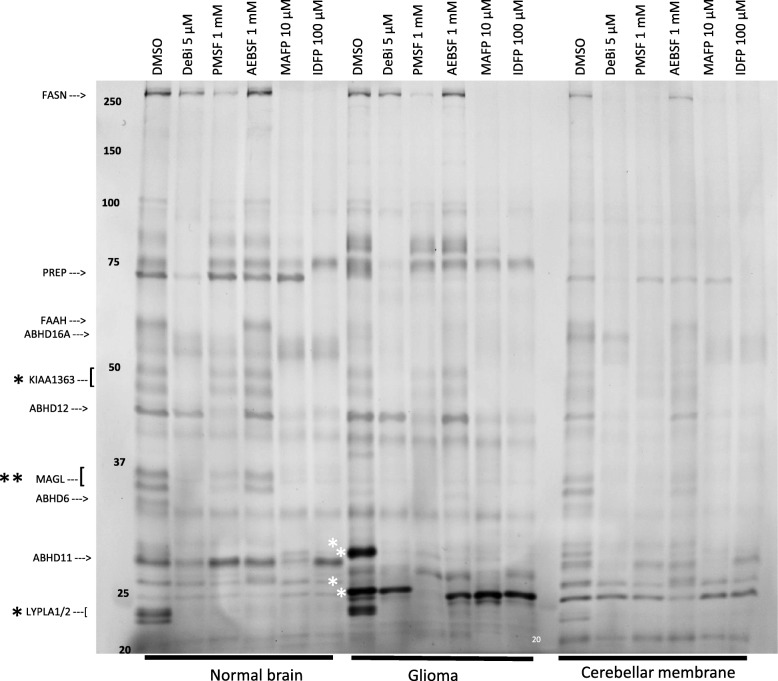
Background: Serine hydrolases (SHs) are a functionally diverse family of enzymes playing pivotal roles in health and disease and have emerged as important therapeutic targets in many clinical conditions. Activity-based protein profiling (ABPP) using fluorophosphonate (FP) probes has been a powerful chemoproteomic approach in studies unveiling roles of SHs in various biological systems. ABPP utilizes cell/tissue proteomes and features the FP-warhead, linked to a fluorescent reporter for in-gel fluorescence imaging or a biotin tag for streptavidin enrichment and LC-MS/MS-based target identification. Existing ABPP approaches characterize global SH activity based on mobility in gel or MS-based target identification and cannot reveal the identity of the cell-type responsible for an individual SH activity originating from complex proteomes.

Results: Here, by using an activity probe with broad reactivity towards the SH family, we advance the ABPP methodology to glioma brain cryosections, enabling for the first time high-resolution confocal fluorescence imaging of global SH activity in the tumor microenvironment. Tumor-associated cell types were identified by extensive immunohistochemistry on activity probe-labeled sections. Tissue-ABPP indicated heightened SH activity in glioma vs. normal brain and unveiled activity hotspots originating from tumor-associated neutrophils (TANs), rather than tumor-associated macrophages (TAMs). Thorough optimization and validation was provided by parallel gel-based ABPP combined with LC-MS/MS-based target verification.

Conclusions: Our study advances the ABPP methodology to tissue sections, enabling high-resolution confocal fluorescence imaging of global SH activity in anatomically preserved complex native cellular environment. To achieve global portrait of SH activity throughout the section, a probe with broad reactivity towards the SH family members was employed. As ABPP requires no a priori knowledge of the identity of the target, we envisage no imaginable reason why the presently described approach would not work for sections regardless of species and tissue source.

Keywords: Activity-based protein profiling (ABPP); Brain cryosection; Glioblastoma multiforme (GBM); Immunohistochemistry; Neutrophil serine protease (NSP); Serine hydrolase activity; TAMRA-FP probe; Tumor-associated neutrophils.


Conflict of interest statement

Competing InterestsThe authors declare that they have no competing interests.

Figures

Fig. 1
Fig. 1
Distribution and overall characteristics of TAMRA-FP signal in glioma and control brain. Note relatively intense and heterogeneously distributed TAMRA-FP fluorescence over the glioma as compared to most regions of the healthy brain. Nuclear DAPI staining indicates dense cell population in glioma as compared to most regions of the healthy brain. Note relatively intense TAMRA-FP and DAPI signal also in cell-dense hippocampal pyramidal cell layer (py) and granular layer of dentate gyrus (GrDG) as well as relatively weak TAMRA-FP signal in white matter tracts of corpus callosum (cc) (a), suggesting that the method offers sufficient sensitivity to enable imaging of TAMRA-FP fluorescence not only in the tumor, but also in various regions of the healthy brain. Building on tissue-ABPP images of gliomas from different animals, a common pattern of TAMRA-FP labeling was established: TAMRA-FP hotspots (b), characterized by intense and widely-distributed non-nuclear TAMRA-FP signal over the glioma, originating from evenly distributed individual cells. We define the second pattern as TAMRA-FP hotspot clusters (c), characterized by intense non-nuclear TAMRA-FP signal originating from cell clusters. In healthy cortical region shown for comparison (d), TAMRA-FP signal is less intense and localizes mainly to cytosol and plasma membrane. The section illustrated here for TAMRA-FP and DAPI staining was further immunostained for the phagocyte marker CD11b/c and is presented again (Figure S18). 3D-animation of merged TAMRA-FP-DAPI fluorescence throughout the section thickness in TAMRA-FP hotspots (green lining) and TAMRA-FP hotspot clusters (yellow lining) is shown in Supplementary Video 1
Fig. 2
Fig. 2
Competitive tissue-ABPP demonstrating that TAMRA-FP reports SH activity in glioma brain sections. Sections were pretreated for 1 h with the indicated concentrations of various serine-nucleophile targeting inhibitors and processed for TAMRA-FP labeling and confocal fluorescence imaging as detailed in Materials and Methods. Sections from two individual rats bearing the tumor were used and images from two independent experiments are shown as separate panels with DMSO control included in both experiments. Desthiobiotin-FP (5 μM) efficiently inhibits TAMRA-FP labeling throughout the brain section. However, a weak residual signal persists as spots over the glioma tissue. Note absence of fluorescence in the section processed without TAMRA-FP, indicating no detectable autofluorescence in the Cy3-window used for TAMRA-FP imaging. Note that PMSF (1 mM), in contrast to AEBSF (1 mM), and IDFP (100 μM) both efficiently inhibit TAMRA-FP labeling throughout the brain sections, yet leaving some residual activity over the tumor. Note in particular that MAFP (10 μM) efficiently inhibits probe labeling throughout the healthy brain regions but only modestly inhibited labeling of TAMRA-FP hotspots in the tumor. However, partial MAFP-sensitivity of TAMRA-FP signal was evident in tumors regions showing moderate probe fluorescence. The scale bars represent 1 mm. Images were adjusted for brightness and contrast
Fig. 3
Fig. 3
Comparative and competitive gel-based ABPP reveals distinct SH activity profiles of glioma and healthy brain. Rat cerebellar membranes were included as an additional control to facilitate SH band comparison and identification. Proteomes (1 mg/ml) were treated for 1 h with DMSO or the indicated concentrations of the SH inhibitors, after which TAMRA-FP labelling was conducted for 1 h as detailed in Materials and Methods. The reaction was quenched and 10 μg protein was loaded per lane and separated by SDS-PAGE. TAMRA-FP labeled bands appear as black after in-gel imaging. Position of molecular weight markers (in kDa) is indicated on the gel. Based on previous studies from this and other laboratories [, –27] many healthy brain-resident SHs were identified, as indicated at left. Note comparable activities of KIAA1363 and LYPLA1/2 doublets (black asterisk) in control brain and glioma. Note also prominent activity of MAGL doublet (double black asterisk) in control brain as opposed to hardly detectable activity in glioma. Note that the glioma contains two prominent SH bands, migrating at ~ 25 and ~ 30 kDa (white asterisks). It is noteworthy that the respective SH bands show low-to-non-detectable activity in control brain. Inhibitor profiling reveals that the glioma 30 kDa band is sensitive to all tested inhibitor whereas the ~ 25 kDa band is fully sensitive to PMSF and less so to deshiobiotin-FP (DeBi), but resistant to other tested inhibitors, including MAFP. The gel is representative of two independent ABPP runs with similar outcome. ABHD1, ABHD6, ABHD11, ABHD12, ABHD16A, α/β-hydrolase domain-containing 1, 6, 11, 12 and 16A; FAAH, fatty acid amide hydrolase; FASN, fatty acid synthase; KIAA1363, also known as AADACL1 or NCEH1 (neutral cholesteryl ester hydrolase 1); LYPLA1/2, lysophospholipase A1/A2 (also known as acyl-protein thioesterases 1/2 or APT1/APT2); MAGL, monoacylglycerol lipase; PREP, prolyl oligopeptidase, also known as POP. Image was adjusted for brightness and contrast
Fig. 4
Fig. 4
Confocal imaging of SH activity in relation to myeloperoxidase (MPO), a marker for neutrophils. Sections went through the tissue-ABPP protocol to label SHs (red) and were thereafter immunostained for MPO (yellow), followed by DAPI staining to visualize nuclei (blue). Panel a shows overall staining pattern throughout the coronal section plane. A control section undergoing identical staining protocol with no primary antibody is illustrated at top. Panel b shows staining pattern in glioma region characterized by intense SH activity originating from individual cells (TAMRA-FP hotspots). Panel c shows staining pattern in glioma region characterized by intense SH activity originating from cell clusters (TAMRA-FP hotspot clusters). Panel d shows staining pattern in healthy brain (cortex). Note detectable expression of MPO in the glioma (a). Note MPO-positive cells in the region of TAMRA-FP hotspots and marked co-localization of MPO with the TAMRA-FP signal (b). Note abundance of MPO-positive cells within TAMRA-FP hotspot clusters, as well as close match of MPO staining with TAMRA-FP hotspot clusters (c). MPO is not visible in control cortical region (d). Primary antibody rabbit anti-MPO (Abcam, cat# ab9535), dilution 1:25, secondary antibody Goat anti-rabbit IgG-Alexa Fluor 647 conjugate, dilution 1:100. Sections were from female rat 11. Scale bars: 1 mm in a, 20 μm in b-d. Images were adjusted for brightness and contrast
Fig. 5
Fig. 5
Confocal imaging of SH activity in relation to neutrophil elastase (NE). Sections went through the tissue-ABPP protocol to label SHs (red) and were thereafter immunostained for NE (yellow), followed by DAPI staining to visualize nuclei (blue). Panel a shows overall staining pattern throughout the coronal section plane. A control section undergoing identical staining protocol with no primary antibody was not available for this experiment. Panel b shows staining pattern in glioma region characterized by intense SH activity originating from individual cells (TAMRA-FP hotspots). Panel c shows staining pattern in glioma region characterized by intense SH activity originating from cell clusters (TAMRA-FP hotspot clusters). Panel d shows staining pattern in healthy brain (cortex). Note detectable expression of NE throughout the glioma (a). Note in particular NE-positive cells in the region of TAMRA-FP hotspots and close match of NE-positive cells with the TAMRA-FP signal (b). Note strong NE-immunostaining of the TAMRA-FP hotspot clusters and close match of NE staining within the TAMRA-FP hotspot cluster (c). NE is not visible in control cortical region (d). Primary antibody rabbit anti-NE (Abcam, cat# ab21595), dilution 1:500, secondary antibody Goat anti-rabbit IgG-Alexa Fluor 647 conjugate, dilution 1:100. Sections were from female rat 11. Scale bars: 1 mm in a, 20 μm in b-d. Images were adjusted for brightness and contrast
Fig. 6
Fig. 6
High-resolution imaging of TAMRA-FP hotspots and TAMRA-FP hotspot clusters and their inhibitor sensitivity in glioma. In a, the upper panel shows hematoxin-eosin (H&E) stained section. For images in the lower panel, sections went through the tissue-ABPP protocol to label SHs (red), followed by DAPI staining to visualize nuclei (blue). Note presence of multi-nucleated cells with TAN-like morphology in regions of TAMRA-FP hotspots and TAMRA-FP hotspot clusters. In b, sections were pretreated with DMSO or with the SH inhibitors PMSF (1 mM) or deshiobiotin-FP (DeBi-FP, 5 μM) for 1 h at RT, after which they went through the tissue-ABPP protocol to label SHs (red), followed by DAPI staining to visualize nuclei (blue). Note that throughout the examined regions, TAMRA-FP labeling is sensitive to the inhibitors. Scale bars 10 μm (a) and 20 μm (b). Images were adjusted for brightness and contrast
Fig. 7
Fig. 7
LC-MS/MS-based identification of the ~ 25–30 kDa SH bands in rat glioma and bone marrow-derived mononuclear cells. Gel-ABPP was conducted using rat glioma homogenates (rGlioma) or lysates of rat bone marrow-derived mononuclear cells (rBM) as detailed in Materials and Methods. To facilitate SDS-PAGE separation of proteins with similar size, the proteomes (4 mg/ml) were deglycosylated (+) or underwent control treatment () prior to TAMRA-FP labeling. For deglycosylation, samples were treated with Protein Deglycosylation Mix II (New England Biolabs, Cat#P6044S) for 1 h at RT as per kit instructions, after which TAMRA-FP labeling was conducted using routine gel-ABPP protocol. Following in-gel fluorescence imaging, gel-pieces encompassing the SH bands of interest (numbered 1-15x) were cut and subjected to LC-MS/MS analysis, as described in Materials and Methods. Gel-pieces marked with x were cut as one sample containing also the numbered gel-piece and split thereafter, yielding two separate samples that were subjected to LC-MS/MS. The SHs identified from the glioma samples (1-7x) are listed in the middle (blue-white table) and those identified from rBM (8-15x) at right (yellow-white table). Note presence of the NSPs CTSG (Ctsg), NE (elastase 2, Elane) and PR3 (Prtn3) in the ~ 25 kDa gel-pieces of both proteomes. Note also presence of platelet-activating factor acetylhydrolases 1b2 and 1b3 (PAFAH1b3 and PAFAH1b2) in the 25–30 kDa gel-pieces of both proteomes. The complete LC-MS/MS data listing all identified proteins and additionally data on BT4C glioma cells is available as Supplementary File 1

References

    1. Long JZ, Cravatt BF. The metabolic serine hydrolases and their functions in mammalian physiology and disease. Chem Rev. 2011;111:6022–6063. doi: 10.1021/cr200075y. - DOI - PMC - PubMed
    1. Simon GM, Cravatt BF. Activity-based proteomics of enzyme superfamilies: serine hydrolases as a case study. J Biol Chem. 2010;285:11051–11055. doi: 10.1074/jbc.R109.097600. - DOI - PMC - PubMed
    1. Bachovchin DA, Cravatt BF. The pharmacological landscape and therapeutic potential of serine hydrolases. Nat Rev Drug Discov. 2012;11:52–68. doi: 10.1038/nrd3620. - DOI - PMC - PubMed
    1. Liu Y, Patricelli MP, Cravatt BF. Activity-based protein profiling: the serine hydrolases. Proc Natl Acad Sci U S A. 1999;96:14694–14699. doi: 10.1073/pnas.96.26.14694. - DOI - PMC - PubMed
    1. Jessani N, Niessen S, Wei BQ, Nicolau M, Humphrey M, Ji Y, et al. A streamlined platform for high-content functional proteomics of primary human specimens. Nat Methods. 2005;2:691–697. doi: 10.1038/nmeth778. - DOI - PubMed