. 2020 Mar 1:2020:3692937.
doi: 10.1155/2020/3692937. eCollection 2020.

Generation of Otic Lineages from Integration-Free Human-Induced Pluripotent Stem Cells Reprogrammed by mRNAs

Affiliations

Generation of Otic Lineages from Integration-Free Human-Induced Pluripotent Stem Cells Reprogrammed by mRNAs

Sarah L Boddy et al. Stem Cells Int. .

Abstract

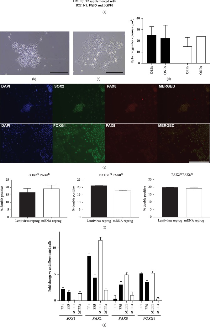
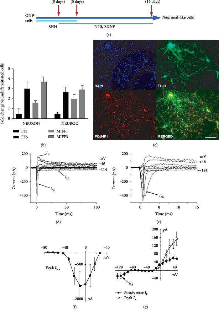
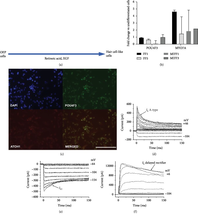
Damage to the sensory hair cells and the spiral ganglion neurons of the cochlea leads to deafness. Induced pluripotent stem cells (iPSCs) are a promising tool to regenerate the cells in the inner ear that have been affected by pathology or have been lost. To facilitate the clinical application of iPSCs, the reprogramming process should minimize the risk of introducing undesired genetic alterations while conferring the cells the capacity to differentiate into the desired cell type. Currently, reprogramming induced by synthetic mRNAs is considered to be one of the safest ways of inducing pluripotency, as the transgenes are transiently delivered into the cells without integrating into the genome. In this study, we explore the ability of integration-free human-induced pluripotent cell lines that were reprogrammed by mRNAs, to differentiate into otic progenitors and, subsequently, into hair cell and neuronal lineages. hiPSC lines were induced to differentiate by culturing them in the presence of fibroblast growth factors 3 and 10 (FGF3 and FGF10). Progenitors were identified by quantitative microscopy, based on the coexpression of otic markers PAX8, PAX2, FOXG1, and SOX2. Otic epithelial progenitors (OEPs) and otic neuroprogenitors (ONPs) were purified and allowed to differentiate further into hair cell-like cells and neurons. Lineages were characterised by immunocytochemistry and electrophysiology. Neuronal cells showed inward Na+ (I Na) currents and outward (I k) and inward K+ (I K1) currents while hair cell-like cells had inward I K1 and outward delayed rectifier K+ currents, characteristic of developing hair cells. We conclude that human-induced pluripotent cell lines that have been reprogrammed using nonintegrating mRNAs are capable to differentiate into otic cell types.


Conflict of interest statement

MNR is the founder of Rinri Therapeutics.

Figures

Figure 1
Figure 1
(a) Diagram showing the protocol for the generation of otic progenitors. Colonies of ONPs (b) and OEPs (c), displaying their typical morphology. Scale bar, 200 μm. (d) Bar chart displaying the relative yields of OEPs and ONPs per reprogramming condition (dark bars are lentivirus-reprogrammed lines, while open bars are for mRNA-reprogrammed ones). (e) Top: ONP colony stained for SOX2 and PAX8. Bottom: otic epithelial progenitors showing coexpression of FOXG1 and PAX8. Images shown are from the FF1 cell line. Scale bar, 400 μm. (f) Bar charts displaying percentages of double-positive otic progenitors, expressing high levels of SOX2/PAX8, FOXG1/PAX8, and PAX2/PAX8. (g) qPCR of otic progenitors showing relative levels of expression for SOX2, PAX2, PAX8, and FOXG1. Data shown as fold change against undifferentiated cells from the same starting population.
Figure 2
Figure 2
(a) Diagram showing the protocol for the generation of spiral ganglion-like neurons. (b) qPCR showing relative expression levels of NEUROG and NEUROD. Data shown as fold change against undifferentiated cells from the same initial population. (c) ONPs differentiating into neurons express markers such as TUJ1 and POU4F1, scale bar is 200 μm. Immunofluorescence images shown are from cell line FF5. (d) Electrophysiological properties of differentiating neuronal-like cells. Depolarizing and hyperpolarizing voltage steps in 10 mV nominal increments from a holding potential of -84 mV revealed Na+ currents (INa; (d), expanded in (e)) in 3 out of 3 neuronal-like cells, small outward K+ currents (IK; (d)) in 3 out of 3 cells and a small inward K+ current (IK1) in 1 out of 3 cells (d). The peak I-V curve for INa indicates that INa activated at potentials near -40 mV and reached a maximum size of 1328 ± 634 pA near -10 mV (n = 3; (f)). The steady state I-V curve for IK was also generated from a holding potential of -84 mV, and the current size at 0 mV and measured at 160 ms was 32 ± 10 pA, n = 3 (g). The small inward K+ current (IK1) was evident for membrane potentials negative to -80 mV (g).
Figure 3
Figure 3
(a) Diagram showing the protocol for the generation of hair cell-like cells. (b) qPCR showing relative expression levels of the hair cell markers POU4F3 and MYO7A. Data shown as fold change against undifferentiated cells from the same initial population. (c) ATOH1 and POU4F3 proteins are coexpressed by some cells. Immunofluorescence images shown are from cell line MIFF1. Scale bar, 200 μm (d). Electrophysiological properties of differentiating hair cell-like cells. To investigate the presence of outward and inward K+ currents, cells were recorded using the same voltage protocol described in Figure 2. Outward K+ currents were observed in 10 out of 12 hair cell-like cells; these currents were either an inactivating A-type K+ current (266 ± 62 pA at 0 mV, n = 7 out of 10 cells (d)) or a delayed rectifier current (476 ± 174 pA at 0 mV, n = 3 out of 10 cells (f)). Inward K+ currents (IK1: −307 ± 79 pA near -124 mV) were observed in 4 out of 12 hair cell-like cells ((e) holding potential -64 mV, same as the cell in (d)).

Similar articles

Cited by

References

    1. Li H., Roblin G., Liu H., Heller S. Generation of hair cells by stepwise differentiation of embryonic stem cells. Proceedings of the National Academy of Sciences of the United States of America. 2003;100(23):13495–13500. doi: 10.1073/pnas.2334503100. - DOI - PMC - PubMed
    1. Oshima K., Shin K., Diensthuber M., Peng A. W., Ricci A. J., Heller S. Mechanosensitive hair cell-like cells from embryonic and induced pluripotent stem cells. Cell. 2010;141(4):704–716. doi: 10.1016/j.cell.2010.03.035. - DOI - PMC - PubMed
    1. Ronaghi M., Nasr M., Ealy M., et al. Inner ear hair cell-like cells from human embryonic stem cells. Stem Cells and Development. 2014;23(11):1275–1284. doi: 10.1089/scd.2014.0033. - DOI - PMC - PubMed
    1. Koehler K. R., Mikosz A. M., Molosh A. I., Patel D., Hashino E. Generation of inner ear sensory epithelia from pluripotent stem cells in 3D culture. Nature. 2013;500(7461):217–221. doi: 10.1038/nature12298. - DOI - PMC - PubMed
    1. Chen W., Johnson S. L., Marcotti W., Andrews P. W., Moore H. D., Rivolta M. N. Human fetal auditory stem cells can be expanded in vitro and differentiate into functional auditory neurons and hair cell-like cells. Stem Cells. 2009;27(5):1196–1204. doi: 10.1002/stem.62. - DOI - PubMed

LinkOut - more resources