. 2020 Feb 29:2020:2390125.
doi: 10.1155/2020/2390125. eCollection 2020.

Antiangiogenesis Efficacy of Ethanol Extract from Amomum tsaoko in Ovarian Cancer through Inducing ER Stress to Suppress p-STAT3/NF-kB/IL-6 and VEGF Loop

Affiliations

Antiangiogenesis Efficacy of Ethanol Extract from Amomum tsaoko in Ovarian Cancer through Inducing ER Stress to Suppress p-STAT3/NF-kB/IL-6 and VEGF Loop

Cheng Chen et al. Evid Based Complement Alternat Med. .

Abstract

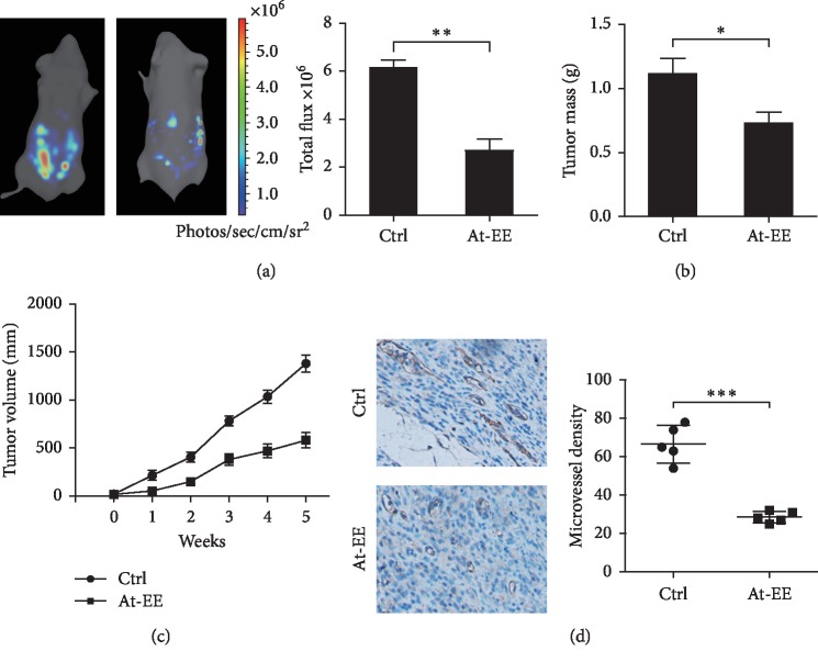
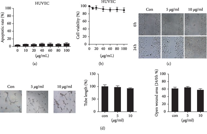
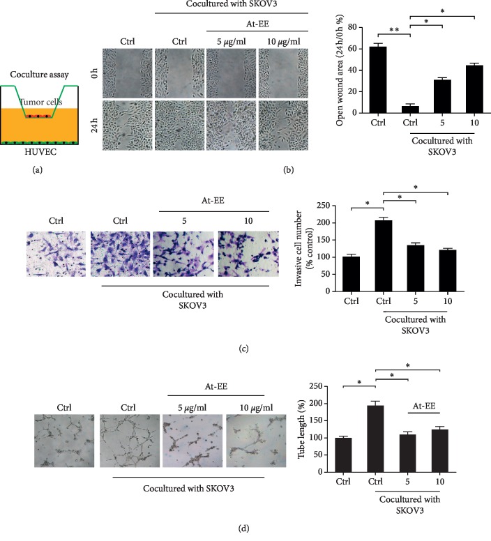
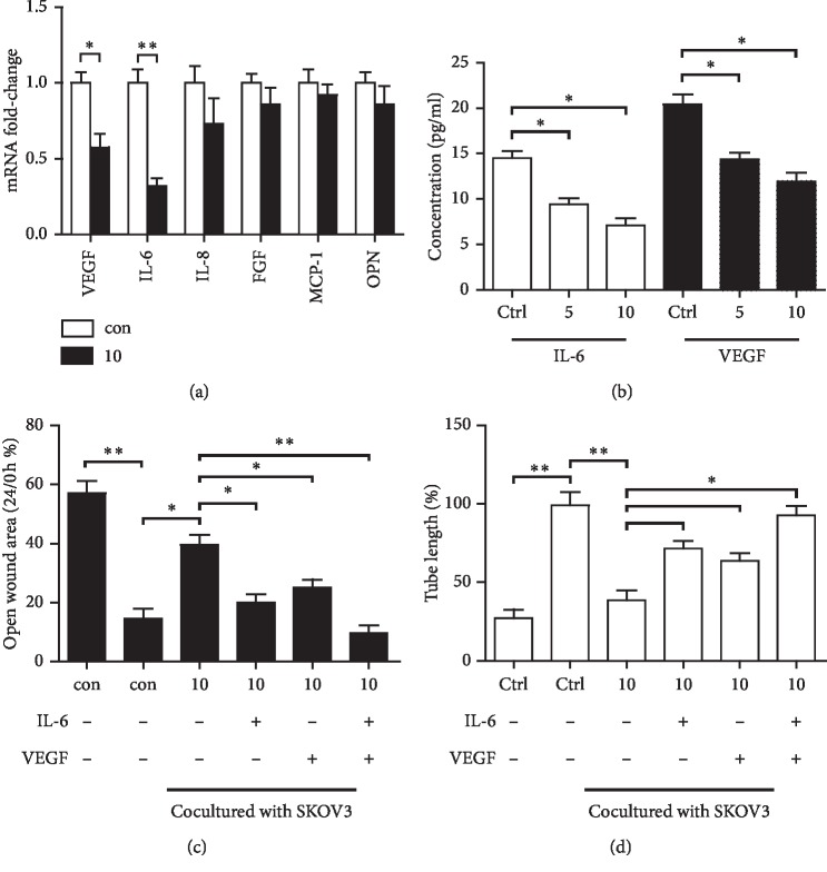
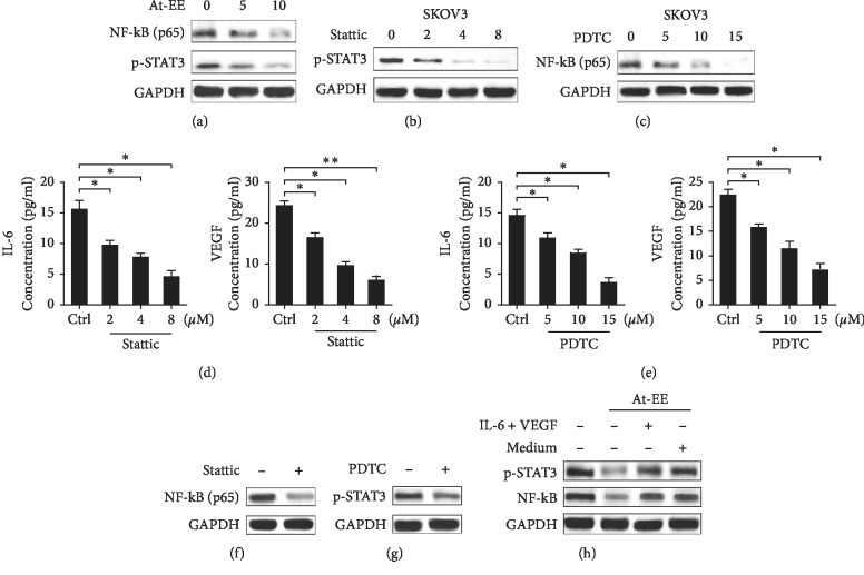
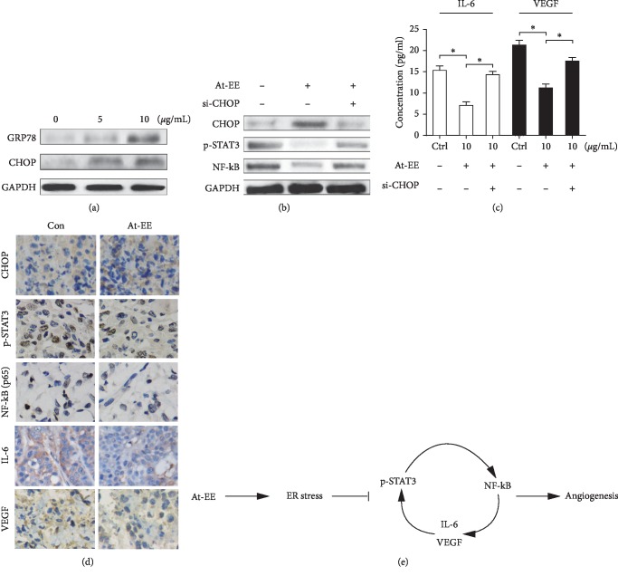
Natural plants are considered as a huge treasure for anticancer. Amomum tsaoko, a plant of Zingiberaceae, is used widely as a food and traditional medicine in East Asia. In previous studies, Amomum tsaoko has antitumor effect on liver cancer cells, but the mechanism is not clear. Here, we demonstrated that ethanol extract from Amomum tsaoko (At-EE) could inhibit ovarian cancer and decrease angiogenesis in vivo. At-EE did not influence vascular endothelial cells directly, but decreased IL-6 and VEGF secreted by ovarian cancer cells to inhibit angiogenesis through inhibition of p-STAT3 and NF-kB activation. In addition, we demonstrated that p-STAT3 and NF-kB could adjust each other and IL-6 and VEGF also mediate p-STAT3 and NF-kB too, which created a loop. In addition, At-EE interrupted p-STAT3/NF-kB/IL-6 and VEGF loop through induced ER stress. These results reveal that p-STAT3/NF-kB/IL-6 and VEGF is a cascade amplification loop in ovarian cancer for angiogenesis, and induced ER stress can interrupt it. Taken together, this work explored the anticancer activities of Amomum tsaoko, which could be a potential therapeutic candidate in the treatment of ovarian cancer.


Conflict of interest statement

The authors declare that they have no conflicts of interest.

Figures

Figure 1
Figure 1
At-EE had antitumor efficacy in ovarian cancer. (a, b) 3 × 106 cells were injected into the ovarian bursa of BALB/c nude mice, which were randomized into control group and At-EE group. (a) 4 weeks later, cancer cells in the abdominal cavity were counted by in vivo bioluminescence imaging. (b) After the experiment, the mice were sacrificed, and the tumors were resected. (c) 5 × 106 cells were inoculated into ovarian bursa of BALB/c nude mice, which were randomized into control group and At-EE group. (d) The expression of CD31 was detected by immunohistochemical staining. After finishing the experiment, tumor tissues from each mouse were resected for measurement. The results were similar in at least three independent experiments. p < 0.05. ∗∗p < 0.01.
Figure 2
Figure 2
At-EE did not influence HUVEC cells directly. (a–d) HUVEC cells were treated with DMSO or At-EE. (a) The apoptotic rate was assessed by flow cytometry. (b) The cell viability rate was analyzed by MTT assay. (c) Wound healing assays were done for the mobility of HUVEC cells. (d) In vitro angiogenesis evaluation was done for HUVECs treated by At-EE. The results were similar in at least three independent experiments. p < 0.05. ∗∗p < 0.01.
Figure 3
Figure 3
At-EE decreased ovarian cancer-mediated angiogenesis. (a–d) HUVEC cells were treated by condition media from ovarian cancer cells treated by At-EE or DMSO. (a) Representative diagram of the coculture assay. (b) Wound healing assays were done for the mobility of HUVEC cells. (c) Cell invasion assays were done for metastasis of HUVEC cells. (d) In vitro angiogenesis evaluation was done for HUVEC cells. The results were similar in at least three independent experiments. p < 0.05. ∗∗p < 0.01.
Figure 4
Figure 4
At-EE decreased IL-6 and VEGF production of ovarian cancer to inhibit angiogenesis. (a) SKOV3 was treated by At-EE for 48 h. Then, the mRNAs of IL-8, VEGF, FGF, MCP-1, OPN, and IL-6 were detected by real-time PCR. (b) SKOV3 was treated by At-EE for 48 h. Then, IL-6 and VEGF in conditioned media were detected by ELISA kits. (c) Wound healing assays were done for the mobility of HUVECs treated by condition media from ovarian cancer cells treated by At-EE in the presence or absence of IL-6, VEGF, or IL-6 and VEGF. (d) In vitro angiogenesis evaluation was done for HUVECs treated by condition media from ovarian cancer cells treated by At-EE in the presence or absence of IL-6, VEGF, or IL-6 and VEGF. The results were similar in at least three independent experiments. p < 0.05. ∗∗p < 0.01.
Figure 5
Figure 5
At-EE suppressed NF-kB/p-STAT3/IL-6 and VEGF loop. (a) Cells were treated with At-EE at 5 or 10 μg/ml for 48 h, and NF-kB and p-STAT3 were analyzed by western blot. (b) Cells were incubated with stattic at 2, 4, or 8 μM for 48 h, and p-STAT3 was analyzed by western blot. (c) Cells were incubated with PDTC at 5, 10, or 15 μM for 48 h, and NF-kB was analyzed by western blot. (d) SKOV3 was treated by stattic for 48 h. Then, IL-6 and VEGF in conditioned media were detected by ELISA kits. (e) SKOV3 was treated by PDTC for 48 h. Then, IL-6 and VEGF in conditioned media were detected by ELISA kits. (f) Cells were incubated with stattic, and NF-kB was analyzed by western blot. (g) Cells were incubated with PDTC, and p-STAT3 was analyzed by western blot. (h) Cells were treated with condition media form ovarian cancer cells treated by At-EE in presence or absence of IL-6 and VEGF, and NF-kB and p-STAT3 were analyzed by western blot. The results were similar in at least three independent experiments. p < 0.05. ∗∗p < 0.01.
Figure 6
Figure 6
At-EE induced ER stress to suppress NF-kB/p-STAT3/IL-6 and VEGF loop. (a) Cells were treated with At-EE at 5 or 10 μg/ml for 48 h, and GRP78 and CHOP were analyzed by western blot. (b) Cells or cells transfected with specific CHOP siRNA were treated by At-EE or DMSO, and NF-kB and p-STAT3 were analyzed by western blot. (c) Cells or cells transfected with specific CHOP siRNA were treated by At-EE or DMSO, and IL-6 and VEGF in conditioned media were detected by ELISA kits. (d) The expressions of p-STAT3, NF-kB, CHOP, VEGF, and IL-6 were detected by immunohistochemical staining. (e) Schematic model illustrating the potential pathway associated with At-EE inhibiting ovarian cancer cells. The results were similar in at least three independent experiments. p < 0.05. ∗∗p < 0.01.

Similar articles

Cited by

References

    1. Liu Y., Gong W., Yang Z. Y., et al. Quercetin induces protective autophagy and apoptosis through ER stress via the p-STAT3/Bcl-2 axis in ovarian cancer. Apoptosis. 2017;22(4):544–557. doi: 10.1007/s10495-016-1334-2. - DOI - PubMed
    1. Goff B. Measuring ovarian cancer care: why are we still failing? Gynecologic Oncology. 2015;136(1):1–2. doi: 10.1016/j.ygyno.2014.12.008. - DOI - PubMed
    1. Pradeep S., Kim S. W., Wu S. Y., et al. Hematogenous metastasis of ovarian cancer: rethinking mode of spread. Cancer Cell. 2014;26(1):77–91. doi: 10.1016/j.ccr.2014.05.002. - DOI - PMC - PubMed
    1. Mayer R. H., Castrogiovanni C., Weiss A., et al. Anti-angiogenic effects of crenolanib are mediated by mitotic modulation independently of PDGFR expression. British Journal of Cancer. 2019;121(2):139–149. doi: 10.1038/s41416-019-0498-2. - DOI - PMC - PubMed
    1. Olugbami J. O., Damoiseaux R., France B., et al. A comparative assessment of antiproliferative properties of resveratrol and ethanol leaf extract of Anogeissus leiocarpus (DC) Guill and Perr against HepG2 hepatocarcinoma cells. BMC Complementary and Alternative Medicine. 2017;17(1):p. 381. doi: 10.1186/s12906-017-1873-2. - DOI - PMC - PubMed

LinkOut - more resources