. 2020 Mar 3:2020:5108298.
doi: 10.1155/2020/5108298. eCollection 2020.

Aloe-Emodin Induces Breast Tumor Cell Apoptosis through Upregulation of miR-15a/miR-16-1 That Suppresses BCL2

Affiliations

Aloe-Emodin Induces Breast Tumor Cell Apoptosis through Upregulation of miR-15a/miR-16-1 That Suppresses BCL2

Xuefeng Jiang et al. Evid Based Complement Alternat Med. .

Abstract

Purpose: Aloe-emodin (AE) is a natural compound derived from aloe vera and palmatum rhubarb and shows anticancer activities in various cancers. Bcl-2 family is the main regulator of cell death or cell survival. This study describes the effects of AE on proliferation of breast tumor (BT) cells.

Methods: MCF-10A, MCF-10AT, MCF-7, and MDA-MB-231 cell lines were exposed to AE. Cell proliferation and apoptosis were assessed by CCK-8 and flow cytometry. Protein levels were measured by Western blotting. The levels of mRNA and miRNA were examined by RT-PCR. Bioinformatics was applied to screen miRNAs that bind to 3'-UTR of mRNA.

Results: The results showed that AE selective activity inhibited the proliferation and induced apoptosis of MCF-10AT and MCF-7 cells but exhibited no significant inhibition in MCF10A and MDA-MB-231 cells. Mechanistically, AE dose-dependently decreased the protein expression of Bcl-2 and Bcl-xl, while it increased Bax protein expression in MCF-10AT and MCF-7 cells. The levels of Bcl-xl and Bax mRNA were altered by AE treatment, which was consistent with the protein expression results. However, Bcl-2 mRNA levels were not affected in either cell line, suggesting that AE may modulate the protein translation of Bcl-2 through miRNAs. In all candidate miRNAs that bind to 3'-UTR of Bcl-2, miR-15a and miR-16-1 were dose-dependently downregulated by AE. Moreover, inhibition of miR-15a/16-1 could eliminate the inhibition of MCF-10AT and MCF-7 cells growth by AE and could reverse the downregulation of AE-induced Bcl-2 protein level.

Conclusion: Our research provides an important basis that AE induces BT cell apoptosis through upregulation of miR-15a/miR-16-1 that suppresses BCL2.


Conflict of interest statement

The authors declare no conflicts of interest regarding the publication of this paper.

Figures

Figure 1
Figure 1
Effects of AE on cells proliferation. (a) MCF-10A, MCF-10AT, MCF-7, and MDA-MB-231 cells were treated with increasing doses of AE (0–100 μM) for 72 h incubation. MCF-10AT (b) and MCF-7 (c) cells were treated with increasing doses of AE (0–100 μM) for 24 h, 48 h, and 72 h incubation. The data of cells viability were shown as mean ± SD of three independent experiments, P < 0.05.
Figure 2
Figure 2
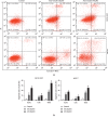
Effect of AE on apoptosis of MCF-10AT and MCF-7. (a) The cell apoptosis rates for AE-treated MCF-10AT and MCF-7 cells were assessed by FITC Annexin V/PI staining. (b) The MCF-10AT and MCF-7 cells in (a) apoptosis rates were quantified. The cell apoptosis rates were shown as the mean ± SD of three independent experiments, P < 0.05.
Figure 3
Figure 3
Effect of AE on the expression of Bcl-2, Bcl-xl, and Bax in MCF-10AT and MCF-7 cells. (a) Western blotting for Bcl-2, Bcl-xl, and Bax protein in MCF-10AT and MCF-7 cells treated with AE. (b) Relative proteins expression of Bcl-2, Bcl-xl, and Bax and the ratio of Bcl-2/Bax in (a). (c) The relative mRNAs expression of Bcl-2, Bcl-xl, and Bax and the ratio of Bcl-2/Bax after AE treatment in MCF-10AT and MCF-7 cells. The data were shown as mean ± SD of three independent experiments, P < 0.05.
Figure 4
Figure 4
miR-15a and miR-16-1 bind to 3′-UTR of Bcl-2 mRNA to suppress its protein translation. (a) RT-qPCR for miR-15a and miR-16-1 levels in MCF-10AT and MCF-7 cells treated with AE. (b) The expression of Bcl-2 protein in MCF-10AT and MCF-7 cells transfected with miR-15a or miR-16-1 mimics. The data were shown as mean ± SD of three independent experiments, P < 0.05.
Figure 5
Figure 5
AE inhibits MCF-10AT and MCF-7 cells growth through upregulation of miR-15a/16-1 that suppresses Bcl-2. (a) Inhibition of miR-15a/16-1 eliminates the inhibition of AE on MCF-10AT and MCF-7 cells growth. (b) The expression of Bcl-2 protein in MCF-10AT and MCF-7 cells with different treatments were measured by Western blot. The data were shown as mean ± SD of three independent experiments, P < 0.05.
Figure 6
Figure 6
Schematic diagram of the mechanism underlying this study. AE induces BT cell apoptosis through regulation of miR-15a/16-1/Bcl-2 signaling.

Similar articles

Cited by

References

    1. Bray F., Ferlay J., Soerjomataram I., Siegel R. L., Torre L. A., Jemal A. Global cancer statistics 2018: GLOBOCAN estimates of incidence and mortality worldwide for 36 cancers in 185 countries. CA: A Cancer Journal for Clinicians. 2018;68(6):394–424. doi: 10.3322/caac.21492. - DOI - PubMed
    1. Fan L., Strasser-Weippl K., Li J.-J., et al. Breast cancer in China. The Lancet Oncology. 2014;15(7):e279–e289. doi: 10.1016/s1470-2045(13)70567-9. - DOI - PubMed
    1. Waks A. G., Winer E. P. Breast cancer treatment. JAMA. 2019;321(3):288–300. doi: 10.1001/jama.2018.19323. - DOI - PubMed
    1. Bartel D. P. MicroRNAs: target recognition and regulatory functions. Cell. 2009;136(2):215–233. doi: 10.1016/j.cell.2009.01.002. - DOI - PMC - PubMed
    1. Baer C., Claus R., Plass C. Genome-wide epigenetic regulation of miRNAs in cancer. Cancer Research. 2013;73(2):473–477. doi: 10.1158/0008-5472.can-12-3731. - DOI - PubMed