Review
. 2020 Mar 13:14:8.
doi: 10.1186/s13036-020-0227-7. eCollection 2020.

Alginate-based hydrogels as drug delivery vehicles in cancer treatment and their applications in wound dressing and 3D bioprinting

Affiliations
Review

Alginate-based hydrogels as drug delivery vehicles in cancer treatment and their applications in wound dressing and 3D bioprinting

Farhad Abasalizadeh et al. J Biol Eng. .

Erratum in

Abstract

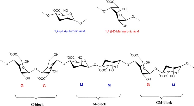
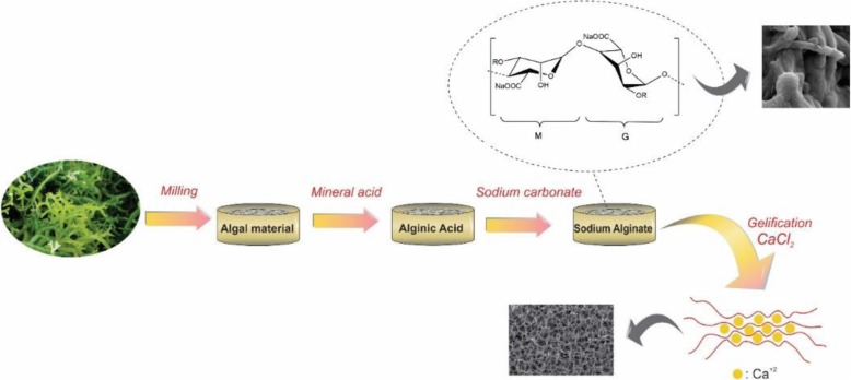
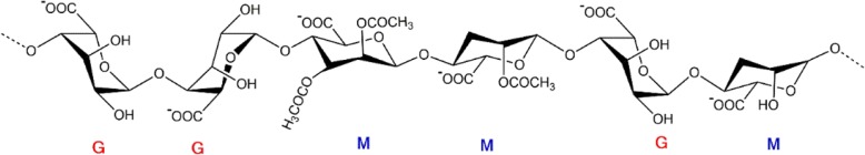
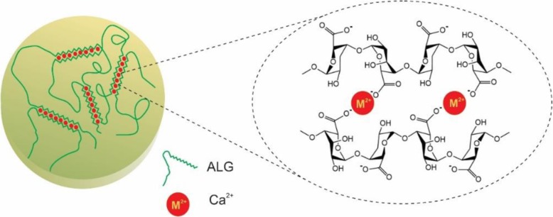
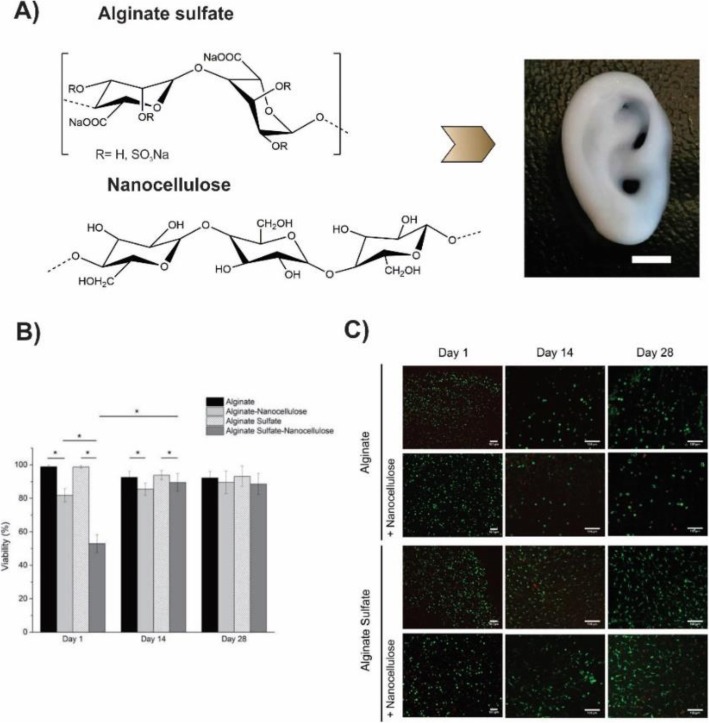
Hydrogels are a three-dimensional and crosslinked network of hydrophilic polymers. They can absorb a large amount of water or biological fluids, which leads to their swelling while maintaining their 3D structure without dissolving (Zhu and Marchant, Expert Rev Med Devices 8:607-626, 2011). Among the numerous polymers which have been utilized for the preparation of the hydrogels, polysaccharides have gained more attention in the area of pharmaceutics; Sodium alginate is a non-toxic, biocompatible, and biodegradable polysaccharide with several unique physicochemical properties for which has used as delivery vehicles for drugs (Kumar Giri et al., Curr Drug Deliv 9:539-555, 2012). Owing to their high-water content and resembling the natural soft tissue, hydrogels were studied a lot as a scaffold. The formation of hydrogels can occur by interactions of the anionic alginates with multivalent inorganic cations through a typical ionotropic gelation method. However, those applications require the control of some properties such as mechanical stiffness, swelling, degradation, cell attachment, and binding or release of bioactive molecules by using the chemical or physical modifications of the alginate hydrogel. In the current review, an overview of alginate hydrogels and their properties will be presented as well as the methods of producing alginate hydrogels. In the next section of the present review paper, the application of the alginate hydrogels will be defined as drug delivery vehicles for chemotherapeutic agents. The recent advances in the application of the alginate-based hydrogels will be describe later as a wound dressing and bioink in 3D bioprinting.

Keywords: 3D bioprinting; Alginate hydrogels; Cancer; Drug delivery; Wound dressing.


Conflict of interest statement

Competing interestsThe authors declare that they have no competing interests.

Figures

Fig. 1
Fig. 1
The conformation of monomers and blocks distribution of alginate salt
Fig. 2
Fig. 2
A typical process for the extraction of sodium alginate from brown algae followed by gellification in the presence of CaCl2
Fig. 3
Fig. 3
Chemical structure and conformation of guluronic acid (G) and mannuronic acid (M) residue in bacterial alginate
Fig. 4
Fig. 4
Egg-box structure for alginate gelation as a result of ionic interaction between alginate and a divalent cation
Fig. 5
Fig. 5
Schematic showing of covalent crosslinking of alginate using adipic acid dihydrazide as cross-linker
Fig. 6
Fig. 6
Schematic of the temperature-dependent property of NH2-PNIPAM-g-Alg polymer. PNIPAM = poly(N-isopropylacrylamide)
Fig. 7
Fig. 7
Schematic showing of construction of cell crosslinked hydrogel of ligand modified alginate
Fig. 8
Fig. 8
Schematic representation of photocrosslinking of methacrylated alginate
Fig. 9
Fig. 9
Release and cytotoxicity assessment of EPI using EPI & AG-G5 nanogels as a pH-responsive carrier. a Schematic representation of EPI & AG-G5 nanogels synthesis. b Time-dependent cumulative release profiles of EPI from nanogels at two pH 7.4 and 5.5. c viability assessment of MCF-7 cells after 48 h incubation with the appropriate amount of nanogels equivalent to EPI concentrations. Statistical significance was carried out by Two-way ANOVA with Tukey’s multiple comparisons test between groups using GraphPad Prism 6.0 software. Statistically significant values were denoted by * (p < 0.05), ** (p < 0.005), and *** (p < 0.001). Statistically insignificant values were represented by ‘ns’ [101]. Matai, I. and P. Gopinath, Chemically cross-linked hybrid nanogels of alginate and PAMAM dendrimers as efficient anticancer drug delivery vehicles. ACS Biomaterials Science & Engineering. 2016, 2(2):213–223. Copyright (2020)
Fig. 10
Fig. 10
a Schematic representation of Alginate-Gelatin composite generated with the Schiff- base reaction. b Illustration of the self-healing process in the Oxidized Alginate-Gelatin type B (OxA-GB) hydrogel. The bottom left image depicts the crosslinked hydrogel cutting in three pieces. The bottom right image shows the stretching of the hydrogel after the recoupling of the pieces (optimum healing time: 7 days). c Schematic representation of the OxA crosslinking with gelatin in the presence of borax [64]. Pettignano, A., et al., Self-healing alginate–gelatin biohydrogels based on dynamic covalent chemistry: elucidation of key parameters. Materials Chemistry Frontiers. 2017, 1(1):73–79. Copyright (2020)
Fig. 11
Fig. 11
Schematic of DOX loaded hybrid cross-linked alginate/liposome past on tongue tissue and displaying deferent hybrid systems of alginate with Rhodamine labeled liposomes (pink) after 2 h release. a past b cross-linked past [100]. Shtenberg, Y., et al., Mucoadhesive alginate pastes with embedded liposomes for local oral drug delivery. International journal of biological macromolecules. 2018, 111:62–69. Copyright (2020)
Fig. 12
Fig. 12
Cytotoxicity assessment of DOX using dual-crosslinked Alg-MA sub-microspheres as chemotherapeutic delivery vehicles. a Chemical structure of dual-crosslinked Alg-MA hydrogel networks. b Schematic representation of microsphere fabrication techniques. Premixing of Alg-MA solutions with or without DOX was followed by water/oil emulsion at room temperature generated microspheres. Alg-MA sub-microspheres were photo-crosslinked upon the exposure to visible or UV light, respectively, and further dual-crosslinked in the presence of 1 M CaCl2. c MTT-based assay of DOX loaded dual-crosslinked Alg-MA sub-microspheres to quantify the cell proliferation over a 5-day period. A549 activity was recorded as the mitochondrial activity and normalized to the non-modified cell controls. Various formulations and concentrations (10–100 μg/mL) of the sub-microspheres were assessed: green photo-crosslinked (Green), green + Ca2+ dual-crosslinked (Green+C), UV photo-crosslinked (UV), UV + Ca2+ dual-crosslinked (UV + C). DOX was added exogenously (Free DOX) to the cell culture medium at the various concentrations to test the effects of the intracellular versus extracellular DOX delivery [107]. Fenn, S.L., et al., Dual-cross-linked methacrylated alginate sub-microspheres for intracellular chemotherapeutic delivery. ACS applied materials & interfaces. 2016, 8(28):17775–17,783, Copyright (2020)
Fig. 13
Fig. 13
a Representative photographs of wound healing mice skin at different times (day 0, day 3, day 7, day 9, and day 12) for untreated wound (no dressing), NaAlg/PVPI and Product A (A commercial Povidone Iodine Non-Adherent Dressing product) as control sample are shown (scale bar 5 nm) b Days of wound healing in mice untreated (white bars), and treated with the NaAlg/PVPI (black bars) or with Product A dressing (grey bars). *p < 0.05 and ***p < 0.001, compared to the untreated mice [117]. Summa, M., et al., A biocompatible sodium alginate/povidone iodine film enhances wound healing. European Journal of Pharmaceutics and Biopharmaceutics. 2018, 122:17–24. Copyright (2020)
Fig. 14
Fig. 14
A) Different types of bioprinting techniques and their application in organ systems. (a) Inkjet bioprinting method (b) Laser-assisted bioprinting method (c) Extrusion bioprinting method (d) Bio-electrospraying/Cell electrospinning [124]. B) The illustration of the bioink based on the alginate (composed of the cells, alginate hydrogel, and—optionally—functional peptides to improve the cell’s biological function) [126]. Republished with permission of ref. [124], Hong, N., et al., 3D bioprinting and its in vivo applications. Journal of Biomedical Materials Research Part B: Applied Biomaterials. 2018, 106(1): 444–459, Copyright (2020)
Fig. 15
Fig. 15
A) Schematic illustration of the alginate-chitosan (Al Ch) polyionic complex hydrogel as bioink in 3D bioprinting. B) Morphological characterization of 3D bioprinted Al Ch a) 3D model of nose b) 3D printed nose, constructed by Al1Ch1.2 bioink and SEM micrograph of 3D printed Al1Ch1.0 bioink with different angles between the filaments (C1, C2) 45°, (d1, d2) 60°, (e1, e2) 90° (1,2 respectively represent front and side of the scaffold) biocompatibility of the 3D bioprinted AlCh polyionic hydrogel. C) Human adipose-derived stem cells (hASCs) were used to test the biocompatibility of the scaffold. Photographs of the inverted fluorescence microscope which represent a) live b) dead c) merged cells on the 3rd day. As shown in the pictures, the live cells distributed uniformly on the hydrogel while little or no dead cells existed. d) proliferation of hASCs distributed on the 3D bioprinted hydrogel. It showed that hASCs could proliferate during the time [127]. Liu, Q., et al., Preparation and Properties of 3D Printed Alginate–Chitosan Polyion Complex Hydrogels for Tissue Engineering. Polymers. 2018, 10(6):664.Copyright (2020)
Fig. 16
Fig. 16
a Chemical structure of the alginate sulfate and nanocellulose. Incorporation of the two biomaterials generates an ideal bioink which is appropriate for the 3D bioprinting of the complex constructs. Here a miniature size eare was 3D bioprinted (scale bar is 5 mm). b and c Viability assessment of chondrocytes and Live/dead staining of bovine chondrocytes respectively, encapsulated in alginate and alginate sulfate with or without nanocelloluse after 1, 14 and 28 days of culture. The scale bar is 100 μm [128]
Fig. 17
Fig. 17
a Chemical structure of the alginate sulfate and nanocellulose. Incorporation of the two biomaterials generates an ideal bioink which is appropriate for the 3D bioprinting of complex constructs. Here a miniature size eare was 3D bioprinted (scale bar is 5 mm). b and c Viability assessment of chondrocytes and Live/dead staining of bovine chondrocytes respectively, encapsulated in alginate and alginate sulfate with or without nanocelloluse after 1, 14 and 28 days of culture. The scale bar is 100 μm
Fig. 18
Fig. 18
Patient-Specific Platelet-Rich Plasma (PRP) bioink using 3D bioprinting of alginate scaffold a Schematic of PRP extraction and its incorporation with alginate to form patient-specific bioink b Schematic of proposed bioprinting process c PRP incorporated alginate scaffold containing fluorescence particles. d Images of different PRP-alginate constructs. In the production of these constructs 0.04% (w/v) CaCl2, 50 U ml− 1 PRP, and 1% (w/v) alginate was used. e, f The fabricated constructs could easily be removed from the substrate without losing their integrity. g Metabolic activity of mesenchymal stem cells (MSCs) treated with alginate and alginate/PRP over 5 days without any growth factor. h Metabolic activity of human umbilical vein endothelial cells (HUVECs) treated with alginate and alginate/PRP over 3 days without any growth factor. (*P < 0.05; **P < 0.01, ***P < 0.001) [129]. Faramarzi, N., et al., Patient-Specific Bioinks for 3D Bioprinting of Tissue Engineering Scaffolds. Advanced healthcare materials, 2018. 7(11), Copyright (2020)

References

    1. Zhu J, Marchant RE. Design properties of hydrogel tissue-engineering scaffolds. Expert Rev Med Devices. 2011;8(5):607–626. doi: 10.1586/erd.11.27. - DOI - PMC - PubMed
    1. Kumar Giri T, et al. Alginate based hydrogel as a potential biopolymeric carrier for drug delivery and cell delivery systems: present status and applications. Curr Drug Deliv. 2012;9(6):539–555. doi: 10.2174/156720112803529800. - DOI - PubMed
    1. Kulkarni RV, Sa B. Enteric delivery of ketoprofen through functionally modified poly(acrylamide-grafted-xanthan)-based pH-sensitive hydrogel beads: preparation, in vitro and in vivo evaluation. J Drug Target. 2008;16(2):167–177. doi: 10.1080/10611860701792399. - DOI - PubMed
    1. Kulkarni RV, et al. Interpenetrating network hydrogel membranes of sodium alginate and poly(vinyl alcohol) for controlled release of prazosin hydrochloride through skin. Int J Biol Macromol. 2010;47(4):520–527. doi: 10.1016/j.ijbiomac.2010.07.009. - DOI - PubMed
    1. Blandino A, Macias M, Cantero D. Glucose oxidase release from calcium alginate gel capsules. Enzym Microb Technol. 2000;27(3–5):319–324. doi: 10.1016/S0141-0229(00)00204-0. - DOI - PubMed

LinkOut - more resources