Review
. 2020 Mar 1:2020:3579143.
doi: 10.1155/2020/3579143. eCollection 2020.

Oxidative Stress in Radiation-Induced Cardiotoxicity

Affiliations
Review

Oxidative Stress in Radiation-Induced Cardiotoxicity

Zhang Ping et al. Oxid Med Cell Longev. .

Abstract

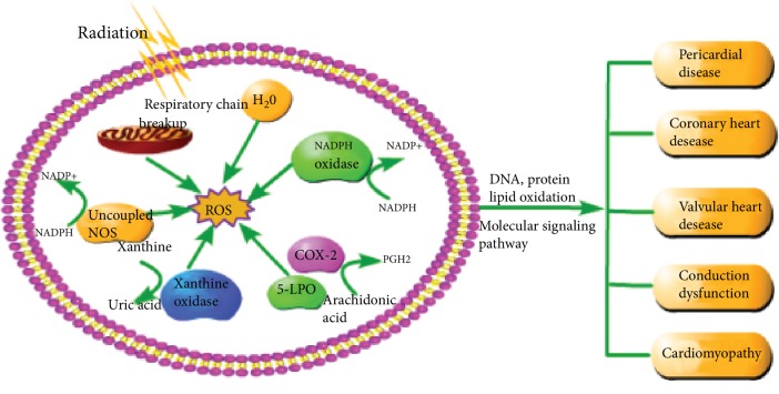
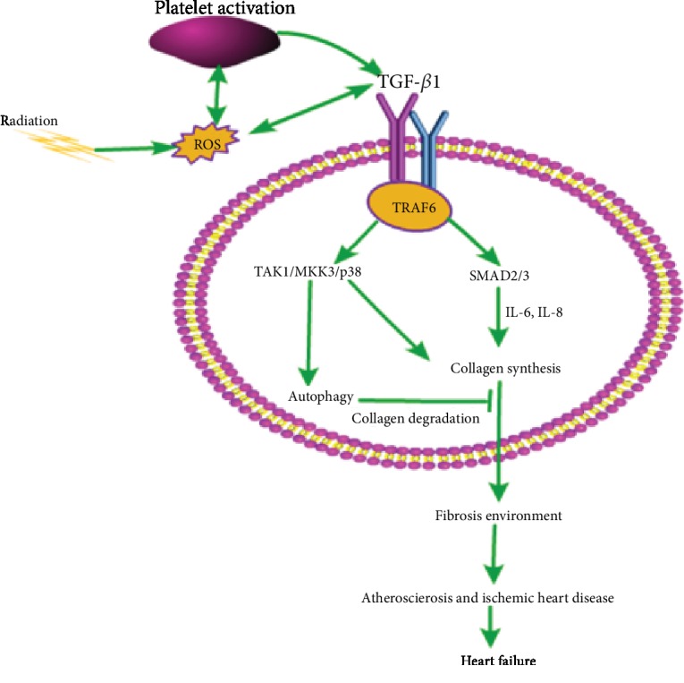
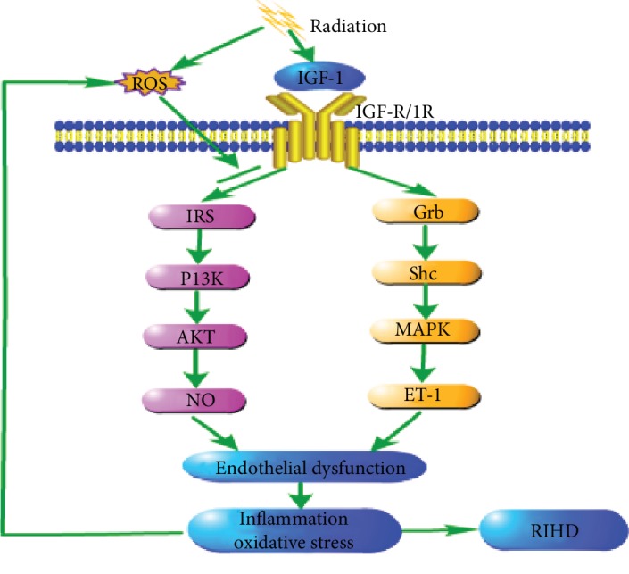
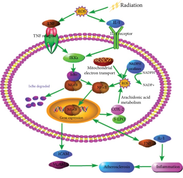
There is a distinct increase in the risk of heart disease in people exposed to ionizing radiation (IR). Radiation-induced heart disease (RIHD) is one of the adverse side effects when people are exposed to ionizing radiation. IR may come from various forms, such as diagnostic imaging, radiotherapy for cancer treatment, nuclear disasters, and accidents. However, RIHD was mainly observed after radiotherapy for chest malignant tumors, especially left breast cancer. Radiation therapy (RT) has become one of the main ways to treat all kinds of cancer, which is used to reduce the recurrence of cancer and improve the survival rate of patients. The potential cause of radiation-induced cardiotoxicity is unclear, but it may be relevant to oxidative stress. Oxidative stress, an accumulation of reactive oxygen species (ROS), disrupts intracellular homeostasis through chemical modification and damages proteins, lipids, and DNA; therefore, it results in a series of related pathophysiological changes. The purpose of this review was to summarise the studies of oxidative stress in radiotherapy-induced cardiotoxicity and provide prevention and treatment methods to reduce cardiac damage.


Conflict of interest statement

The authors declare that they have no conflict of interest.

Figures

Figure 1
Figure 1
Formation of ROS after radiation and the general manifestation of cardiotoxicity. As described in the article, the sources of ROS are varied, even under the radiation conditions. IR can directly cause the respiratory chain of mitochondria to breakup and cause the decomposition of water molecules, leading to respiratory chain dysfunction and ROS production, reducing antioxidant capacity. NADPH oxidase is a family of multisubunit complex enzymes that catalyze the conversion of oxygen into O2 by using NADPH as an electron source, which is present in vascular endothelia cells, smooth muscle cells, fibroblasts, and cardiomyocytes. The mechanism of uncoupled NOSs is similar to NADPH oxidase. Xanthine oxidase catalyzes the conversion of xanthine to uric acid while H2O2 and O2 are generated at the same time. In addition, COX-2 and 5-LPO produce PGH2 accompanied by ROS formation during catalytic arachidonic acid metabolism. ROS can interact with macrobiomolecules (DNA, protein, and lipid), causing oxidation of DNA, proteins, and lipids and cause cardiac damage through some signaling pathways, which are described in article above. The cardiac damage includes pericardial disease, coronary heart disease, heart valve disease, conduction disorders, and cardiomyopathy.
Figure 2
Figure 2
Oxidative stress in TGF-β1-mediated fibrosis. Radiation induces reactive oxygen species (ROS), and ROS can activate TGF-β1, resulting in the generation of thrombin and the activation of platelets to promote further production of ROS and TGF-β1. TGF-β1 binds to Type I (TbRI) and Type II (TbRII) transmembrane receptors, activating the Smad pathway and the TAK1/MKK3/p38 pathway through the specific nuclear signaling transduction molecule, TRAF6. The Smad pathway requires a number of inflammatory cytokines, in particular IL-6 and IL-8, to mediate collagen synthesis. TGF-β1 activates the TAK1/MKK3/p38 signaling pathway and downregulates autophagy-mediated collagen degradation, which may further promote the synthesis of collagen, form a fibrotic environment, cause atherosclerosis and ischemic heart disease, and may even lead to heart failure.
Figure 3
Figure 3
Radiation induced IGF-1 selective resistance. Insulin-like growth factor-1 (IGF-1) is structurally similar to insulin. It can activate the IRS/PI3K/Akt pathway (PI3K dependent) and the Grb/Shc/MAPK pathway (PI3K independent) through IGF-R, IR, or the hybrid receptor. However, radiation can inhibit the anti-inflammatory and antioxidant stress of the IRS/PI3K/Akt signaling pathway but not inhibit the inflammatory MAPK kinase pathway, which leads to the transformation of the IGF-1 signal to the oxidation and inflammatory environment, which leads to selective resistance of IGF-1. ROS can also cause selective inhibition of IRS/PI3K/Akt pathway and even enhance the MAPK signaling pathway. IGF-1 selective resistance causes an imbalance between NO and ET-1 production, resulting in endothelial dysfunction, resulting in oxidative stress and inflammatory level and further leads to the occurrence and development of radiation-induced heart diseases.
Figure 4
Figure 4
The relationship between oxidative stress and NF-κB. In normal cases, NF-κB combines with the inhibitor protein (IKB) to form a protein complex and remain in an inactive state. However, when the cells are exposed to ionizing radiation, oxidative stress reactions, such as NADPH oxidase activity, improved and mitochondrial electron transport is impaired and occurs rapidly, and reactive oxygen species is excessively generated in response to oxidative stress, which stimulates the inflammatory cells to produce IL-1 and TNF. IL-1 and TNF bind to the IL-1 receptor and the TNF receptor, respectively, to activate the downstream NF-κB signal. The IKB protein is phosphorylated and NF-κB is activated and translocated to the nucleus, which binds to the various promoter regions of its target gene and induces the transcription of a corresponding inflammation-related gene. The activated NF-κB induced the expression of COX-2 and 5-LPO, resulting in the generation of ROS, the formation of a positive feedback loop, and increased levels of inflammation and oxidative stress, and the intracellular ROS can activate NF-κB directly. In addition, the activation of NF-κB also results in an increase in the expression of intercellular adhesion molecules (ICAM) and vascular cell adhesion molecules (VCAM). In addition, NF-κB upregulated the expression of cytokines such as IL-1 and TNF. These cytokines increase inflammation not only by attracting white blood cells but also by activating NF-κB. Thus, the inflammation state of atherosclerosis may again form a positive self-regulatory loop, forming proinflammatory and fibrotic environments, leading to tissue remodeling, myocardial fibrosis, and atherosclerosis.

References

    1. Armanious M. A., Mohammadi H., Khodor S., Oliver D. E., Johnstone P. A., Fradley M. G. Cardiovascular effects of radiation therapy. Current Problems in Cancer. 2018;42(4):433–442. doi: 10.1016/j.currproblcancer.2018.05.008. - DOI - PubMed
    1. Puukila S., Lemon J. A., Lees S. J., Tai T. C., Boreham D. R., Khaper N. Impact of ionizing radiation on the cardiovascular system: a review. Radiation Research. 2017;188(4.2):539–546. doi: 10.1667/RR14864.1. - DOI - PubMed
    1. Weintraub N. L., Jones W. K., Manka D. Understanding radiation-induced vascular disease. Journal of the American College of Cardiology. 2010;55(12):1237–1239. doi: 10.1016/j.jacc.2009.11.053. - DOI - PMC - PubMed
    1. Russell N. S., Hoving S., Heeneman S., et al. Novel insights into pathological changes in muscular arteries of radiotherapy patients. Radiotherapy and Oncology. 2009;92(3):477–483. doi: 10.1016/j.radonc.2009.05.021. - DOI - PubMed
    1. Carr Z. A., Land C. E., Kleinerman R. A., et al. Coronary heart disease after radiotherapy for peptic ulcer disease. International Journal of Radiation Oncology, Biology, Physics. 2004;61(3):842–850. - PubMed

MeSH terms

LinkOut - more resources