

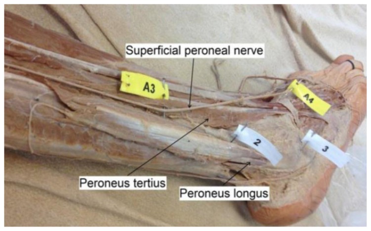
Superficial peroneal nerve compression due to peroneus brevis muscle herniation
- PMID: 32190181
- PMCID: PMC7060001
- DOI: 10.3941/jrcr.v13i11.3757
Superficial peroneal nerve compression due to peroneus brevis muscle herniation
Abstract
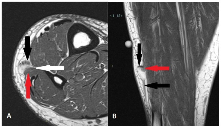
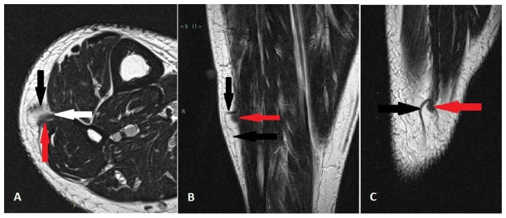
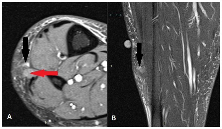
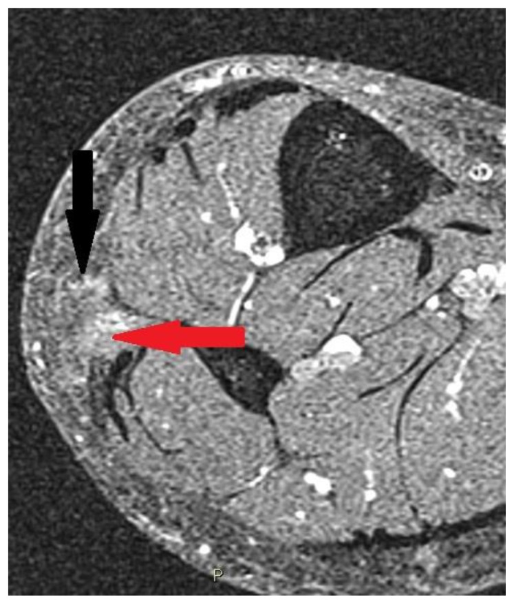
Muscle hernias of the extremities most commonly occur in the leg, between the knee and ankle. Symptomatic muscle hernias in the leg are rare cause of chronic leg pain and neuropathy, and not routinely encountered in surgical practice. Although this condition is especially an esthetic problem, with palpable subcutaneous soft tissue mass, it can lead to spontaneous pain, cramp, local tenderness or potentially neuropathic symptoms. Moreover, among leg muscles involved in this process, peroneus brevis is less frequent than tibialis anterior. Magnetic Resonance Imaging is the method of choice in establishing the diagnosis. Symptomatic cases can be treated surgically in different ways, the preferred one is nerve releasing with fasciotomy. The purpose of this case report is to present the Magnetic Resonance findings of a superficial nerve compression due to a peroneus brevis muscle herniation.
Keywords: Fasciotomy; Magnetic Resonance Imaging; Muscle Hernia; Nerve Releasing; Peripheral Neuropathy.
Copyright Journal of Radiology Case Reports.
Figures
References
-
- Paolasso I, Cambise C, Coraci D, et al. Tibialis anterior muscle herniation with superficial peroneal nerve involvement: ultrasound role for diagnosis and treatment. Clin Neurol Neurosurg. 2016 Dec;151:6–8. - PubMed
-
- Oktay B, Tevfik Y, Murat G, Mehmet K. Muscle herniation of the extremities: two case report. Prog Orthop SCI. 2016;2(1):4–7.
-
- Toms AF, Rushton LA, Kennedy NR. Muscle herniation of the peroneus longus muscle triggering superficial fibular nerve paresthesia. Sonography. 2017;5:36–40.
Publication types
MeSH terms
LinkOut - more resources
Full Text Sources
Medical