

Cleft foot: A case report and review of literature
- PMID: 32190557
- PMCID: PMC7063450
- DOI: 10.5312/wjo.v11.i2.129
Cleft foot: A case report and review of literature
Abstract
Background: Cleft foot is a very rare congenital anomaly, which is characterized by central rays deficiency of the foot. It is also known as split foot or ectrodactyly of the foot, and it is very often combined with splitting of the hands. The defect develops due to insufficient activity of the median apical ectodermal ridge, which leads to an increase in cell death or a decrease in cell proliferation. Due to the rarity of the pathology, there are few papers on the surgical treatment of this congenital foot disease, and publications to date concern the treatment of children.
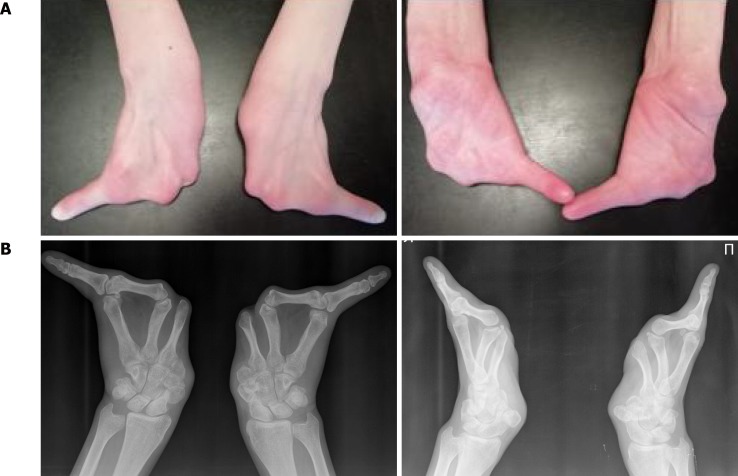
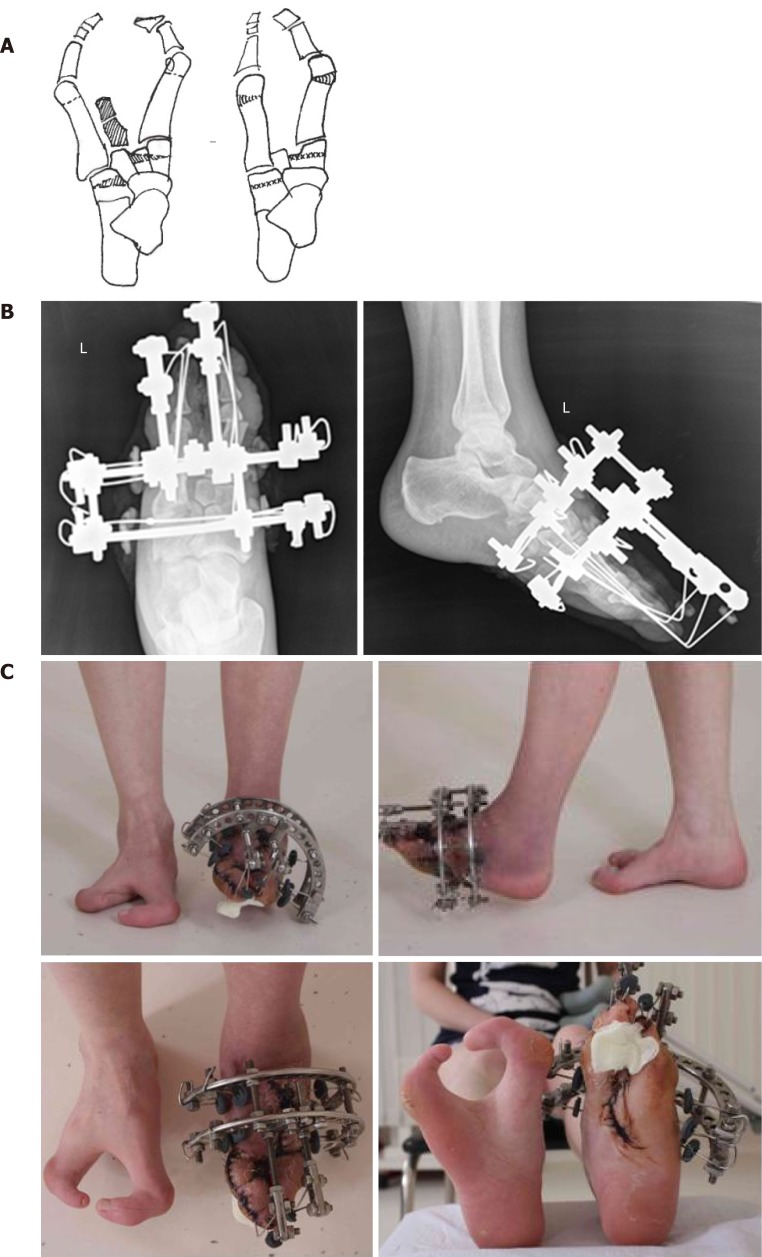
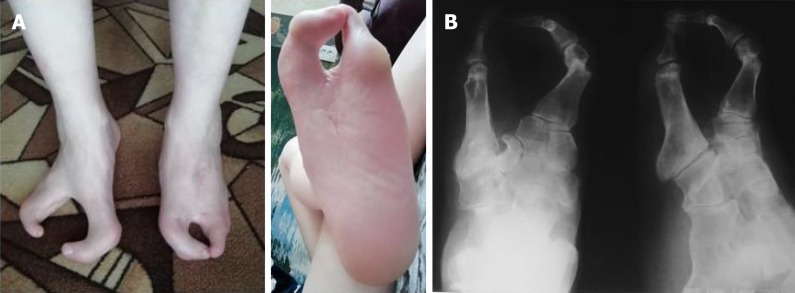
Case summary: We present a clinical case of congenital splitting of the feet and hands in a 31-year-old woman and a long-term result of foot treatment using the minimal arrangement of the Ilizarov apparatus. The patient had paternal inheritance of the trait. After the surgical treatment, cosmetic view and functional condition of the foot were improved and persisted two years after intervention. There were no complications in the treatment process.
Conclusion: The possibility of dosed control and stable fixation of the foot rays made it possible to create favorable conditions for the healing of the central wound and the closure of the segment splitting without complications. The long-term outcome of the treatment of foot congenital splitting using the proposed Ilizarov apparatus arrangement has shown its effectiveness. Our approach should be considered as an option of treatment in similar cases.
Keywords: Case report; Cleft foot; Congenital malformation; Ectrodactyly; Ilizarov; Split foot.
©The Author(s) 2020. Published by Baishideng Publishing Group Inc. All rights reserved.
Conflict of interest statement
Conflict-of-interest statement: There is no conflict of interest.
Figures

References
-
- Hartsinck G. Beschryving van Guiana of de wilde Just iin Zuid America. Vol 2. Amsterdam: Gerrit Teilenburg; 1770.
-
- Blauth W, Borisch NC. Cleft feet. Proposals for a new classification based on roentgenographic morphology. Clin Orthop Relat Res. 1990;258:41–48. - PubMed
-
- Basel D, Kilpatrick MW, Tsipouras P. The expanding panorama of split hand foot malformation. Am J Med Genet A. 2006;140:1359–1365. - PubMed
-
- Durmaz MS, Demirtaş H, Hattapoğlu S, Kara T, Göya C, Adin ME. Bilateral cleft foot: Radiographic and prenatal ultrasound features of two siblings with a review of literature. Medicina (Kaunas) 2016;52:257–261. - PubMed
Publication types
LinkOut - more resources
Full Text Sources
