. 2020 Apr 8;142(14):6554-6568.
doi: 10.1021/jacs.9b11622. Epub 2020 Mar 30.

Quantitative Control of Gene-Engineered T-Cell Activity through the Covalent Attachment of Targeting Ligands to a Universal Immune Receptor

Quantitative Control of Gene-Engineered T-Cell Activity through the Covalent Attachment of Targeting Ligands to a Universal Immune Receptor

Nicholas G Minutolo et al. J Am Chem Soc. .

Abstract

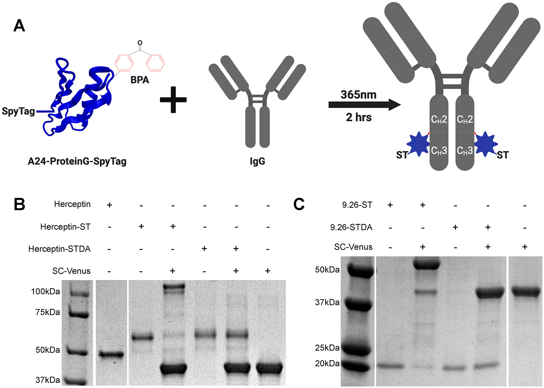
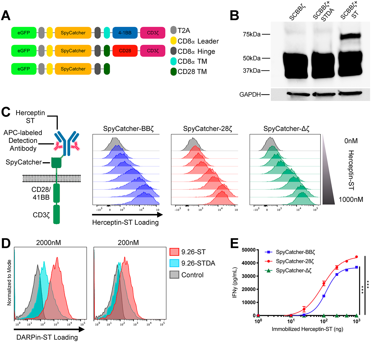
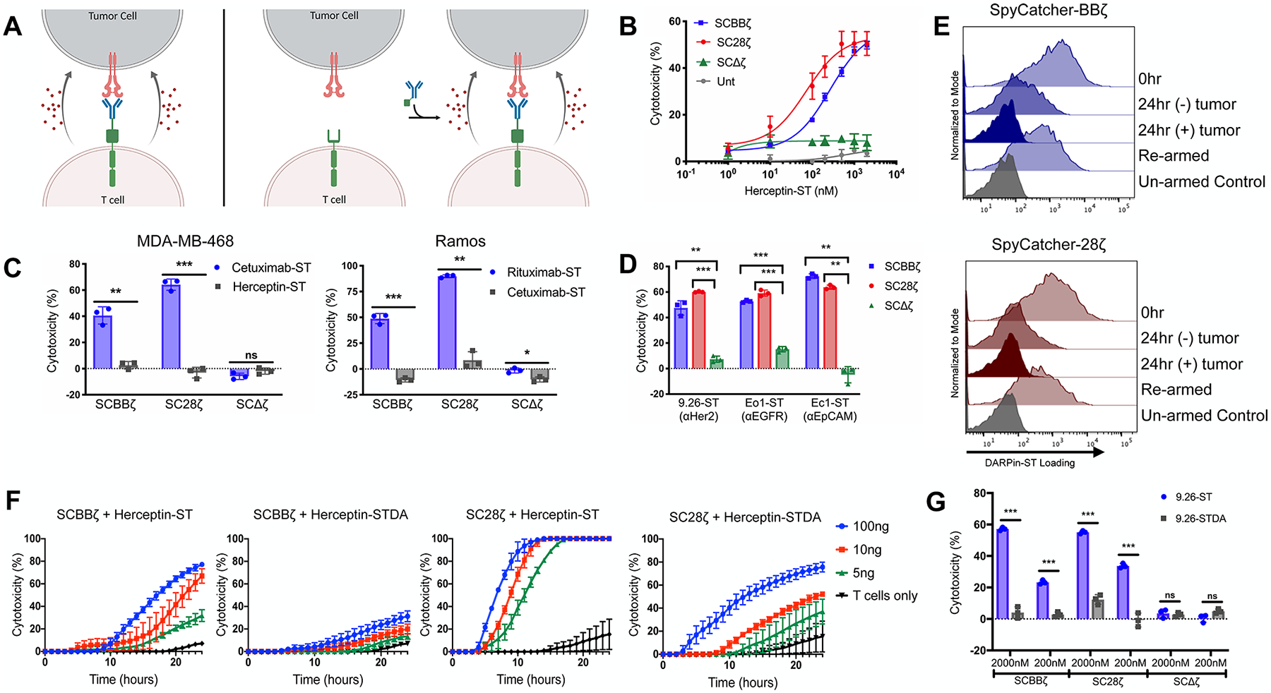
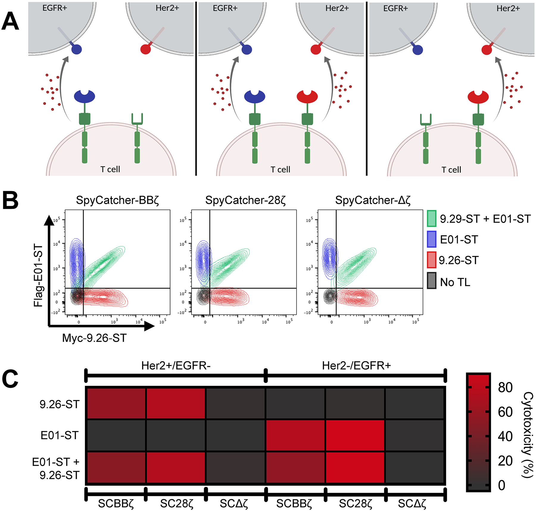
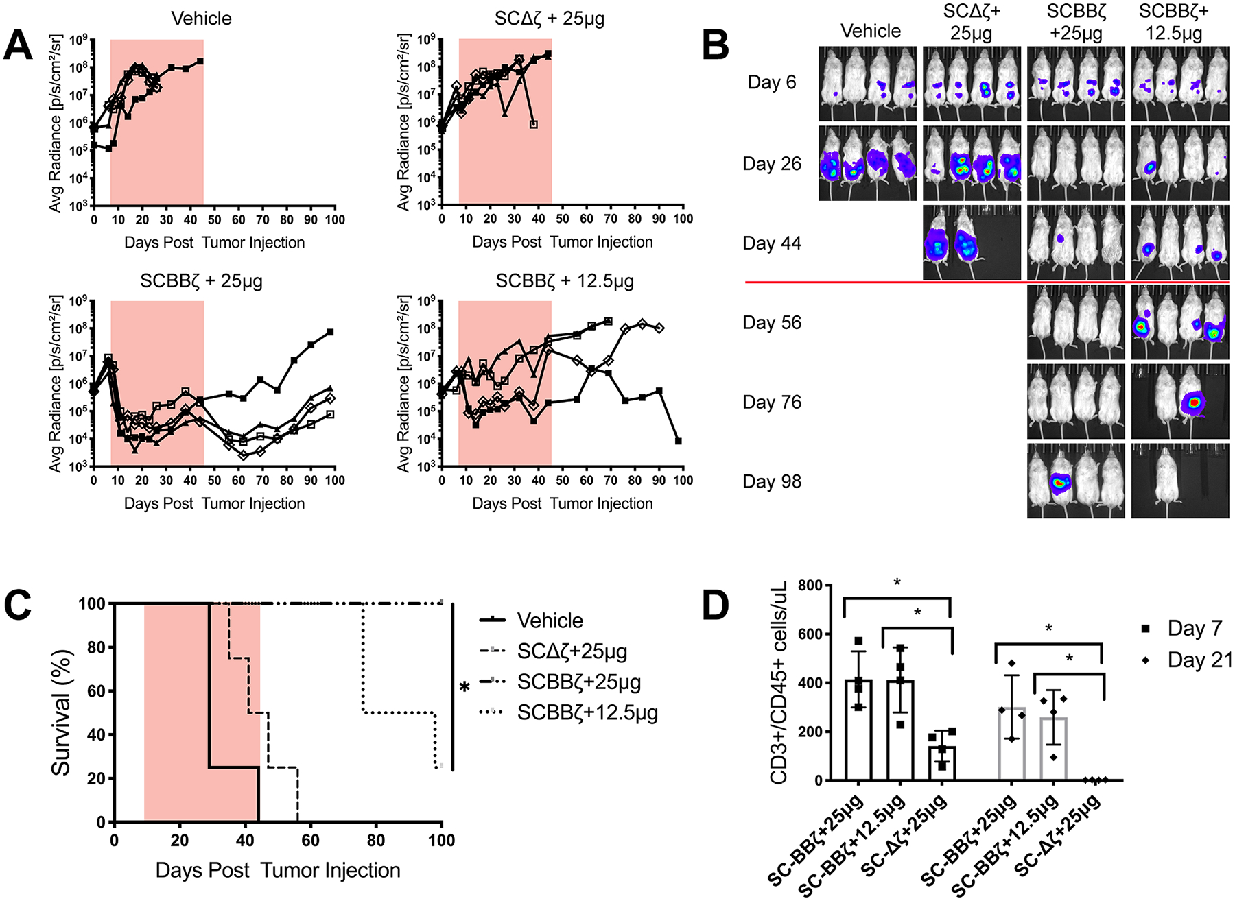
Universal immune receptors represent a rapidly emerging form of adoptive T-cell therapy with the potential to overcome safety and antigen escape challenges faced by conventional chimeric antigen receptor (CAR) T-cell therapy. By decoupling antigen recognition and T-cell signaling domains via bifunctional antigen-specific targeting ligands, universal immune receptors can regulate T-cell effector function and target multiple antigens with a single receptor. Here, we describe the development of the SpyCatcher immune receptor, the first universal immune receptor that allows for the post-translational covalent attachment of targeting ligands at the T-cell surface through the application of SpyCatcher-SpyTag chemistry. The SpyCatcher immune receptor redirected primary human T cells against a variety of tumor antigens via the addition of SpyTag-labeled targeting ligands, both in vitro and in vivo. SpyCatcher T-cell activity relied upon the presence of both target antigen and SpyTag-labeled targeting ligand, allowing for dose-dependent control of function. The mutational disruption of covalent bond formation between the receptor and the targeting ligand still permitted redirected T-cell function but significantly compromised antitumor function. Thus, the SpyCatcher immune receptor allows for rapid antigen-specific receptor assembly, multiantigen targeting, and controllable T-cell activity.


Conflict of interest statement

The authors declare the following competing financial interest(s): Daniel Powell and Andrew Tsourkas have a patent filed on this technology.

Figures

Figure 1.
Figure 1.
Development of SpyCatcher immune receptor targeting ligands. (A) Schematic representation of Protein G-ST cross-linking to clinical-grade human IgGs. (B) Cross-linking of Herceptin with Protein G-ST or Protein G-STDA, followed by subsequent reaction with SpyCatcher-Venus analyzed by SDS-Page gel under reducing conditions with coomassie staining. (C) DARPin9.26-ST and DARPin9.26-STDA reacted with SpyCatcher-Venus analyzed by SDS-Page gel with Coomassie staining.
Figure 2.
Figure 2.
SpyCatcher immune receptor is expressed and capable of covalent loading with SpyTag-labeled ligands. (A) Schematic of the lentiviral SpyCatcher immune receptor constructs. (B) SpyCatcher immune receptor-expressing SKOV3 cells were incubated in culture medium containing 2000 nM RFP-ST or RFP-STDA. Covalent bond formation between the two components was detected via SDS-Page gel under reducing conditions and Western blot staining for total CD3ζ protein. (C) SpyCatcher T cells were armed with varying amounts of Herceptin-ST for 1 h at 37 °C. Herceptin-ST loading onto the SpyCatcher immune receptor was detected by staining with APC polyclonal antihuman IgG and flow cytometric analysis. (D) Comparison of covalent (DARPin9.26-ST) vs noncovalent (DARPin9.26-ST) maximal loading of the SpyCatcher immune receptor at various concentrations. (E) SpyCatcher T cells were incubated in wells containing various amounts of immobilized Herceptin-ST for 16 h. Supernatant was harvested and analyzed for IFNγ by ELISA. Error bars represent the mean ± standard deviation. Data points are the averages of three replicates. A representative T-cell donor is shown. *P < 0.05, **P < 0.01, and ***P < 0.001.
Figure 3.
Figure 3.
SpyCatcher T cells are capable of in vitro lytic function against a host of different antigen-expressing tumor lines. (A) Schematic representation of armed SpyCatcher immune receptor lysis (left) and on-demand lysis (right). (B) SpyCatcher or untransduced (Unt) T cells were armed with various concentrations of Herceptin-ST and cocultured in the presence of SKOV3 (Her2+) tumor cells. (C) SpyCatcher T cells were armed with antigen-specific and nonspecific IgGs and cocultured with either MDA-MB-468 (EGFR+/Her2-) or Ramos (CD20+/EGFR-) tumor cells. (D) SpyCatcher T cells armed with antigen-specific DARPins were coculture with SKOV3, MDA-MB-468, or A1847 (EpCAM+) tumor cells expressing luciferase. (E) DARPin9.26-ST armed SpyCatcher T cells were incubated with or without SKOV3 tumor cells (E/T = 3:1) for 24 h, removed from culture, and incubated in media ± 2000 nM DARPin9.26-ST. Receptor arming was analyzed via anti-myc staining and flow cytometric analysis. (F) Unarmed SpyCatcher T cells were incubated with SKOV3 tumor cells for 4 h followed by the addition of Herceptin-ST or Herceptin-STDA (time = 0). (G) SpyCatcher T cells were armed with either DARPin9.26-ST or DARPin9.26-STDA at various concentrations and cocultured with SKOV3 tumor cells expressing luciferase. (B, C: MDA-MB-468, F) Lysis was measured using real-time cell analysis. (C: Ramos, D, G) Residual luciferase expression was calculated after 20 h. All cocultures were carried out using a 7:1 E/T ratio. Error bars represent the mean ± standard deviation. Data points are averages of three replicates. A representative T-cell donor is shown. *P < 0.05, **P < 0.01, and ***P < 0.001.
Figure 4.
Figure 4.
Simultaneous arming of SpyCatcher T cells with two targeting ligands generates a single cell product capable of dual antigen targeting. (A) Schematic representation of single vs dual targeting ligand loading. (B) SpyCatcher T cells were armed with either 1000 nM myc-9.26-ST (αHer2; red), 1000 nM Flag-E01-ST (αEGFR; blue), or both simultaneously at 1000 nM each (green). Receptor loading was detected with a combination of fluorescently conjugated anti-myc and anti-flag antibodies and assessed via flow cytometry. (C) Single- or dual-armed SpyCatcher T cells were cocultured with Ramos cells expressing either Her2 or EGFR and luciferase. Residual luciferase expression was calculated after 20 h. All cocultures were carried out using a 7:1 E/T ratio. Data points are averages of three replicates. A representative T-cell donor is shown.
Figure 5.
Figure 5.
SpyCatcher-BBζ T cells prevent tumor growth in vivo. NSG mice (n = 4 per group) were injected intraperitoneally with 1 × 10⁶ SKOV3 tumor cells expressing luciferase on day 0, followed by 12.5 × 10⁶ Herceptin-ST armed SpyCatcher-BBζ T cells on day 7. Herceptin-ST was administered on day 8, followed by injections every 3 days during the dosing window, indicated by either an orange box (A) or red line (B). Injection amounts indicate the dose per mouse. (A) Tumor growth was monitored by luminescence and plotted as the average radiance for each individual mouse. (B) Luminescence images of treated mice. (C) Survival curve for mice treated in (A). (D) TruCount analysis of total human T cells (CD3+/CD45+) on days 7 and 21 post-T-cell injection. Error bars represent the mean ± standard deviation (D). *P < 0.05, **P < 0.01, and ***P < 0.001.

References

    1. Porter DL; Levine BL; Kalos M; Bagg A; June CH Chimeric Antigen Receptor-Modified T Cells in Chronic Lymphoid Leukemia. N. Engl. J. Med 2011, 365 (8), 725–733. - PMC - PubMed
    1. Kalos M; Levine BL; Porter DL; Katz S; Grupp SA; Bagg A; June CH T Cells with Chimeric Antigen Receptors Have Potent Antitumor Effects and Can Establish Memory in Patients with Advanced Leukemia. Sci. Transl. Med 2011, 3 (95), 95ra73. - PMC - PubMed
    1. Grupp SA; Kalos M; Barrett D; Aplenc R; Porter DL; Rheingold SR; Teachey DT; Chew A; Hauck B; Wright FJ; Milone MC; Levine BL; June CH Chimeric Antigen Receptor–Modified T Cells for Acute Lymphoid Leukemia. N. Engl. J. Med 2013, 368 (16), 1509–1518. - PMC - PubMed
    1. Maude SL; Frey N; Shaw PA; Aplenc R; Barrett DM; Bunin NJ; Chew A; Gonzalez VE; Zheng Z; Lacey SF; Mahnke YD; Melenhorst JJ; Rheingold SR; Shen A; Teachey DT; Levine BL; June CH; Porter DL; Grupp SA Chimeric Antigen Receptor T Cells for Sustained Remissions in Leukemia. N. Engl. J. Med 2014, 371 (16), 1507–1517. - PMC - PubMed
    1. Schuster SJ; Svoboda J; Chong EA; Nasta SD; Mato AR; Anak Ö; Brogdon JL; Iulian P-M; Bhoj V; Landsburg D; Wasik M; Levine BL; Lacey SF; Melenhorst JJ; Porter DL; June CH Chimeric Antigen Receptor T Cells in Refractory B-Cell Lymphomas. N. Engl. J. Med 2017, 377 (26), 2545–2554. - PMC - PubMed

Publication types