Case Reports
. 2020 May 1;138(5):569-574.
doi: 10.1001/jamaophthalmol.2020.0427.

Genomic and Transcriptomic Tumor Heterogeneity in Bilateral Retinoblastoma

Affiliations
Case Reports

Genomic and Transcriptomic Tumor Heterogeneity in Bilateral Retinoblastoma

Ursula Winter et al. JAMA Ophthalmol. .

Erratum in

  • Error in Author Name.
    [No authors listed] [No authors listed] JAMA Ophthalmol. 2020 Jul 1;138(7):803. doi: 10.1001/jamaophthalmol.2020.1841. JAMA Ophthalmol. 2020. PMID: 32437508 Free PMC article. No abstract available.

Abstract

Importance: Comprehensive understanding of the genomic and gene-expression differences between retinoblastoma tumors from patients with bilateral disease may help to characterize risk and optimize treatment according to individual tumor characteristics.

Objective: To compare the genomic features between each eye and a specimen from an orbital relapse in patients with bilateral retinoblastoma.

Design, setting, and participants: In this case, 2 patients with retinoblastoma underwent upfront bilateral enucleation. Tumor samples were subjected to genomic and gene-expression analysis. Primary cell cultures were established from both of the tumors of 1 patient and were used for gene-expression studies.

Main outcomes and measures: Whole-exome sequencing was performed on an Illumina platform for fresh tumor samples and DNA arrays (CytoScan or OncoScan) were used for paraffin-embedded samples and cell lines. Gene-expression analysis was performed using Agilent microarrays. Germinal and somatic alterations, copy number alterations, and differential gene expression were assessed.

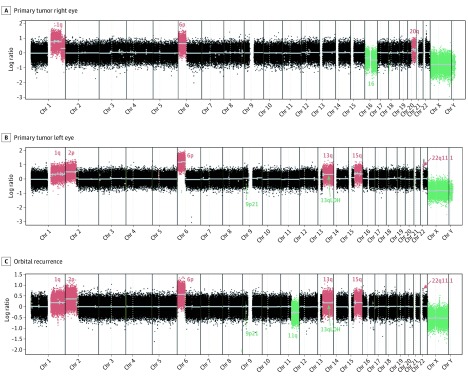
Results: After initial bilateral enucleation, patient 1 showed massive choroidal and laminar optic nerve infiltration, while patient 2 showed choroidal and laminar optic nerve invasion. Patient 1 developed left-eye orbital recurrence and bone marrow metastasis less than 1 year after enucleation. Both ocular tumors showed gains on 1q and 6p but presented other distinct genomic alterations, including an additional gain in 2p harboring the N-myc proto-oncogene (MYCN) in the left tumor and orbital recurrence. Similar copy number alterations between the orbital recurrence and the left eye supported the origin of the relapse, with an additional 11q loss only detected in the orbital relapse. Specimens from patient 2 showed common copy number gains and losses, but further evolution rendered a 2p gain spanning MYCN in the left tumor. For this patient, microarray expression analysis showed differential expression of the MYCN and the forkhead box protein G1 (FOXG1) gene pathways between the left and right tumors.

Conclusions and relevance: Differential genomic and gene expression features were observed between tumors in 2 patients with bilateral disease, confirming intereye heterogeneity that might be considered if targeted therapies are used in such patients. Chromosomal alteration profile supported the origin of the orbital recurrence from the homolateral eye in 1 patient. Loss in chromosome 11q may have been associated with extraocular relapse in this patient.


Conflict of interest statement

Conflict of Interest Disclosures: Dr Sendoya reported nonfinancial support from Fundación Instituto Leloir and personal fees from National Scientific and Technical Research Council (CONICET) during the conduct of the study. Dr Mena reported personal fees from Fundacion Natali Dafne Flexer during the conduct of the study. Dr Doz reported personal fees from Bayer, Bristol-Myers Squibb, Celgene, Loxo Oncology, Roche, Servier, and Tesaro outside the submitted work. Dr Llera reported nonfinancial support from Amazon Web Services and grants from National Agency for Science and Technology Promotion (Argentina) during the conduct of the study. Dr Schaiquevich reported grants from National Institute of Cancer, Ministry of Health, Argentina; Fondation Nelia et Amadeo Barletta; National Agency for Science and Technology Promotion, Argentina; National Agency for Science and Technology Promotion, Argentina; Fund for Ophthalmic Knowledge; and Fundación Natalie Dafne Flexer de Ayuda al Niño con Cancer, Argentina, during the conduct of the study. No other disclosures were reported.

Figures

Figure 1.
Figure 1.. Images at Presentation
A and B, Patient 1’s right and left eyes at presentation. C and D, Clinical presentation of the right and left eyes of patient 2.
Figure 2.
Figure 2.. Chromosomal Copy Number Alterations of Patient 1
A, Right-eye tumor. B, Left-eye tumor. C, Orbital recurrence. Each dot represents a log ratio of tumor signal compared with the normal reference. Losses and gains in chromosomal regions correspond with negative and positive values, respectively. The y-axis depicts the log ratio, and chromosomal positions are represented in the x-axis (Chr indicates chromosome). The orbital recurrence shows a similar profile to the left eye, with an additional loss at 11q.
Figure 3.
Figure 3.. Copy Number Alterations in Samples From Patient 2
A, Right-eye tumors. B, Left-eye tumors. C and D, Matched primary cell lines. Similar chromosomal alterations were detected between eye tumors, except for a 2p gain encompassing the MYCN gene locus. The chromosomal profiles of both HPG-RBT-12R (C) and HPG-RBT-12L (D) cells were similar to the matched original tumors, with no additional alterations. Chr indicates chromosome.

References

    1. Abramson DH, Notterman RB, Ellsworth RM, Kitchin FD. Retinoblastoma treated in infants in the first six months of life. Arch Ophthalmol. 1983;101(9):1362-1366. doi:10.1001/archopht.1983.01040020364004 - DOI - PubMed
    1. Thériault BL, Dimaras H, Gallie BL, Corson TW. The genomic landscape of retinoblastoma: a review. Clin Exp Ophthalmol. 2014;42(1):33-52. doi:10.1111/ceo.12132 - DOI - PMC - PubMed
    1. Kapatai G, Brundler M-A, Jenkinson H, et al. . Gene expression profiling identifies different sub-types of retinoblastoma. Br J Cancer. 2013;109(2):512-525. doi:10.1038/bjc.2013.283 - DOI - PMC - PubMed
    1. Kooi IE, Mol BM, Massink MPG, et al. . Somatic genomic alterations in retinoblastoma beyond RB1 are rare and limited to copy number changes. Sci Rep. 2016;6(1):25264. doi:10.1038/srep25264 - DOI - PMC - PubMed
    1. Pérez V, Sampor C, Rey G, et al. . Treatment of nonmetastatic unilateral retinoblastoma in children. JAMA Ophthalmol. 2018;136(7):747-752. doi:10.1001/jamaophthalmol.2018.1501 - DOI - PMC - PubMed

Publication types