

Evolutionary History and Activity of RNase H1-Like Proteins in Arabidopsis thaliana
- PMID: 32191307
- PMCID: PMC7295395
- DOI: 10.1093/pcp/pcaa040
Evolutionary History and Activity of RNase H1-Like Proteins in Arabidopsis thaliana
Abstract
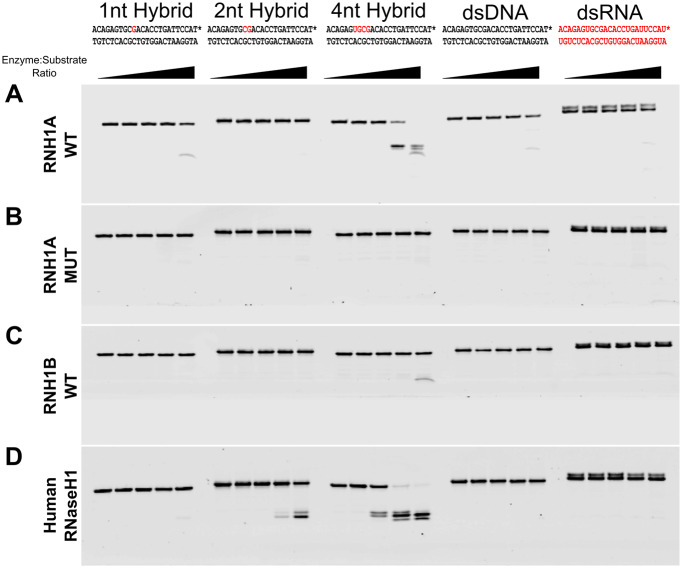
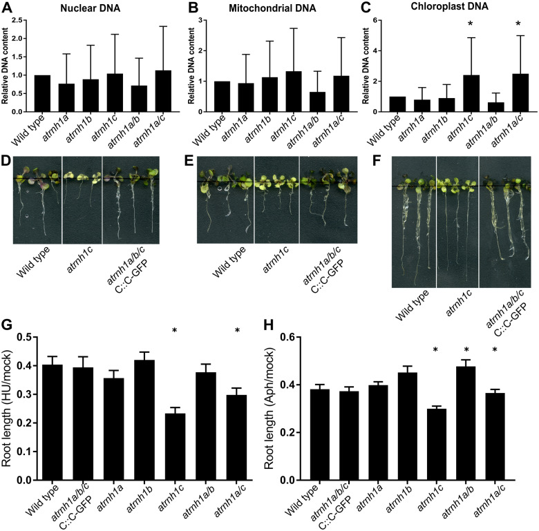
RNase H1 is an endonuclease specific toward the RNA strand of RNA:DNA hybrids. Members of this protein family are present in most living organisms and are essential for removing RNA that base pairs with DNA. It prevents detrimental effects of RNA:DNA hybrids and is involved in several biological processes. Arabidopsis thaliana has been previously shown to contain three genes encoding RNase H1 proteins that localize to three distinct cellular compartments. We show that these genes originate from two gene duplication events. One occurred in the common ancestor of dicots and produced nuclear and organellar RNase H1 paralogs. Second duplication occurred in the common ancestor of Brassicaceae and produced mitochondrial- and plastid-localized proteins. These proteins have the canonical RNase H1 activity, which requires at least four ribonucleotides for endonucleolytic digestion. Analysis of mutants in the RNase H1 genes revealed that the nuclear RNH1A and mitochondrial RNH1B are dispensable for development under normal growth conditions. However, the presence of at least one organellar RNase H1 (RNH1B or RNH1C) is required for embryonic development. The plastid-localized RNH1C affects plastid DNA copy number and sensitivity to replicative stress. Our results present the evolutionary history of RNH1 proteins in A. thaliana, demonstrate their canonical RNase H1 activity and indicate their role in early embryonic development.
Keywords: Arabidopsis thaliana; RNase H; organellar genomes.
© The Author(s) 2020. Published by Oxford University Press on behalf of Japanese Society of Plant Physiologists. All rights reserved. For permissions, please email: journals.permissions@oup.com.
Figures


Similar articles
-
Mitochondrial RNase H1 activity regulates R-loop homeostasis to maintain genome integrity and enable early embryogenesis in Arabidopsis.PLoS Biol. 2021 Aug 3;19(8):e3001357. doi: 10.1371/journal.pbio.3001357. eCollection 2021 Aug. PLoS Biol. 2021. PMID: 34343166 Free PMC article.
-
A 125 kDa RNase E/G-like protein is present in plastids and is essential for chloroplast development and autotrophic growth in Arabidopsis.J Exp Bot. 2008;59(10):2597-610. doi: 10.1093/jxb/ern126. Epub 2008 May 31. J Exp Bot. 2008. PMID: 18515828 Free PMC article.
-
Multiple ribonuclease H-encoding genes in the Caenorhabditis elegans genome contrasts with the two typical ribonuclease H-encoding genes in the human genome.Mol Biol Evol. 2002 Nov;19(11):1910-9. doi: 10.1093/oxfordjournals.molbev.a004015. Mol Biol Evol. 2002. PMID: 12411600
-
The Jekyll and Hyde character of RNase H1 and its multiple roles in mitochondrial DNA metabolism.DNA Repair (Amst). 2019 Dec;84:102630. doi: 10.1016/j.dnarep.2019.06.001. Epub 2019 Jun 4. DNA Repair (Amst). 2019. PMID: 31178343 Review.
-
RNases H: Structure and mechanism.DNA Repair (Amst). 2019 Dec;84:102672. doi: 10.1016/j.dnarep.2019.102672. Epub 2019 Jul 20. DNA Repair (Amst). 2019. PMID: 31371183 Review.
Cited by
-
Regulation of Heat Stress in Physcomitrium (Physcomitrella) patens Provides Novel Insight into the Functions of Plant RNase H1s.Int J Mol Sci. 2022 Aug 17;23(16):9270. doi: 10.3390/ijms23169270. Int J Mol Sci. 2022. PMID: 36012542 Free PMC article.
-
Uncovering the genetic basis for enhanced mushroom flavor in Quercus fabri through genome sequencing and metabolic profiling.Hortic Res. 2025 Jul 9;12(9):uhaf156. doi: 10.1093/hr/uhaf156. eCollection 2025 Sep. Hortic Res. 2025. PMID: 40861034 Free PMC article.
-
RnhP is a plasmid-borne RNase HI that contributes to genome maintenance in the ancestral strain Bacillus subtilis NCIB 3610.Mol Microbiol. 2021 Jan;115(1):99-115. doi: 10.1111/mmi.14601. Epub 2020 Sep 25. Mol Microbiol. 2021. PMID: 32896031 Free PMC article.
-
How RNases Shape Mitochondrial Transcriptomes.Int J Mol Sci. 2022 May 30;23(11):6141. doi: 10.3390/ijms23116141. Int J Mol Sci. 2022. PMID: 35682820 Free PMC article. Review.
-
Ribonucleotide and R-Loop Damage in Plastid DNA and Mitochondrial DNA during Maize Development.Plants (Basel). 2023 Sep 2;12(17):3161. doi: 10.3390/plants12173161. Plants (Basel). 2023. PMID: 37687407 Free PMC article.
References
-
- Arudchandran A., Cerritelli S., Narimatsu S., Itaya M., Shin D.-Y., Shimada Y., et al. (2000) The absence of ribonuclease H1 or H2 alters the sensitivity of Saccharomyces cerevisiae to hydroxyurea, caffeine and ethyl methanesulphonate: implications for roles of RNases H in DNA replication and repair. Genes Cells 5: 789–802. - PubMed
MeSH terms
Substances
Grants and funding
LinkOut - more resources
Full Text Sources
Molecular Biology Databases
Miscellaneous
