. 2020 Jul 1;130(7):3637-3653.
doi: 10.1172/JCI134424.

Oncogene-independent BCR-like signaling adaptation confers drug resistance in Ph-like ALL

Affiliations

Oncogene-independent BCR-like signaling adaptation confers drug resistance in Ph-like ALL

Christian Hurtz et al. J Clin Invest. .

Abstract

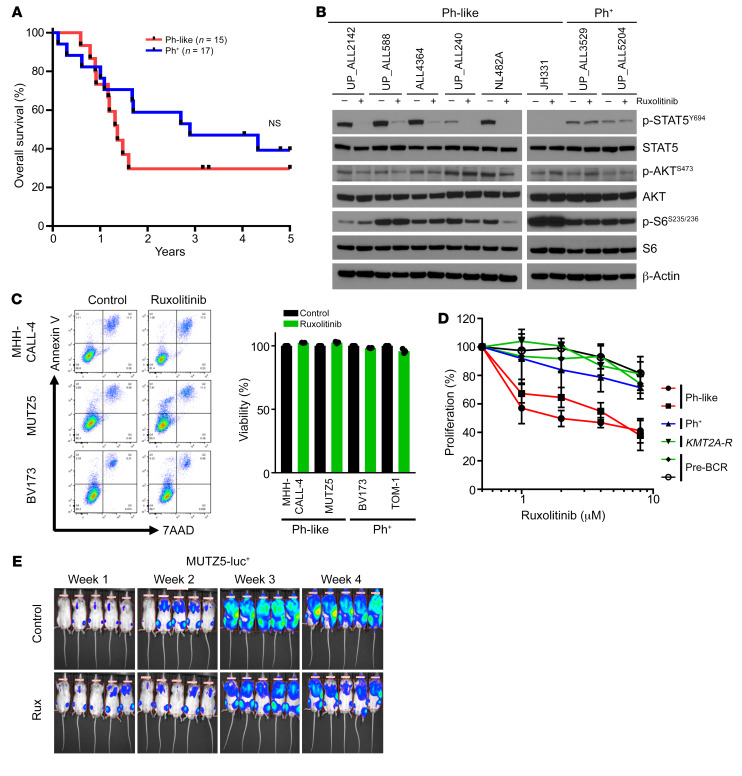
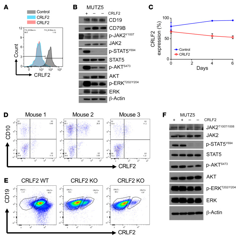
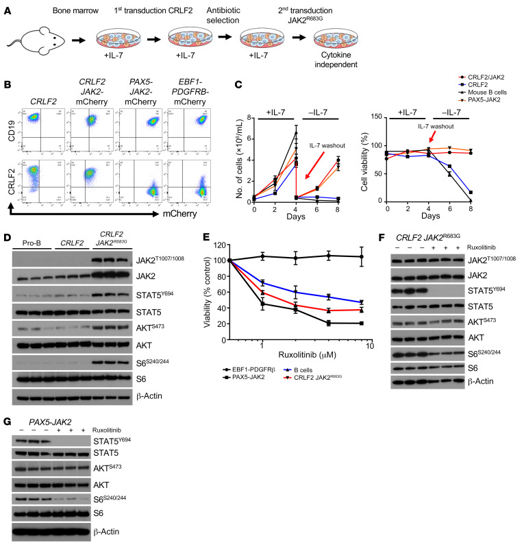
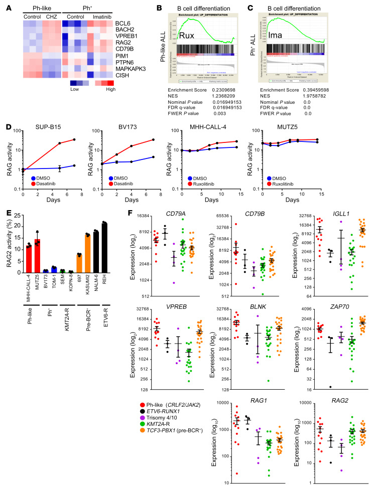
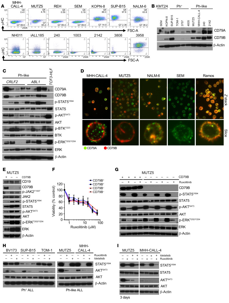
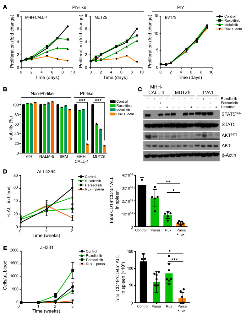
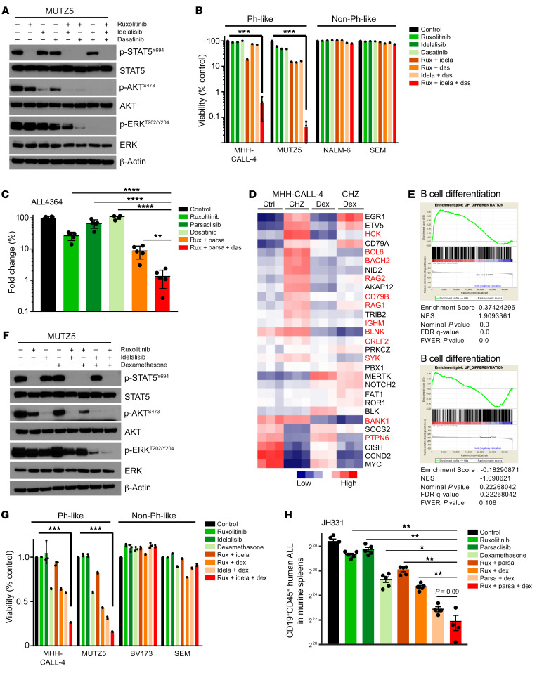
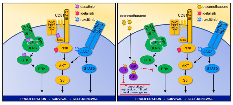
Children and adults with Philadelphia chromosome-like B cell acute lymphoblastic leukemia (Ph-like B-ALL) experience high relapse rates despite best-available conventional chemotherapy. Ph-like ALL is driven by genetic alterations that activate constitutive cytokine receptor and kinase signaling, and early-phase trials are investigating the potential of the addition of tyrosine kinase inhibitors (TKIs) to chemotherapy to improve clinical outcomes. However, preclinical studies have shown that JAK or PI3K pathway inhibition is insufficient to eradicate the most common cytokine receptor-like factor 2-rearranged (CRLF2-rearranged) Ph-like ALL subset. We thus sought to define additional essential signaling pathways required in Ph-like leukemogenesis for improved therapeutic targeting. Herein, we describe an adaptive signaling plasticity of CRLF2-rearranged Ph-like ALL following selective TKI pressure, which occurs in the absence of genetic mutations. Interestingly, we observed that Ph-like ALL cells have activated SRC, ERK, and PI3K signaling consistent with activated B cell receptor (BCR) signaling, although they do not express cell surface μ-heavy chain (μHC). Combinatorial targeting of JAK/STAT, PI3K, and "BCR-like" signaling with multiple TKIs and/or dexamethasone prevented this signaling plasticity and induced complete cell death, demonstrating a more optimal and clinically pragmatic therapeutic strategy for CRLF2-rearranged Ph-like ALL.

Keywords: Hematology; Leukemias; Oncology; Protein kinases; Signal transduction.


Conflict of interest statement

Conflict of interest: AEP receives research funding from Astellas Pharmaceuticals, Daiichi Sankyo, Novartis, and FujiFilm and is a consultant/advisory board member for Astellas, Daiichi Sankyo, Novartis, Arog, Pfizer, Takeda, Agios, and AbbVie. MC receives research funding from Astellas Pharmaceuticals and Incyte Corp. and is a consultant/advisory board member for Janssen Pharmaceuticals. SKT receives research funding from Incyte Corp. and Gilead Sciences and is a scientific advisory board member for Aleta Biotherapeutics.

Figures

Figure 1
Figure 1. Poor clinical outcomes and inadequate treatment effects of JAK inhibitor monotherapy in Ph-like ALL.
(A) Kaplan-Meier survival analysis of adult patients with Ph+ or Ph-like ALL treated at the University of Pennsylvania for whom outcome data were available (n = 49). (B) Western blot analysis of indicated proteins in 6 Ph-like ALL PDX cases and 2 Ph+ ALL PDX cases. (C) Ph-like ALL cell lines and Ph+ ALL cell lines were treated with 1 μM ruxolitinib for 72 hours (n = 3 independent experiments), and viability was assessed via flow cytometry. (D) B-ALL cell lines were treated with increasing concentrations of ruxolitinib for 72 hours (n = 3 independent experiments). Cell proliferation and viability were measured via XTT assay. (E) One million luciferase-labeled MUTZ5 cells were injected via tail vein into NSG mice and treated with control or ruxolitinib chow for 28 days. Data are represented as individual values with mean ± SEM bars. Significance for A was calculated by the log-rank (Mantel-Cox) test.
Figure 2
Figure 2. CRLF2 signaling is not required for survival of Ph-like ALL cells.
(A) MUTZ5 cells were electroporated with nontargeting control or ribonucleoproteins targeting CRLF2. CRLF2 deletion was validated via flow cytometry. (B) Western blot analysis of CRLF2+ and CRLF2-null MUTZ5 cells for the indicated proteins. (C) CRLF2-deleted (null or KO) MUTZ5 cells mixed with native CRLF2+ MUTZ5 control cells were monitored in vitro over time for outgrowth of CRLF2+ cells (n = 3 independent experiments). (D) Flow cytometry analysis of human CRLF2-deleted MUTZ5 cells in peripheral blood of engrafted NSG mice at 2 months after injection (n = 3). (E and F) End-study analysis of flow cytometry–sorted CRLF2-deleted (–) and CRLF2+ (+) MUTZ5 cells harvested from engrafted murine spleens by flow cytometry with CD19 and CRLF2/TSLPR surface staining (E) and Western blotting of the indicated proteins (F). Data are represented as individual values with mean ± SEM bars.
Figure 3
Figure 3. Genetic murine model recapitulates human Ph-like ALL signaling phenotype.
(A) Schematic illustrating the transduction procedure to generate murine Ph-like ALL models. (B) Flow cytometry analysis of CD19, CRLF2/TSLPR, and mCherry staining was performed on murine Ph-like ALL cells to confirm expression. (C) The left graph shows cell proliferation of the indicated murine cells in the presence of IL-7 and after IL-7 washout. The right graph shows the corresponding cell viability (n = 3 independent experiments). (D) Western blot analysis of the indicated target genes in IL-7–dependent pro-B cells, CRLF2+ cells, and CRLF2+ JAK2R683G-transduced (mCRLF2-JAK2) cells. (E) Viability analysis was performed on the indicated cell types with increasing concentrations of ruxolitinib for 72 hours (n = 3 independent experiments). (F and G) Western blot analysis of the indicated proteins in CRLF2/JAK2–transformed (F) and PAX5-JAK2–transformed (G) murine cells treated with 1 μM DMSO or ruxolitinib for 1 hour. Data are represented as individual values with mean ± SEM bars.
Figure 4
Figure 4. JAK inhibition induces partial differentiation of Ph-like ALL cells.
(A) Meta-analysis of gene expression data comparing MHH-CALL-4 cells treated with the type 2 JAK2 inhibitor CHZ868 with Ph+ ALL cells (BV173, NALM-1, SUP-B15, and TOM-1) treated with imatinib (12, 38). (B and C) Gene set enrichment analysis (GSEA) plots demonstrate enrichment of B cell differentiation gene sets in MHH-CALL-4 cells treated with ruxolitinib 1 μM for 12 hours (n = 3) (B) and Ph+ ALL treated with imatinib (38) (C); statistical analysis is shown. (D) Ph+ ALL cell lines and Ph-like ALL cell lines were lentivirally transduced with a RAG enzyme activity reporter construct. Ph+ and Ph-like ALL cells were treated with 100 nM dasatinib and 1 μM ruxolitinib, respectively. RAG enzyme activity was measured at the indicated times (n = 3 independent experiments). (E) Steady-state RAG enzyme activity was measured via flow cytometry in B-ALL cell lines (n = 3 independent experiments). (F) Supervised analysis of publicly available gene expression data from children with CRLF2-R/JAK2-mutant Ph-like ALL (n = 12), ETV6-RUNX1 ALL (n = 3), hyperdiploid ALL with trisomy 4 and 10 (n = 4), KMT2A-R ALL (n = 21), and TCF3-PBX1 ALL (n = 23) from the National Cancer Institute TARGET database. Data are represented as individual values with mean ± SEM bars.
Figure 5
Figure 5. Expression of BCR signaling molecules in Ph-like ALL cells.
(A) Flow cytometry analysis of immunoglobulin μ-heavy chain (μHC) expression in B-ALL cell lines (top) and Ph-like patient samples (bottom). The NALM-6 cell line and a primary pre-BCR+ B-ALL patient specimen (3958) were used as positive controls. (B) Western blot analysis of CD79A and CD79B expression in B-ALL cell lines and patient samples. (C) Western blot analysis of BCR signaling molecules in 8 Ph-like ALL PDX specimens and 1 TCF3-HLF ALL PDX case. (D) CD79A (green) and CD79B (red) expression was assessed via immunofluorescence microscopy in Ph-like ALL cell lines, pre-BCR+/BCR+ ALL positive controls, and a KMT2A-R B-ALL negative control. Original magnifications: ×63 for upper panel, ×158 for lower panel. (E) Western blot analysis of indicated proteins in control (+) and CD79B-deleted (–) MUTZ5 cells. (F) Sensitivity analysis of CD79B+ and CD79B MUTZ5 cells to increasing concentrations of ruxolitinib (n = 3 independent experiments per sample). (G) Western blot analysis of CD79B+ and CD79B MUTZ5 cells 3 weeks after electroporation treated with and without 1 μM ruxolitinib for 2 hours. (H) Ph+ ALL and Ph-like ALL cells were treated with 1 μM ruxolitinib for 1 hour or 1 μM idelalisib for 1 hour, and p-STAT5 and p-AKT levels were measured. (I) MUTZ5 and MHH-CALL-4 cells were treated with 1 μM ruxolitinib or 1 μM idelalisib for 72 hours alone or in combination, and protein expression of p-STAT5Y694 and p-AKTS473 was measured using the total proteins as controls. Data are represented as individual values with mean ± SEM bars.
Figure 6
Figure 6. Combined JAK and PI3K inhibition induces cell death and halts proliferation.
(A) Indicated B-ALL cell lines were treated with DMSO, 1 μM JAKi ruxolitinib (rux), 1 μM PI3Kδi idelalisib (idela), or a combination of ruxolitinib and idelalisib (1 μM each) for 9 days (n = 1 for each cell line). Drugs and cell culture media were replaced every 3 days. Cell density was counted at indicated time points. (B) Indicated cell lines were subjected to cell viability analysis via flow cytometry after exposure to DMSO, 1 μM ruxolitinib, 1 μM idelalisib, or a combination of ruxolitinib and idelalisib (1 μM each) for 7 days (n = 3 independent experiments). (C) Western blot analysis of p-STAT5Y694 and p-AKTS473 levels in CRLF2-R (MUTZ5, MHH-CALL-4) or ABL1-R (TVA1) Ph-like ALL cells treated with 1 μM ruxolitinib, 1 μM INCB05465, and/or 100 nM dasatinib. (D and E) Two CRLF2-R Ph-like ALL PDX models (ALL4364 [n = 5 mice] and JH331 [n = 7 mice]) were randomized to treatment with vehicle (control), 1 mg/kg PI3Kδi parsaclisib (parsa, formerly INCB050465) orally twice daily, 2 g/kg ruxolitinib rodent chow continuously provided, or both inhibitors for the specified times with flow cytometric quantification of human ALL in murine peripheral blood (line graphs) and in end-study spleens (bar graphs). Data are represented as individual values with mean ± SEM bars. *P < 0.05; **P < 0.01; ***P < 0.001 by unpaired t test (B) or ANOVA with Dunnett’s post-test for multiple comparisons (D and E).
Figure 7
Figure 7. Triple targeting of kinase signaling is required to induce Ph-like ALL cell death.
(A) MUTZ5 cells were treated with 1 μM ruxolitinib (rux), 1 μM idelalisib (idela), and/or 1 μM SRC/ABLi dasatinib (das) in the indicated combinations for 1.5 hours before protein analysis. (B) Flow cytometric cell viability analysis of B-ALL cell lines treated with 1 μM ruxolitinib, 1 μM idelalisib, and/or 1 μM dasatinib for 7 days (n = 3 independent experiments). (C) A CRLF2-R Ph-like ALL PDX model (ALL4364) was treated with vehicle (control), 2 g/kg ruxolitinib chow continuously provided, 1 mg/kg parsaclisib orally twice daily, 10 mg/kg dasatinib orally twice daily, or multiple inhibitors for 14 days, with flow cytometric (FC) quantification of human ALL cells in end-study murine spleens (n = 5 mice per group). (D) Supervised meta-analysis of gene expression data of MHH-CALL-4 cells treated with 500 nM JAK2i CHZ868, 120 nM dexamethasone, or both drugs for 12 hours (12). (E) Top: GSEA plots for B cell differentiation gene sets in CHZ868- versus vehicle-treated MHH-CALL-4 cells. Bottom: Cotreatment of CHZ868 with dexamethasone. (F) MUTZ5 cells were treated with 1 μM ruxolitinib, 1 μM idelalisib, and/or 10 nM dexamethasone for 1.5 hours before Western blot analysis. (G) FC cell viability analysis of B-ALL cell lines treated with 1 μM ruxolitinib, 1 μM idelalisib, and/or 10 nM dexamethasone for 72 hours (n = 3 independent experiments) in the indicated combinations. (H) A CRLF2-R Ph-like ALL PDX model (JH331) was treated with vehicle, 2 g/kg ruxolitinib chow, 1 mg/kg parsaclisib (parsa) orally twice daily, 1 mg/kg dexamethasone (dex) i.p. once daily, or multiple drugs for 14 days, with FC quantification of human ALL cells in murine end-study spleens. Data are represented as individual values with mean ± SEM bars. *P < 0.05; **P < 0.01; ***P < 0.001; ****P < 0.0001 by unpaired t test (B and G) or ANOVA with Dunnett’s post-test for multiple comparisons (C and H).
Figure 8
Figure 8. “Optimally lethal” combination therapeutic strategies for Ph-like ALL.

References

    1. Herold T, et al. Adults with Philadelphia chromosome-like acute lymphoblastic leukemia frequently have IGH-CRLF2 and JAK2 mutations, persistence of minimal residual disease and poor prognosis. Haematologica. 2017;102(1):130–138. doi: 10.3324/haematol.2015.136366. - DOI - PMC - PubMed
    1. Jain N, et al. Ph-like acute lymphoblastic leukemia: a high-risk subtype in adults. Blood. 2017;129(5):572–581. doi: 10.1182/blood-2016-07-726588. - DOI - PMC - PubMed
    1. Roberts KG, et al. High frequency and poor outcome of Philadelphia chromosome-like acute lymphoblastic leukemia in adults. J Clin Oncol. 2017;35(4):394–401. doi: 10.1200/JCO.2016.69.0073. - DOI - PMC - PubMed
    1. Kruth KA, et al. Suppression of B-cell development genes is key to glucocorticoid efficacy in treatment of acute lymphoblastic leukemia. Blood. 2017;129(22):3000–3008. doi: 10.1182/blood-2017-02-766204. - DOI - PMC - PubMed
    1. Roberts KG, et al. Outcomes of children with BCR-ABL1–like acute lymphoblastic leukemia treated with risk-directed therapy based on the levels of minimal residual disease. J Clin Oncol. 2014;32(27):3012–3020. doi: 10.1200/JCO.2014.55.4105. - DOI - PMC - PubMed

Publication types

MeSH terms