Clinical Trial
. 2020 Jul 1;130(7):3543-3559.
doi: 10.1172/JCI135953.

Longitudinal study reveals HIV-1-infected CD4+ T cell dynamics during long-term antiretroviral therapy

Affiliations
Clinical Trial

Longitudinal study reveals HIV-1-infected CD4+ T cell dynamics during long-term antiretroviral therapy

Annukka Ar Antar et al. J Clin Invest. .

Abstract

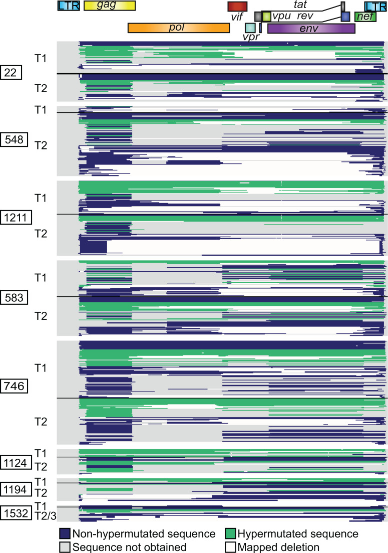
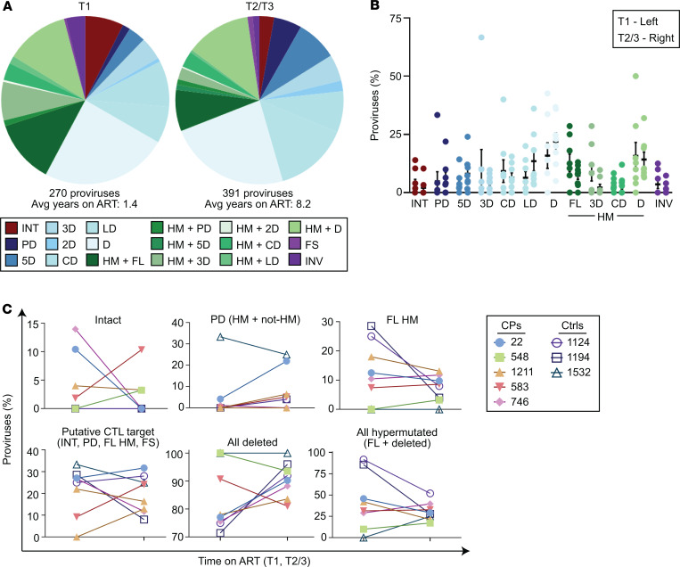
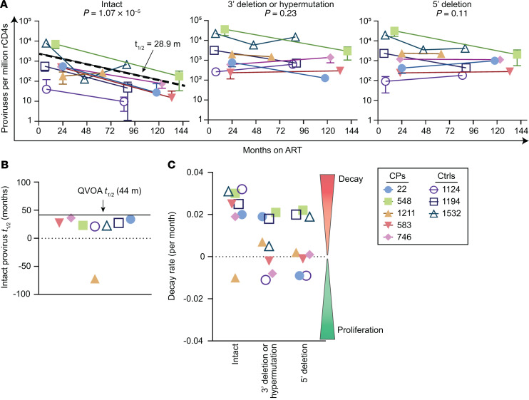
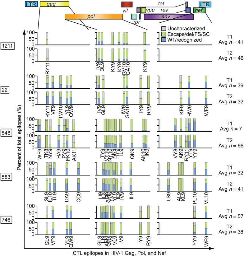
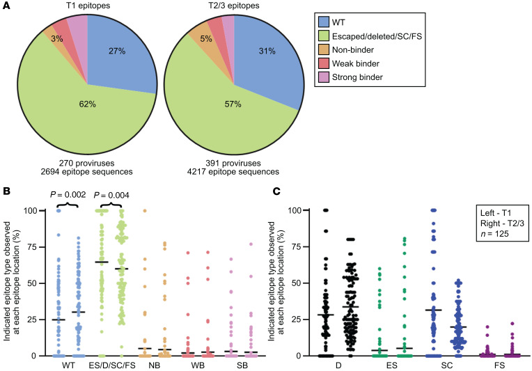
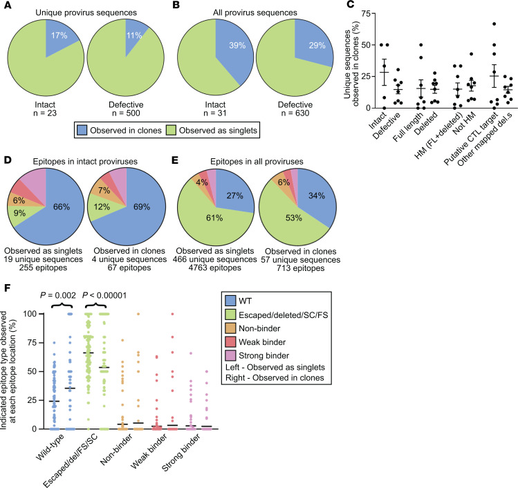
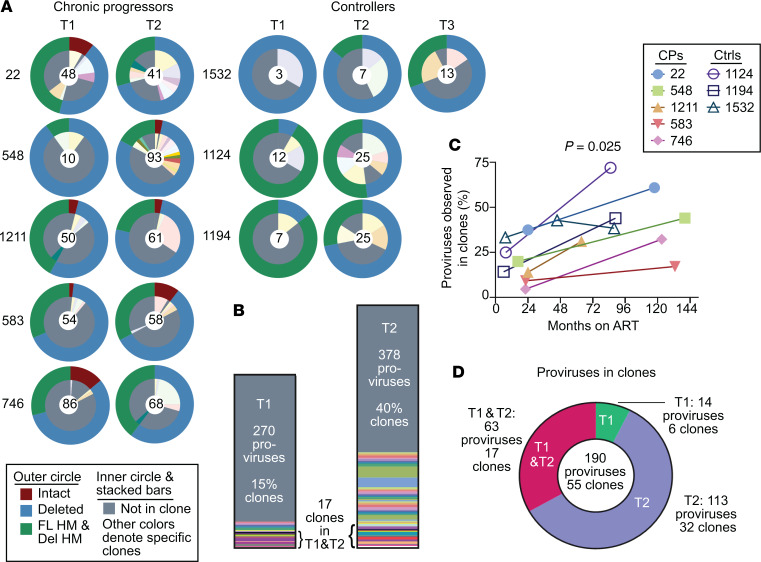
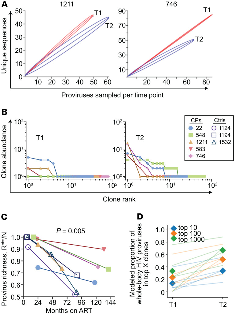
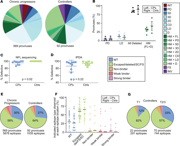
Proliferation of CD4+ T cells harboring HIV-1 proviruses is a major contributor to viral persistence in people on antiretroviral therapy (ART). To determine whether differential rates of clonal proliferation or HIV-1-specific cytotoxic T lymphocyte (CTL) pressure shape the provirus landscape, we performed an intact proviral DNA assay (IPDA) and obtained 661 near-full-length provirus sequences from 8 individuals with suppressed viral loads on ART at time points 7 years apart. We observed slow decay of intact proviruses but no changes in the proportions of various types of defective proviruses. The proportion of intact proviruses in expanded clones was similar to that of defective proviruses in clones. Intact proviruses observed in clones did not have more escaped CTL epitopes than intact proviruses observed as singlets. Concordantly, total proviruses at later time points or observed in clones were not enriched in escaped or unrecognized epitopes. Three individuals with natural control of HIV-1 infection (controllers) on ART, included because controllers have strong HIV-1-specific CTL responses, had a smaller proportion of intact proviruses but a distribution of defective provirus types and escaped or unrecognized epitopes similar to that of the other individuals. This work suggests that CTL selection does not significantly check clonal proliferation of infected cells or greatly alter the provirus landscape in people on ART.

Keywords: AIDS/HIV; Adaptive immunity; Antigen presentation; T cells.


Conflict of interest statement

Conflict of interest: The intact proviral DNA assay is the subject of patent application 16/078760. RFS is a consultant on cure-related HIV research for Merck and AbbVie. This research was funded in part from an unrestricted research grant to YCH and RFS from Gilead. JDS is a consultant on cure-related HIV research for Southern Research and Gilead. The following patent disclosures are unrelated to the present study: RFS is an inventor on patent 7,468,274 and also on patent applications 15/528230, 15/552452, and 15/568893. SGD is an inventor on patent application 15/867829. FMH is an inventor on patent applications 13/891970, 12/677278, and 11/880126.

Figures

Figure 1
Figure 1. Landscape of HIV-1 provirus genomes at 2 widely spaced time points.
Six hundred sixty-one individual provirus genomes were obtained by near-full-length sequencing from peripheral resting CD4+ T cells from 8 participants who maintained suppressed viral loads on ART for years. Each horizontal bar represents a single provirus genome. The inner PCR primers corresponding to Gag and Env were the most likely to amplify, being the shortest. Therefore, nucleotides in the Gag and Env regions are more likely to be sequenced using this method than nucleotides in other regions. Areas that are gray correspond to regions in the provirus for which sequence was not obtained and may or may not contain a deletion. Areas that are white correspond to mapped deletions. T1, T2, and T3: time points 1, 2, and 3.
Figure 2
Figure 2. The distribution of provirus types appears similar over long periods of time on suppressive ART by near-full-length sequencing.
(A) Summary of 270 proviral sequences (time point 1, T1) and 391 proviral sequences (T2/3) from 8 individuals maintained on long-term suppressive ART. Defective proviruses are categorized by type of defect. (B) Percentage of intact and various types of defective proviruses relative to total proviruses from each individual at the indicated time point, with mean and SEM shown in black. Left, T1; Right, T2/3. A and B share a color-coded figure legend. (C) Percentage of each indicated provirus type or grouping among total proviruses at the indicated time point for each individual. No intact proviruses were observed in the controllers on ART. (B and C) Paired-sample t tests with Bonferroni’s correction were applied. No provirus type or grouping change over time was statistically significant. CPs, chronic progressors; Ctrls, controllers; INT, intact; PD, deletion within packaging signal region; 5D, deletion within 5′ half of HIV-1 genome; 3D, deletion within 3′ half of HIV-1 genome; 2D, two or more deletions; CD, deletion spans center of genome and less than 75% of genome length; LD, deletion of more than 75% of HIV-1 genome length; D, unmapped deletion; HM, hypermutated; FL, full-length; FS, intact save for 1 or more frameshifts affecting genes required for replication; INV, deleted with inverted sequence.
Figure 3
Figure 3. Longitudinal analysis of provirus frequency by IPDA reveals slow decay of intact proviruses over time on ART.
IPDA analysis was performed on resting CD4+ T cells (rCD4s) from the same samples as those analyzed by near-full-length sequencing. (A) Left: Intact proviruses per million resting CD4+ T cells plotted as a function of months on ART. Log-linear mixed-effects modeling was used to determine the population level half-life for intact DNA (28.9 months) — plotted here as a dashed line — and generate P values for all types of proviruses. Proviruses with 3′ deletions and/or hypermutation (center) and proviruses with 5′ deletions (right) per million resting CD4+ T cells plotted as a function of months on ART. (B) Half-lives (t1/2) of cells carrying intact proviruses assuming exponential decay, excluding any IPDA measurement of zero. The half-life of replication-competent HIV-1 proviruses in people on ART as measured by quantitative viral outgrowth assays (QVOA) is known to be approximately 44 months and is shown in black. (C) IPDA data are plotted in terms of decay rate assuming exponential decay, excluding IPDA measurements of zero. CP, chronic progressors; Ctrls, controllers.
Figure 4
Figure 4. Percentages of unrecognized CTL epitopes in Gag, Pol, and Nef from HIV-1 proviruses do not change dramatically over long periods of time on suppressive ART.
Percentages of wild-type, unrecognized (deleted, escaped, frameshifted, or preceding stop codon), and uncharacterized epitope sequences at each dominant epitope in 5 chronic progressors on suppressive ART at 2 widely spaced time points. Provirus epitope sequences that lay in an area with unavailable sequence information were discarded from the analysis. The average number of total available epitope sequences per participant time point is shown at right. Predicted dominant epitopes in Gag, Pol, and Nef given each participant’s HLA type are shown and labeled. Epitope information can be found in Supplemental Table 2. Del, epitope lay within a mapped deletion; FS, a frameshift mutation precedes the epitope; SC, a stop codon precedes the epitope.
Figure 5
Figure 5. Percentages of unrecognized epitope sequences in Gag, Pol, and Nef from HIV-1 proviruses decrease over time in people on suppressive ART.
(A) Summary pie charts of Gag, Pol, and Nef epitope sequences from 125 predicted dominant epitopes from all 8 participants from widely spaced time points after the initiation of suppressive ART. Unrecognized epitopes include escaped epitopes (ES), epitopes that lie within a deleted region of the provirus (D), and epitopes with a preceding frameshift (FS) mutation or stop codon (SC). Epitope sequences that were uncharacterized in the literature are categorized as nonbinder (NB), weak binder (WB), or strong binder (SB) based on predictive software (NetMHC 4.0) and relevant HLA allele. (B and C) For each of the 125 predicted dominant epitopes, the percentage of the indicated epitope type among the proviruses with epitope sequence available at that site is plotted as 1 dot, with the mean shown in black. In each pair, left denotes time point 1 (T1) and right, T2 and T3. P values were generated to test the hypothesis that the percentage of each type of epitope did not change over time on ART using the nonparametric Wilcoxon’s signed-rank test. Significant results after application of Bonferonni’s correction are shown. (B) Epitope sequences were divided into 5 categories shown here, with all unrecognized epitope sequences grouped together. (C) Epitope sequences were divided into 8 categories, with each type of unrecognized epitope counted individually. Shown here are the 4 unrecognized epitope types. WT, wild-type.
Figure 6
Figure 6. The proportion of intact proviruses observed in large clones is similar to the proportion of defective proviruses observed in large clones.
(A and B) Summary pie charts of all intact (left) and defective (right) proviruses from all participant time points in this study showing proportions observed in clones (blue) versus those observed as singlets (green). In A and CF, each clone family was collapsed into one unique sequence for counting purposes. In B, clone families were not collapsed. (C) Percentage of proviruses of the indicated type observed in clones relative to total proviruses of the indicated type from each individual, with mean and SEM shown. The putative CTL target grouping includes intact, FL HM, PD, and FS. There was no statistically significant difference in each pair when the nonparametric Kruskal-Wallis test was applied. (D) Summary pie charts of Gag, Pol, and Nef epitope sequences from predicted dominant epitopes from intact proviruses that were observed as singlets (left) or observed in clones (right). (E) Summary pie charts of Gag, Pol, and Nef epitope sequences from predicted dominant epitopes from all proviruses observed as singlets (left) or observed in clones (right). (F) Percentage of indicated epitope type relative to total epitope sequences at each of 125 predicted dominant epitopes from 8 individuals, with mean shown in black. Left: Epitope sequences from proviruses observed as singlets. Right: Epitope sequences from proviruses observed in clones. The nonparametric Wilcoxon’s test was applied and significant P values are shown; see also Supplemental Table 3. D, E, and F share a figure legend. Del, epitope within mapped deletion in provirus; SC or FS, stop codon or frameshift observed between relevant gene’s start codon and epitope; HM, hypermutated; FL, full-length.
Figure 7
Figure 7. The proportion of HIV-1 proviruses observed in clones increases with time on suppressive ART.
(A) Summary of provirus type and clonality for all 8 individuals. The outer circles’ colors denote whether the provirus is intact, deleted but not hypermutated, or hypermutated with or without deletions. The inner circle denotes unique provirus sequences (gray) and those observed in clones (other colors). In the center is the number of proviruses sampled per participant time point. (B) Stacked bar chart summary of clonality for all 8 participants. Time point 3 for participant 1532 is not included. Gray color indicates unique provirus sequences. Each color and its height indicate a clone family and the number of proviruses observed in it. (C) Percentage of proviruses observed in clones by months on suppressive ART. Pearson’s r = 0.54. Pearson’s P value shown. (D) Of proviruses observed in clones, pie chart of the percentage found in time point 1 only (green), time point 2 only (purple), or found in both time points (red). T1, time point 1; T2, time point 2; HM, hypermutated; FL, full-length; Del, deleted; CPs, chronic progressors; Ctrls, controllers.
Figure 8
Figure 8. HIV-1 proviruses are concentrated into large clones during ART.
(A) Rarefaction curves of observed provirus richness in 2 chronic progressor participants. See also Supplemental Figure 4. (B) Rank-abundance curves of provirus clones at time point 1 (T1) and T2. (C) Provirus richness divided by the number of sampled proviruses by months on suppressive ART. Pearson’s r = –0.67. Pearson’s P value shown. (D) The data from B were fit into a power-law model, where clone abundance is directly proportional to 1/rankα. See Supplemental Figure 5 for details. Modeled reservoir extrapolations illustrate the proportion of whole-body HIV-1 proviruses contained in the top 10, 100, and 1000 clones, respectively (colors). Proviruses are increasingly concentrated among dominant large clones over time for all CPs (lines) and when averaged across individuals (shaded diamonds). In B and D, analyses exclude controllers given low sampling depth. CPs, chronic progressors; Ctrls, controllers.
Figure 9
Figure 9. Persistence of defective HIV-1 proviruses in controllers on ART is similar to that of chronic progressors on ART.
(A) Summary of provirus types obtained from all time points of the chronic progressors (left) and controllers (right) maintained on long-term suppressive ART. (B) Percentage of various types of defective proviruses relative to total sampled proviruses from 5 chronic progressors (left) and 3 controllers (right), with mean and SEM shown in black. Only 4 categories are shown because only these types are present in all 3 controllers. (C and D) Plots of the percentage of defective proviruses at each participant time point for chronic progressors (left) and controllers (right) on ART by near-full-length sequencing (C) and by IPDA (D). Two-tailed t test P values shown. (E) Summary pie charts of Gag, Pol, and Nef epitope sequences from predicted dominant epitopes from proviruses isolated from chronic progressors and controllers. (F) Percentage of indicated epitope type relative to total epitope sequences at each of 75 (CP, left) or 50 (Ctrl, right) predicted dominant epitopes from 5 CPs and 3 Ctrls, with the mean shown in black. (B and F) No significant differences were observed when the nonparametric Kruskal-Wallis test was applied. (G) Summary pie charts of Gag, Pol, and Nef epitope sequences from Ctrls at time point 1 (T1) and T2/3. EG share a figure legend. CPs, chronic progressors; Ctrls, controllers; INT, intact; PD, deletion in packaging signal or major splice donor site; LD, deletion of more than 75% of HIV-1 genome length; HM, hypermutated; FL, full-length; FS, intact provirus save for 1 frameshift (A) or preceding frameshift mutation (EG); INV, deleted with inverted sequence; Del, deleted; SC, preceding stop codon; NFL, near full-length; IPDA, intact proviral DNA assay.

Similar articles

Cited by

References

    1. Finzi D, et al. Identification of a reservoir for HIV-1 in patients on highly active antiretroviral therapy. Science. 1997;278(5341):1295–1300. doi: 10.1126/science.278.5341.1295. - DOI - PubMed
    1. Chun TW, et al. Presence of an inducible HIV-1 latent reservoir during highly active antiretroviral therapy. Proc Natl Acad Sci U S A. 1997;94(24):13193–13197. doi: 10.1073/pnas.94.24.13193. - DOI - PMC - PubMed
    1. Finzi D, et al. Latent infection of CD4+ T cells provides a mechanism for lifelong persistence of HIV-1, even in patients on effective combination therapy. Nat Med. 1999;5(5):512–517. doi: 10.1038/8394. - DOI - PubMed
    1. Wong JK, et al. Recovery of replication-competent HIV despite prolonged suppression of plasma viremia. Science. 1997;278(5341):1291–1295. doi: 10.1126/science.278.5341.1291. - DOI - PubMed
    1. Davey RT, et al. HIV-1 and T cell dynamics after interruption of highly active antiretroviral therapy (HAART) in patients with a history of sustained viral suppression. Proc Natl Acad Sci U S A. 1999;96(26):15109–15114. doi: 10.1073/pnas.96.26.15109. - DOI - PMC - PubMed

Publication types

MeSH terms

Substances