. 2020 Jul 1;130(7):3437-3452.
doi: 10.1172/JCI135060.

Complement-activated interferon-γ-primed human endothelium transpresents interleukin-15 to CD8+ T cells

Affiliations

Complement-activated interferon-γ-primed human endothelium transpresents interleukin-15 to CD8+ T cells

Catherine B Xie et al. J Clin Invest. .

Abstract

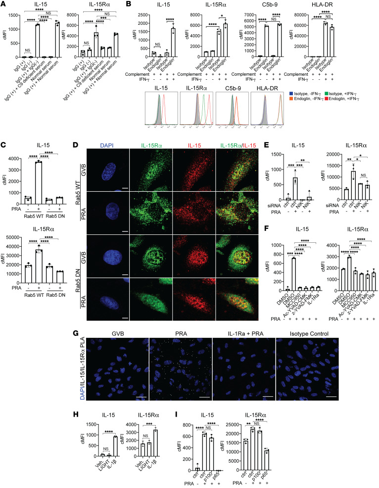
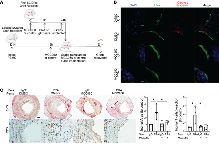
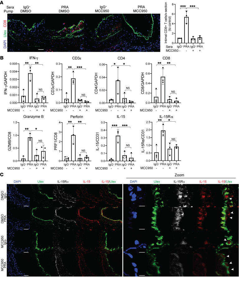
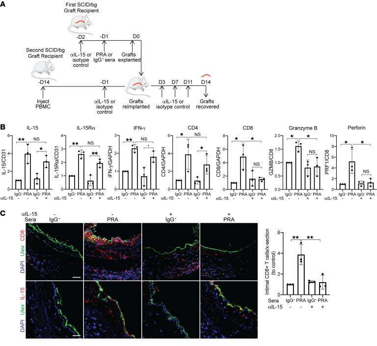
Alloantibodies in presensitized transplant candidates deposit complement membrane attack complexes (MACs) on graft endothelial cells (ECs), increasing risk of CD8+ T cell-mediated acute rejection. We recently showed that human ECs endocytose MACs into Rab5+ endosomes, creating a signaling platform that stabilizes NF-κB-inducing kinase (NIK) protein. Endosomal NIK activates both noncanonical NF-κB signaling to synthesize pro-IL-1β and an NLRP3 inflammasome to process and secrete active IL-1β. IL-1β activates ECs, increasing recruitment and activation of alloreactive effector memory CD4+ T (Tem) cells. Here, we report that IFN-γ priming induced nuclear expression of IL-15/IL-15Rα complexes in cultured human ECs and that MAC-induced IL-1β stimulated translocation of IL-15/IL-15Rα complexes to the EC surface in a canonical NF-κB-dependent process in which IL-15/IL-15Rα transpresentation increased activation and maturation of alloreactive CD8+ Tem cells. Blocking NLRP3 inflammasome assembly, IL-1 receptor, or IL-15 on ECs inhibited the augmented CD8+ Tem cell responses, indicating that this pathway is not redundant. Adoptively transferred alloantibody and mouse complement deposition induced IL-15/IL-15Rα expression by human ECs lining human coronary artery grafts in immunodeficient mice, and enhanced intimal CD8+ T cell infiltration, which was markedly reduced by inflammasome inhibition, linking alloantibody to acute rejection. Inhibiting MAC signaling may similarly limit other complement-mediated pathologies.

Keywords: Adaptive immunity; Immunology; Transplantation; endothelial cells.


Conflict of interest statement

Conflict of interest: The authors have declared that no conflict of interest exists.

Figures

Figure 1
Figure 1. IFN-γ induces human ECs to upregulate expression of intracellular IL-15 and IL-15Rα and surface IL-15Rα.
(A) ECs were treated with IFN-γ for 6 hours, 24 hours, and 48 hours. IL-15 and IL-15Rα transcript levels were assessed by qRT-PCR (n = 4). (B) After IFN-γ treatment for 6 hours, 24 hours, and 48 hours, EC cell lysates were analyzed for IL-15 and IL-15Rα by immunoblotting. Densitometric values of IL-15 and IL-15Rα bands on exposed immunoblots from 3 independent experiments using 3 HUVEC donors were calculated and normalized to the intensity of β-actin bands (n = 3). (C) ECs were treated with IFN-γ for 6 hours, 24 hours, and 48 hours and analyzed for surface IL-15 and IL-15Rα staining by flow cytometry. FACS plots show a representative experiment (n = 3). Data represent mean SEM. *P < 0.05; ***P < 0.001; ****P < 0.0001; 1-way ANOVA and Tukey’s multiple comparisons test. Representative of 3 independent experiments using 3 HUVEC donors.
Figure 2
Figure 2. MAC induces nuclear translocation and coordinate expression of IL-15/IL-15Rα on the cell surfaces of IFN-γ-primed human ECs.
(A) Representative images of confocal microcopy analysis of ECs that were pretreated with IFN-γ for 48 hours before being treated with PRA for 30 minutes or 4 hours, fixed and permeabilized, and stained for intracellular IL-15 and IL-15Rα. Scale bars: 5 μm. (B) IFN-γ–pretreated ECs were treated with either gelatin veronal buffer (GVB) control or PRA sera treatment, fixed and permeabilized prior performing PLA between IL-15 and IL-15Rα. Representative images of confocal microscopy analysis. Scale bars: 30 μm. (C) IFN-γ–pretreated ECs were treated with either GVB or PRA for 30 minutes and 4 hours. Extracts from 4 specific cellular compartments (cytoplasmic [C], membrane [M], soluble nuclear [SN], and chromatin-bound nuclear [CN]) were isolated by stepwise lysis and analyzed for IL-15 and IL-15Rα protein by immunoblotting. (D) ELISA measurements of IL-15 in culture supernatants of IFN-γ–primed ECs treated with PRA sera. (E) ECs were pretreated with IFN-γ for 48 hours before PRA treatment and analysis of surface staining IL-15 and IL-15Rα by flow cytometry (n = 4). (F) Representative images of confocal immunofluorescence analysis of IL-15 and IL-15Rα surface staining on unpermeabilized IFN-γ–pretreated ECs treated with either PRA sera or control GVB. Scale bars for top row: 30 μm; scale bars for “Zoom” bottom row: 5 μm. (G) IFN-γ–pretreated ECs were treated with either GVB control or PRA sera treatment. Proximity ligation assay (PLA) was performed between surface IL-15 and IL-15Rα and analyzed by confocal microscopy. Scale bars: 30 μm. Data represent mean ± SEM. *P < 0.05; **P < 0.01; ***P < 0.001; ****P < 0.0001; 1-way ANOVA and Tukey’s multiple comparisons test in D and unpaired 2-tailed Student’s t test in E. Representative of 3 independent experiments using 3 HUVEC donors.
Figure 3
Figure 3. MAC stabilization of endosomal NIK, activation of NLRP3 inflammasome, and IL-1 signaling drives nuclear translocation and induction of IL-15/IL-15Rα complexes on human EC cell surfaces.
(A) PRA sera was separated into IgG+ and IgG fractions and added to IFN-γ–pretreated ECs alone or in combination with C9-deficient or normal serum before flow cytometry analysis of surface IL-15 and IL-15Rα (n = 3). (B) Unprimed and IFN-γ–primed ECs were incubated with complement-fixing anti–human endoglin IgG2a antibody and human complement before flow cytometry analysis of IL-15, IL-15Rα, HLA-DR, and C5b-9 (n = 3). (C) IFN-γ–primed and stably transduced Rab5-WT or Rab5-DN (S43N) ECs were treated with PRA sera or control GVB and stained for surface IL-15 and IL-15Rα (n = 3). (D) IFN-γ–primed Rab5-WT and Rab5-DN ECs were treated with PRA and stained for intracellular IL-15 and IL-15Rα. Scale bars: 5 μm. (E) IFN-γ–pretreated ECs were transfected with NIK siRNA, treated with PRA, and analyzed for surface IL-15 and IL-15Rα (n = 3). (F) IFN-γ–primed ECs were pretreated with NLRP3 inhibitor MCC950, caspase-1 inhibitors Ac-YVAD-CMK or z-YVAD-FMK, or IL-1 receptor antagonist (IL-1Ra), treated with PRA and analyzed for surface IL-15 and IL-15Rα (n = 3). (G) IFN-γ–primed ECs were pretreated with either vehicle or IL-1Ra before PRA treatment. Proximity ligation assay (PLA) was performed between surface IL-15 and IL-15Rα and analyzed by confocal microscopy. Scale bars: 30 μm. (H) IFN-γ–primed ECs were treated with LIGHT, IL-1β, or mock treatment before analysis of surface IL-15 and IL-15Rα (n = 3). (I) IFN-γ–primed ECs were transfected with control, p100, or p65 siRNA before PRA treatment and flow cytometry analysis for surface IL-15 and IL-15Rα (n = 3). Data represent mean ± SEM. **P < 0.01; ****P < 0.0001; 1-way ANOVA and Tukey’s multiple comparisons test. Representative of 3 independent experiments using 3 HUVEC donors.
Figure 4
Figure 4. Surface IL-15 on MAC-activated ECs is presented in trans and enhances allogeneic CD8+ Tem cell activation, proliferation, and differentiation.
(A) Proliferation of CD4+ Tem cells after coculture for 7 days with IFN-γ–primed ECs pretreated with NLRP3 inflammasome inhibitor MCC950, IL-1 receptor antagonist, anti–IL-15 blocking antibody, or DMSO before PRA sera or vehicle treatment for 6 hours. CFSE dilution was assessed on day 7 by flow cytometry. FACS plots show a representative experiment (n = 3). (B) CD8+ Tem cell proliferation by CFSE dilution, activation by HLA-DR surface expression, and differentiation by granzyme B and perforin expression were assessed after coculture with IFN-γ–primed ECs pretreated with MCC950, z-YVAD-FMK, IL-1Ra, anti–IL-15 blocking antibody, or DMSO before PRA sera or vehicle treatment for 6 hours. Flow cytometry analysis was performed after 7 days of coculture. FACS plots show a representative experiment (n = 3). (C) IFN-γ production by allogeneic memory CD8+ T cells after coculture with IFN-γ–primed ECs transfected with control or p65 siRNA before addition of exogenous IL-1β or mock treatment. IFN-γ production was assessed by ELISA after 24 hours for coculture (72 hours after siRNA transfection). (D) Proliferation, activation, and granzyme B and perforin expression of CFSE-labeled CD8+ Tem cells after coculture with IFN-γ–primed ECs transfected with p65 or control siRNA and PRA or vehicle treatment for 6 hours. Flow cytometry analysis was performed on day 7. Data represent mean ± SEM. **P < 0.01; ***P < 0.001; ****P < 0.0001; 1-way ANOVA and Tukey’s multiple comparisons test. Representative of 3 to 4 independent experiments using 3 HUVEC donors.
Figure 5
Figure 5. MCC950 blocks MAC-induced NLRP3 inflammasome activation by human ECs lining human artery xenografts and reduces the enhanced allogeneic memory T cell infiltration in vivo.
(A) Human coronary artery grafts from a single donor were implanted into a set of 4 SCID/bg immunodeficient mice and quiesced for 7 days before pretreatment with NLRP3 inhibitor MCC950 or control DMSO in PBS before PRA or IgG sera treatment. After 24 hours, grafts were explanted and retransplanted into a second SCID/bg host with circulating allogeneic PBMCs. Osmotic pumps filled with MCC950 or DMSO in PBS were implanted subcutaneously in the second graft recipient at time of retransplantation. Grafts were recovered after 14 days. The experiment was repeated 3 times with different artery donors. (B) Human ECs lining arterial grafts were identified by Ulex staining and analyzed for cleaved caspase-1 staining by immunofluorescence. Scale bars: 50 μm. (C) Neointimal areas of grafts were assessed between treatment groups following EVG staining. Infiltrating intimal CD3+ T cells were identified and quantified following immunohistochemistry staining (n = 3). Scale bars: 50 μm. Data represent mean ± SEM. *P < 0.05, 1-way ANOVA and Tukey’s multiple comparisons test. Results shown are representative of 3 artery grafts from 3 different artery donors.
Figure 6
Figure 6. MCC950 blocks MAC-induced IL-15/IL-15Rα transpresentation by human ECs lining human artery xenografts and reduces the enhanced allogeneic memory CD8+ T cell infiltration, proliferation, and differentiation in vivo.
(A) Artery grafts from Figure 6 were stained for Ulex to detect human endothelium and CD8. Infiltrating intimal CD8+ T cells were identified and quantified following immunofluorescence staining (n = 3). Scale bar: 50 μm. (B) qRT-PCR analysis of IL-15 and IL-15Rα (normalized to CD31); CD3ε, CD4, and CD8 (normalized to GAPDH); and granzyme B and perforin (normalized to CD8) in the grafts (n = 3). (C) Human ECs lining grafts were analyzed for IL-15 and IL-15Rα staining by immunofluorescence. Scale bar: 50 μm. Zoom shows higher magnification of the IL-15 and IL-15Rα staining of the human ECs lining the grafts, indicated by arrowheads, shown in the left panel. Scale bars: 10 μm. Note that IL-15 staining is increased in PRA-treated grafts, a change that is abrogated by MCC950, and that upon enlargement of the images, IL-15 staining is no longer confined to the nucleus of the ECs. Data represent mean ± SEM. *P < 0.05; **P < 0.01; ***P < 0.001; 1-way ANOVA and Tukey’s multiple comparisons test. Results shown are representative of 3 artery grafts from 3 different artery donors.
Figure 7
Figure 7. Anti–IL-15 blocking antibody reduces intimal CD8+ T cell infiltration and expression of effector molecules.
(A) Human coronary artery grafts from the same donor were implanted into sets of 4 immunodeficient mice. Recipients were pretreated with anti–human IL-15 blocking antibody (αIL-15) or control isotype antibody before PRA or control sera treatment. Grafts were retransplanted into a second recipient with circulating allogeneic human PBMCs and similarly treated with anti–IL-15 blocking antibody or isotype control (n = 3). (B) qRT-PCR analysis of IL-15 and IL-15Rα (normalized to CD31); IFN-γ, CD4, CD8 (normalized to GAPDH); and granzyme B and perforin (normalized to CD8) in the grafts. Normalized expression is relative to isotype and IgG control group (n = 3). (C) Immunofluorescence detection of CD8 or IL-15 expression and human endothelium by Ulex in grafts. The intimal infiltrating CD8+ T cells were quantified. Scale bars: 50 μm. Data represent mean SEM. *P < 0.05; **P < 0.01; 1-way ANOVA and Tukey’s multiple comparisons test. Results shown are representative of 3 artery grafts from 3 different artery donors.

References

    1. Abrahimi P, Liu R, Pober JS. Blood vessels in allotransplantation. Am J Transplant. 2015;15(7):1748–1754. doi: 10.1111/ajt.13242. - DOI - PubMed
    1. Biedermann BC, Pober JS. Human vascular endothelial cells favor clonal expansion of unusual alloreactive CTL. J Immunol. 1999;162(12):7022–7030. - PubMed
    1. Dengler TJ, Pober JS. Human vascular endothelial cells stimulate memory but not naive CD8+ T cells to differentiate into CTL retaining an early activation phenotype. J Immunol. 2000;164(10):5146–5155. doi: 10.4049/jimmunol.164.10.5146. - DOI - PubMed
    1. Lakkis FG, Lechler RI. Origin and biology of the allogeneic response. Cold Spring Harb Perspect Med. 2013;3(8):a014993. - PMC - PubMed
    1. Abrahimi P, et al. Blocking MHC class II on human endothelium mitigates acute rejection. JCI Insight. 2016;1(1):e85293. - PMC - PubMed

Publication types

MeSH terms