. 2020 Mar 17;13(3):47.
doi: 10.3390/ph13030047.

In Vitro Anti-Prostate Cancer Activity of Two Ebselen Analogues

Affiliations

In Vitro Anti-Prostate Cancer Activity of Two Ebselen Analogues

Katarzyna B Kaczor-Keller et al. Pharmaceuticals (Basel). .

Abstract

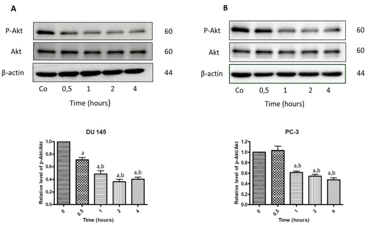
Scientific research has been underway for decades in order to develop an effective anticancer drug, and it has become crucial to find a novel and effective chemotherapeutics in the case of prostate cancer treatment. Ebselen derivatives have been shown to possess a variety of biological activities, including cytostatic and cytotoxic action against tumor cells. In this study, the cytotoxic effect and anticancer mechanism of action of two organoselenium compounds- (N-allyl-1,2-benzisoselenazol-3(2H)-one (N-allyl-BS) and N-(3-methylbutyl)-1,2-benzisoselenazol-3(2H)-one) (N-(3-mb)-BS)-were investigated on two phenotypically different prostate cancer cell lines DU 145 and PC-3. The influence of analyzed compounds on the viability parameter was also assessed on normal prostate cell line PNT1A. The results showed that both organoselenium compounds (OSCs) efficiently inhibited cancer cell proliferation, whereas normal PNT1A cells were less sensitive to the analazyed ebselen analouges. Both OSCs induced G2/M cell cycle arrest and prompted cell death through apoptosis. The detection of cleaved Poly (ADP-ribose) Polymerase (PARP) confirmed this. In addition, N-allyl-BS and N-(3-m)-b-BS increased the level of reactive oxygen species (ROS) formation, however only N-allyl-BS induced DNA damage. Based on our data, we assume that OSCs' anticancer action can be associated with oxidative stress induction and inactivation of the Akt- dependent signalling pathway. In conclusion, our data demonstrate that ebselen derivatives showed strong cytotoxic efficiency towards prostate cancer cells and may be elucidated as a novel, potent anticancer agent.

Keywords: DNA damage 4; Organoselenium compound 1; ROS 3; apoptosis 2; chemoprevention 5.


Conflict of interest statement

The authors declare no conflict of interest. The funders had no role in the design of the study; in the collection, analyses, or interpretation of data; in the writing of the manuscript; or in the decision to publish the results.

Figures

Figure 1
Figure 1
Molecular structure of ebselen, N-allyl-1,2-benzisoselenazol-3(2H)-one (N-allyl-BS), and N-(3-methylbutyl)-1,2-benzisoselenazol-3(2H)-one (N-(3-mb)-BS).
Figure 2
Figure 2
N-allyl-BS and N-(3-mb)-BS respectively inhibit viability of DU 145 (A,B) and PC-3 (C,D) prostate cancer cells in a dose dependent manner, while normal prostate cells PNT1A (E,F) are more resistant. The cells were treated with indicated concentrations of N-allyl-BS or N-(3-mb)-BS for 24 h. Viability was determined using SRB assay. Data are presented as mean ± SE (n = 3); significance of variation was calculated using one-way ANOVA followed by Dunnett’s multiple comparison test. (* P < 0.01) (** P < 0.001).
Figure 3
Figure 3
40 µM N-allyl-BS and N-(3-mb)-BS respectively induce G2/M cell cycle arrest in prostate cancer cells DU145 (A,B), PC-3 (C,D), after 8h treatment. The data are presented as mean ± SE (n = 3). Significance of variations compared with control was calculated using one-way ANOVA followed by Bonferroni’s multiple comparison test. (*P < 0.01).
Figure 4
Figure 4
40 µM N-allyl-BS (A,C) and N-(3-mb)-BS (B,D) respectively induce apoptosis and necrosis of cancer cells after 24 h treatment. Percentage of live, apoptotic and necrotic cells was determined by flow cytometry. The results are presented as a mean ± SE of three independent experiments. Significance of variations was determined by one-way ANOVA followed by Bonferroni’s multiple comparison test. (* P < 0.01).
Figure 5
Figure 5
Western blot analysis of cleaved PARP in DU 145 (A) and PC-3 cells (B) treated with 40 µM N-allyl-BS for indicated time points. β-actin was used as a lane loading control. The results are presented as mean ± SE of three independent experiments. The statistical significance of differences between respective samples was determined by one-way ANOVA followed by Bonferroni’s multiple comparison test, where (A) indicates significant difference between control and treated cells P < 0.05 and (b, c) indicates significant difference between 4, 8, and 24 h of 40 µM N-allyl-BS-treated cells P < 0.05.
Figure 6
Figure 6
Western blot analysis of cleaved PARP in DU 145 (A) and PC-3 cells (B) treated with 40 µM and N-(3-mb)-BS, respectively, for indicated time points. β-actin was used as a lane loading control. The results are presented as mean ± SE of three independent experiments. The statistical significance of differences between respective samples was determined by one-way ANOVA followed by Bonferroni’s multiple comparison test, where (a) indicates significant difference between control and treated N-(3-mb)-BS treated cells P < 0.05.
Figure 7
Figure 7
40 µM N-allyl-BS and N-(3-mb)-BS, respectively, enhance reactive oxygen species (ROS) generation in DU 145 (A,B) and PC-3 (C,D) cancer cells after 2 h of treatment. The amount of ROS generation was assessed using flow cytometry. Results are presented as a mean ± SE of three independent experiments. The significance of differences compared to control was calculated using student T test. (*P < 0.01).
Figure 8
Figure 8
The influence of 40 µM N-allyl-BS on DNA damage in DU 145 (A) and PC-3 (B) cells after 24 h treatment was determined using comet assay. The results are presented asthe percentage of DNA in Tail Intensity ± SE (n = 3). Significance of differences compared with control was calculated by student T test. (* P < 0.05). Representative pictures of DU 145 (C) and PC-3 (D) cells in the upper 90th percentile of tail intensity.
Figure 9
Figure 9
40 µM N-allyl-BS and N-(3-mb)-BS exhibit similar cytotoxic activity towards PC-3 cells both in hypoxic and normoxic conditions (AD). The cells were treated with indicated concentration of N-allyl-BS or N-(3-mb)-BS for 24 h. The viability was assessed using flow cytometry. Data are presented as mean ± SE (n = 3); significant difference was calculated using one-way ANOVA followed by Dunnett’s multiple comparison test (* P < 0.01).
Figure 10
Figure 10
Western blot analysis of p-Akt and Akt kinase in DU145 (A) and PC-3 (B) cells treated treated with 40 µM N-allyl-BS for indicated time points. β-actin was used as a lane loading control. Results are presented as mean ± SE (n = 3). The statistical significance of differences between respective samples was determined by one-way ANOVA followed by Bonferroni’s multiple comparison test, where (a) indicates significant difference between control and treated cells P < 0.01 and (b, c) indicates significant difference between 4, 8, and 24 h of 40 µM N-allyl-BS-treated cells P < 0.01.
Figure 11
Figure 11
Western blot analysis of p-Akt and Akt kinase in DU145 (A) and PC-3 (B) cells treated treated with 40 µM N-(3-mb)-BS for indicated time points. β-actin was used as a lane loading control. Results are presented as mean ± SE (n = 3). The statistical significance of differences between respective samples was determined by one-way ANOVA followed by Bonferroni’s multiple comparison test, where (a) indicates significant difference between control and treated cells P < 0.01 and (b, c) indicates significant difference between 4, 8, and 24 h of 40 µM N-(3-mb)-BS -treated cells P < 0.01.

Similar articles

Cited by

References

    1. Bray F., Ferlay J., Soerjomataram I., Siegel R.L., Torre L.A., Jemal A. Global cancer statistics 2018: Globocan estimates of incidence and mortality worldwide for 36 cancers in 185 countries. CA Cancer J. Clin. 2018;68:394–424. doi: 10.3322/caac.21492. - DOI - PubMed
    1. Venkateswaran V., Klotz L.H., Fleshner N.E. Selenium modulation of cell proliferation and cell cycle biomarkers in human prostate carcinoma cell lines. Cancer Res. 2002;62:2540–2545. - PubMed
    1. Sundaram N., Pahwa A.K., Ard M.D., Lin N., Perkins E., Bowles A.P. Selenium causes growth inhibition and apoptosis in human brain tumor cell lines. J. Neurooncol. 2000;46:125–133. doi: 10.1023/A:1006436326003. - DOI - PubMed
    1. Cho S.D., Jiang C., Malewicz B., Dong Y., Young C.Y., Kang K.S., Lee Y.S., Ip C., Lu J. Methyl selenium metabolites decrease prostate-specific antigen expression by inducing protein degradation and suppressing androgen-stimulated transcription. Mol. Cancer Ther. 2004;3:605–611. - PubMed
    1. Zhao H., Whitfield M.L., Xu T., Botstein D., Brooks J.D. Diverse effects of methylseleninic acid on the transcriptional program of human prostate cancer cells. Mol. Biol. Cell. 2004;15:506–519. doi: 10.1091/mbc.e03-07-0501. - DOI - PMC - PubMed

LinkOut - more resources