Comparative Study
. 2020 Mar 17;20(6):1659.
doi: 10.3390/s20061659.

Diagnosis of Occlusal Caries with Dynamic Slicing of 3D Optical Coherence Tomography Images

Affiliations
Comparative Study

Diagnosis of Occlusal Caries with Dynamic Slicing of 3D Optical Coherence Tomography Images

Minh N Luong et al. Sensors (Basel). .

Abstract

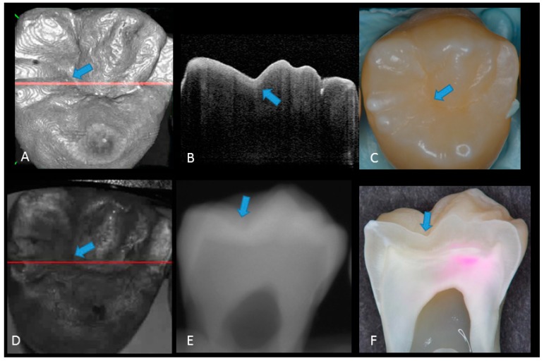
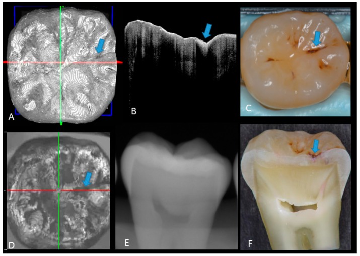
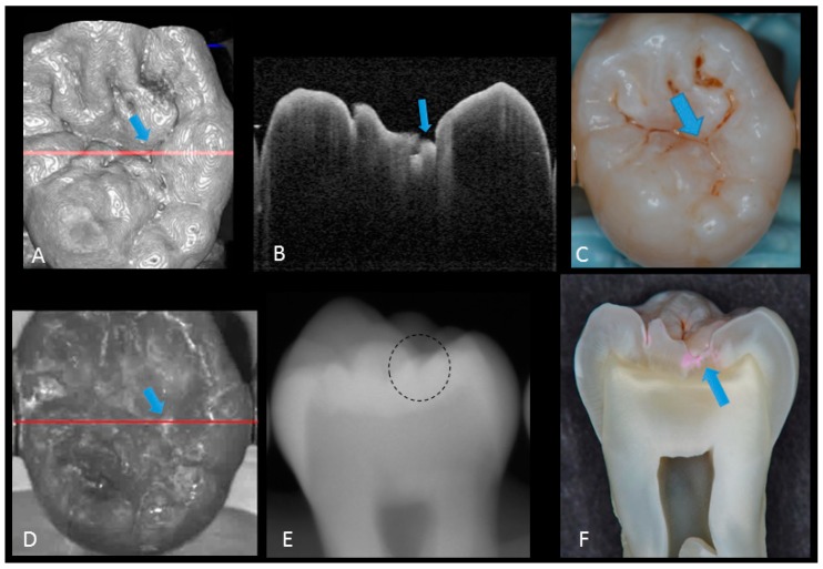
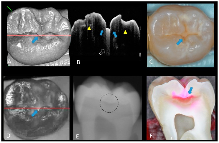
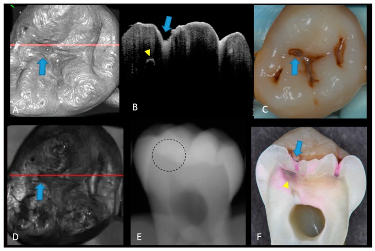
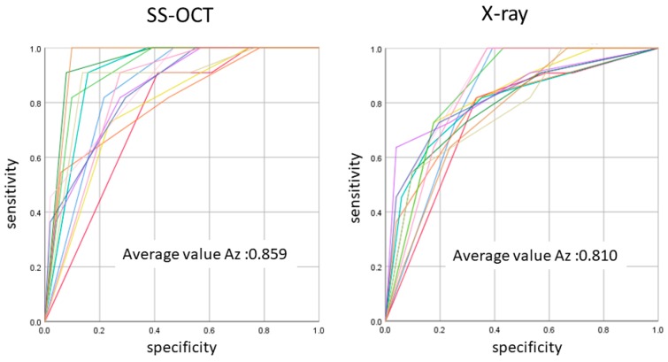
Detecting the extent of occlusal caries is a clinically important but challenging task required for treatment decision making. The aim of this study was to assess the diagnostic power of 3D swept-source optical coherence tomography (OCT) for evaluation of occlusal caries in comparison with X-ray radiography. Extracted human molars not exhibiting American Dental Association (ADA) criteria advanced caries were mounted in a silicone block and digital dental radiographs were captured from the buccal side. Subsequently, occlusal surfaces were scanned with a prototype Yoshida Dental OCT. Thirteen examiners evaluated the presence and extent of caries on radiographs and dynamically sliced 3D OCT video images, using a 4 level scale-0: intact; 1: enamel demineralization without cavitation; 2: enamel caries with cavitation; 3: dentin caries with or without cavitation. Sensitivity, specificity and area under operating characteristic curves (Az) were statistically analyzed (α = 0.05). Reliability analysis showed an excellent agreement among the 13 examiners for both methods. The OCT presented a significantly higher sensitivity and Az value for the detection of caries compared to radiographs (p < 0.05). Radiography showed especially low sensitivity for dentin caries (0-2 versus 3). Dynamic slicing of 3D OCT volumes is a powerful adjunct tool to visual inspection to diagnose the dentin occlusal caries in vitro.

Keywords: dentin; dentino-enamel junction DEJ; enamel; hidden caries; optical coherence tomography; radiograph; receiver operating characteristic (ROC) analysis.


Conflict of interest statement

The authors declare no conflict of interest with regard to the authorship of this manuscript.

Figures

Figure 1
Figure 1
Prototype Yoshida Dental optical coherence tomography (OCT) system. (A) Specimen positioning to image the occlusal surface with the intraoral mirror tip for posterior teeth imaging attached to the handheld probe; (B) schematic diagram OCT system. An interference pattern is produced by splitting the beam of the Micro Electro Mechanical System (MEMS)-based laser source into two arms (i.e., reference arm and sample arm). The backscattered light from the sample is redirected back and recombined with the light from the reference arm in the coupler. The interference signal is transformed into raw A-scan data. Series of A-scans produce raw B-scan (2D) and the composition of 2D leads to the acquisition of 3D images; (C) overview of the OCT system with foot pedal for clinical imaging.
Figure 2
Figure 2
Sound enamel. (A) gray-scale three-dimensional (3D) OCT image; (B) cross-sectional OCT image along the red line in (A) and (D) with intact enamel with bright band across the fissure base extending over the cusps (arrow), suggesting a developmental feature (not acquired caries); (C) photographic presentation of occlusal enamel with seemingly intact fissure (arrow); (D) OCT en face intensity projection; (E) radiograph; (F) cross-sectional photograph with no dye at the observed fissure (arrow) confirming sound surface (score 0). The dynamic slicing 3D video is presented in Supplementary Materials: Video 1.
Figure 3
Figure 3
Enamel caries: (A) 3D OCT image; (B) cross-sectional OCT image along the red line in (A). Arrow shows a wedge-shaped area with increased scattering approximately half the thickness of the enamel, indicating demineralization with no surface breakdown; (C) photographic presentation of occlusal enamel with stained fissure (arrow); (D) OCT en face intensity projection; (E) radiograph showed no visible lesion; (F) cross-section view with shallow enamel dye penetration confirming demineralization without surface breakdown (score 1). The dynamic slicing 3D video is presented in Supplementary Materials: Video 2.
Figure 4
Figure 4
Enamel breakdown: (A) gray-scale three-dimensional (3D) OCT image; (B) cross-sectional OCT image along the red line in (A) and (D). Arrow shows cavitated caries; (C) visual score 2 of enamel cavitation; (D) OCT en face intensity projection; (E) enamel lesion was not distinguished on radiograph due to the superimposition of buccal and lingual cusps; (F) cross-section view with penetration of pink dye enclosed at enamel (score 2). Supplementary Materials: Video 3.
Figure 5
Figure 5
Hidden dentin caries; (A) 3D OCT image; (B) cross-sectional OCT along the red line in (A) and (D). Bold arrows show cavitated caries with discontinued enamel surface and scattering beyond dentino-enamel junction (DEJ) (triangle). Blank arrow suggests strong scattering from enamel into dentin (score 3); (C) photographic presentation of occlusal enamel; (D) OCT en face intensity projection; (E) dashed circle shows suspected radiolucency beyond DEJ on the radiograph; (F) cross-section view with penetration of red dye beyond DEJ, confirming the presence of caries infected dentin (score 3). The dynamic slicing 3D video is presented in Supplementary Materials: Video 5.
Figure 6
Figure 6
Dentin caries: (A) gray-scale three-dimensional (3D) OCT image; (B) cross-sectional OCT image of the lesion along the red line in (A) and (D). A separation of DEJ (triangle) was observed at the bottom at the lesion (arrow) indicating the lateral invasion of bacteria; (C) visual score 3 with visible dentin; (D) OCT en face intensity projection; (E) radiolucent lesion at DEJ; (F) cross-section view with penetration of dark red dye one-third of dentin thickness. The dynamic slicing 3D video is presented in Supplementary Materials: Video 4.
Figure 7
Figure 7
The average Az value of ROC with 13 lines represented by the Az value from thirteen examiners. There was a difference between OCT and radiography in the accuracy of detection in occlusal caries in three cutoff points (p < 0.05).

Similar articles

Cited by

References

    1. Young D.A., Nový B.B., Zeller G.G., Hale R., Hart T.C., Truelove E.L., Ekstrand K.R., Featherstone J.D.B., Fontana M., Ismail A., et al. The American Dental Association Caries Classification System for clinical practice: A report of the American Dental Association Council on Scientific Affairs. J. Am. Dent. Assoc. 2015;146:79–86. doi: 10.1016/j.adaj.2014.11.018. - DOI - PubMed
    1. Jordan R.A., Krois J., Schiffner U., Micheelis W., Schwendicke F. Trends in caries experience in the permanent dentition in Germany 1997–2014, and projection to 2030: Morbidity shifts in an aging society. Sci. Rep. 2019;9:5534. doi: 10.1038/s41598-019-41207-z. - DOI - PMC - PubMed
    1. Kaste L.M., Selwitz R.H., Oldakowski R.J., Brunelle J.A., Winn D.M., Brown L.J. Coronal caries in the primary and permanent dentition of children and adolescents 1–17 years of age: United States, 1988–1991. J. Dent. Res. 1996;75:631–641. doi: 10.1177/002203459607502S03. - DOI - PubMed
    1. Demirci M., Tuncer S., Yuceokur A.A. Prevalence of caries on individual tooth surfaces and its distribution by age and gender in university clinic patients. Eur. J. Dent. 2010;4:270–279. doi: 10.1055/s-0039-1697839. - DOI - PMC - PubMed
    1. Ricketts D., Kidd E., Weerheijm K.L., de Soet H. Hidden caries: What is it? Does it exist? Does it matter? Int. Dent. J. 1997;47:259–265. doi: 10.1002/j.1875-595X.1997.tb00786.x. - DOI - PubMed

MeSH terms