. 2020 Mar 17;21(6):2049.
doi: 10.3390/ijms21062049.

Cryptides Identified in Human Apolipoprotein B as New Weapons to Fight Antibiotic Resistance in Cystic Fibrosis Disease

Affiliations

Cryptides Identified in Human Apolipoprotein B as New Weapons to Fight Antibiotic Resistance in Cystic Fibrosis Disease

Rosa Gaglione et al. Int J Mol Sci. .

Abstract

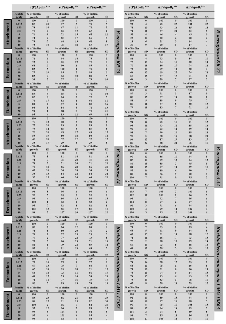
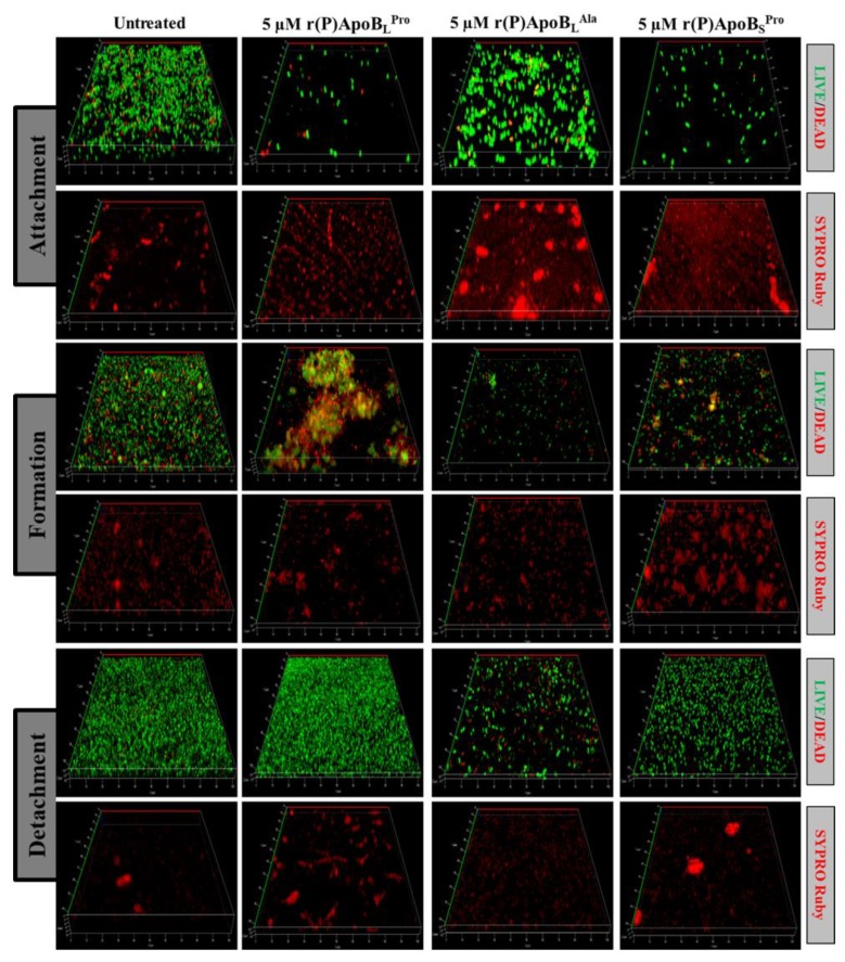
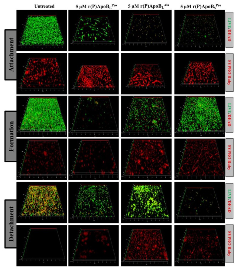
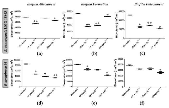
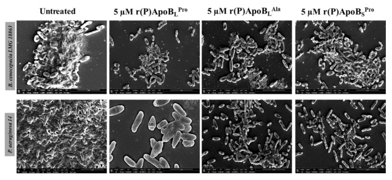
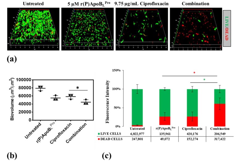
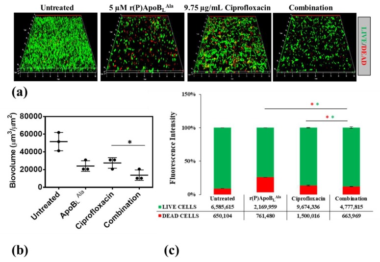
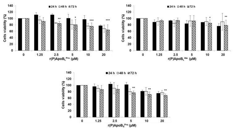
Chronic respiratory infections are the main cause of morbidity and mortality in cystic fibrosis (CF) patients, and are characterized by the development of multidrug resistance (MDR) phenotype and biofilm formation, generally recalcitrant to treatment with conventional antibiotics. Hence, novel effective strategies are urgently needed. Antimicrobial peptides represent new promising therapeutic agents. Here, we analyze for the first time the efficacy of three versions of a cryptide identified in human apolipoprotein B (ApoB, residues 887-922) towards bacterial strains clinically isolated from CF patients. Antimicrobial and anti-biofilm properties of ApoB-derived cryptides have been analyzed by broth microdilution assays, crystal violet assays, confocal laser scanning microscopy and scanning electron microscopy. Cell proliferation assays have been performed to test cryptide effects on human host cells. ApoB-derived cryptides have been found to be endowed with significant antimicrobial and anti-biofilm properties towards Pseudomonas and Burkholderia strains clinically isolated from CF patients. Peptides have been also found to be able to act in combination with the antibiotic ciprofloxacin, and they are harmless when tested on human bronchial epithelial mesothelial cells. These findings open interesting perspectives to cryptide applicability in the treatment of chronic lung infections associated with CF disease.

Keywords: anti-biofilm peptides; antibiotic resistance; antimicrobial peptides; cryptides; cystic fibrosis; host defense peptides; synergistic effects.


Conflict of interest statement

The authors declare no conflict of interest.

Figures

Figure 1
Figure 1
Anti-biofilm activity of r(P)ApoBLPro, r(P)ApoBLAla, and r(P)ApoBSPro peptides on P. aeruginosa RP 73, P. aeruginosa KK 27, P. aeruginosa 14, P. aeruginosa AA2, B. multivorans LMG 17582, and B. cenocepacia LMG 18863 in MHB medium.
Figure 2
Figure 2
Effects of r(P)ApoBLPro, r(P)ApoBLAla, and r(P)ApoBSPro peptides on B. cenocepacia LMG 18863 biofilm attachment, formation and detachment. Biofilm cells were stained by using LIVE/DEAD BacLight bacterial viability kit (Molecular Probes, Eugene, OR, USA) containing 1:1 ratio of Syto-9 (green fluorescence, all cells) and propidium iodide (PI, red fluorescence, dead cells) and FilmTracer™ SYPRO® Ruby biofilm matrix staining (Invitrogen™, F10318). Images are 3D projections of biofilm structure obtained by laser scanning confocal z-stack using Zen Lite 2.3 software. All images were taken under identical conditions.
Figure 3
Figure 3
Effects of r(P)ApoBLPro, r(P)ApoBLAla and r(P)ApoBSPro peptides on P. aeruginosa 14 biofilm attachment, formation and detachment. Biofilm cells were stained by using LIVE/DEAD BacLight bacterial viability kit (Molecular Probes, Eugene, OR, USA) containing 1:1 ratio of Syto-9 (green fluorescence, all cells) and propidium iodide (PI, red fluorescence, dead cells) and FilmTracer™ SYPRO® Ruby biofilm matrix staining (Invitrogen™, F10318). Images are 3D projections of biofilm structure obtained by laser scanning confocal z-stack using Zen Lite 2.3 software. All images were taken under identical conditions.
Figure 4
Figure 4
Analysis of the effects of r(P)ApoBLPro, r(P)ApoBLAla and r(P)ApoBSPro peptides on biofilm attachment (a,d), formation (b, e) and detachment (c,f) in the case of B. cenocepacia LMG 18863 (ac) and P. aeruginosa 14 (df). Biovolume (µm3/µm2) was measured by using Zen Lite 2.3 software. Significant differences were indicated as * p < 0.05 or ** p < 0.01 for treated versus control samples.
Figure 5
Figure 5
Morphological analyses of B. cenocepacia LMG 18863 (top panel) and P. aeruginosa 14 (lower panel) preformed biofilms by SEM. Representative images are shown upon treatment of bacterial biofilm with 5 μM r(P)ApoBLPro, r(P)ApoBLAla, and r(P)ApoBSPro. Bars 5 μm.
Figure 6
Figure 6
Effects of r(P)ApoBLPro, ciprofloxacin and a combination of the two compounds on preformed biofilm (a). Biofilm cells were stained by using LIVE/DEAD BacLight bacterial viability kit (Molecular Probes, Eugene, OR) containing 1:1 ratio of Syto-9 (green fluorescence, all cells) and propidium iodide (PI, red fluorescence, dead cells). Images are 3D projections of biofilm structure obtained by laser scanning confocal z-stack using Zen Lite 2.3 software. All images were taken under identical conditions. Biovolume (µm3/µm2) was measured by using Zen Lite 2.3 software. Significant differences were indicated as * p < 0.05 for treated versus control samples (b). Numbers of live and dead cells were evaluated by using Zen Lite 2.3 software (c).
Figure 7
Figure 7
Effects of r(P)ApoBLAla, ciprofloxacin and a combination of the two compounds on preformed biofilm (a). Biofilm cells were stained by using LIVE/DEAD BacLight bacterial viability kit (Molecular Probes, Eugene, OR) containing 1:1 ratio of Syto-9 (green fluorescence, all cells) and propidium iodide (PI, red fluorescence, dead cells). Images are 3D projections of biofilm structure obtained by laser scanning confocal z-stack using Zen Lite 2.3 software. All images were taken under identical conditions. Biovolume (µm3/µm2) was measured by using Zen Lite 2.3 software. Significant differences were indicated as * p < 0.05 for treated versus control samples (b). Numbers of live and dead cells were evaluated by using Zen Lite 2.3 software (c).
Figure 8
Figure 8
Effects of r(P)ApoBLPro, r(P)ApoBLAla and r(P)ApoBSPro peptides on the viability of BEAS cells. Cell viability was assessed by MTT assays, and expressed as the percentage of viable cells with respect to controls (untreated cells). Error bars indicate standard deviations obtained from at least three independent experiments, each one carried out with triplicate determinations. Significant differences were indicated as * p < 0.05, ** p < 0.01 or *** p < 0.001 for treated versus control samples.

Similar articles

Cited by

References

    1. Farrell P.M. The prevalence of cystic fibrosis in the European Union. J. Cyst. Fibros. 2008;7:450–453. doi: 10.1016/j.jcf.2008.03.007. - DOI - PubMed
    1. Evans C.M., Koo J.S. Airway mucus: The good, the bad, the sticky. Pharmacol Ther. 2009;121:332–348. doi: 10.1016/j.pharmthera.2008.11.001. - DOI - PMC - PubMed
    1. Chace K.V., Flux M., Sachdev G.P. Comparison of physicochemical properties of purified mucus glycoproteins isolated from respiratory secretions of cystic fibrosis and asthmatic patients. Biochemistry. 1985;24:7334–7341. doi: 10.1021/bi00346a047. - DOI - PubMed
    1. Knowles M.R., Durie P.R. What is cystic fibrosis? N. Engl. J. Med. 2002;347:439–442. doi: 10.1056/NEJMe020070. - DOI - PubMed
    1. Ciofu O., Hansen C.R., Høiby N. Respiratory bacterial infections in cystic fibrosis. Curr. Opin. Pulm. Med. 2013;19:251–258. doi: 10.1097/MCP.0b013e32835f1afc. - DOI - PubMed

MeSH terms

Substances