. 2020 Mar 17;21(6):2047.
doi: 10.3390/ijms21062047.

Effect of Streptozotocin-Inducted Diabetes on the Pathophysiology of Enteric Neurons in the Small Intestine Based on the Porcine Diabetes Model

Affiliations

Effect of Streptozotocin-Inducted Diabetes on the Pathophysiology of Enteric Neurons in the Small Intestine Based on the Porcine Diabetes Model

Michał Bulc et al. Int J Mol Sci. .

Abstract

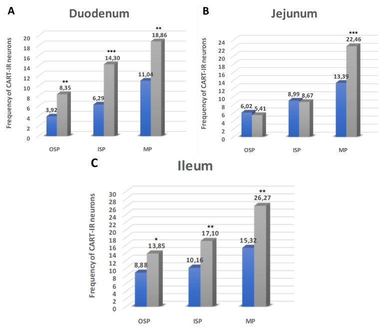
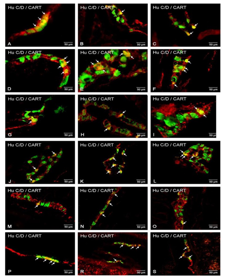
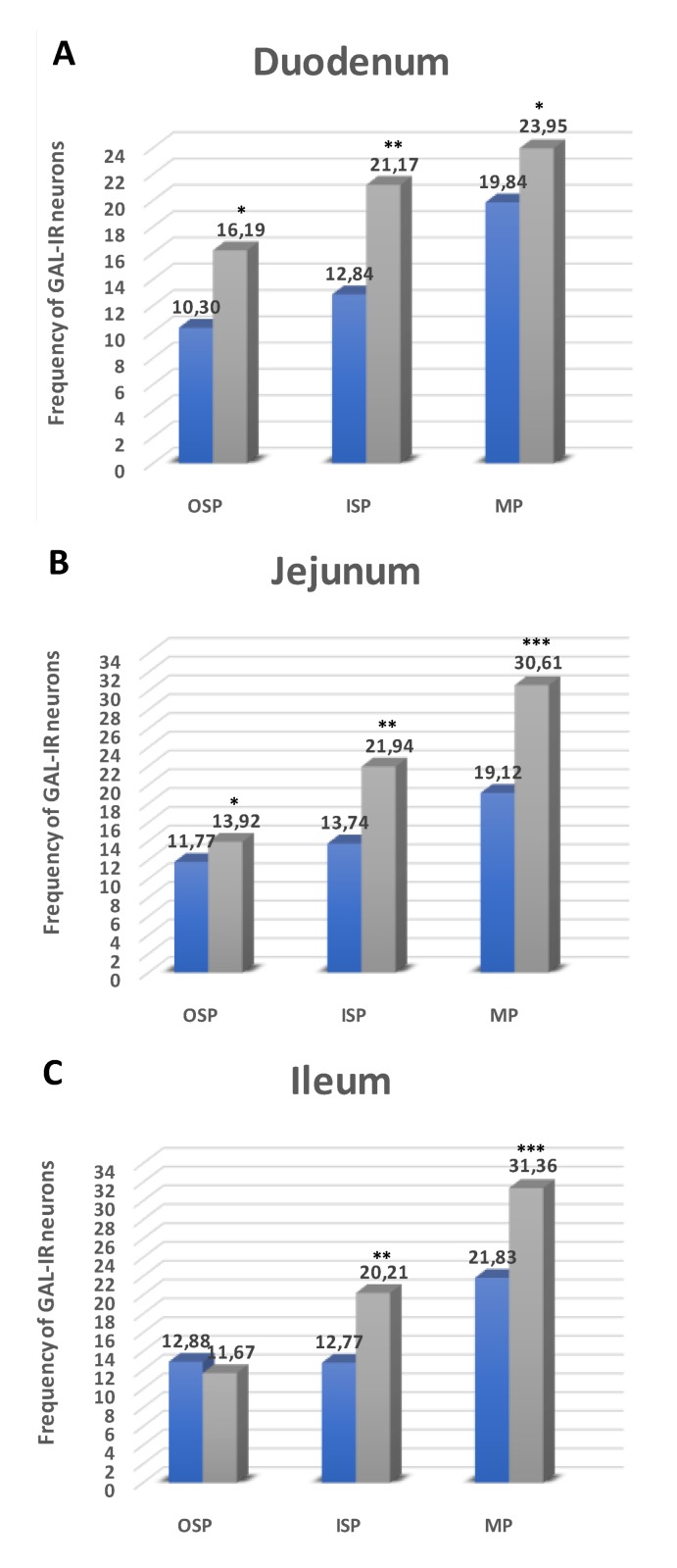
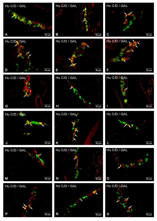
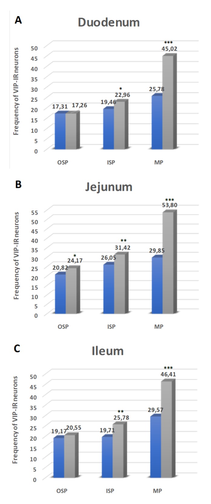
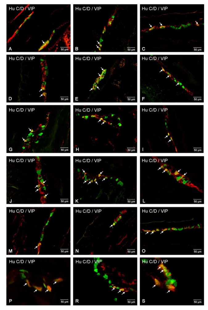
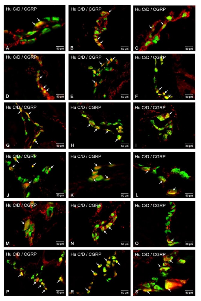
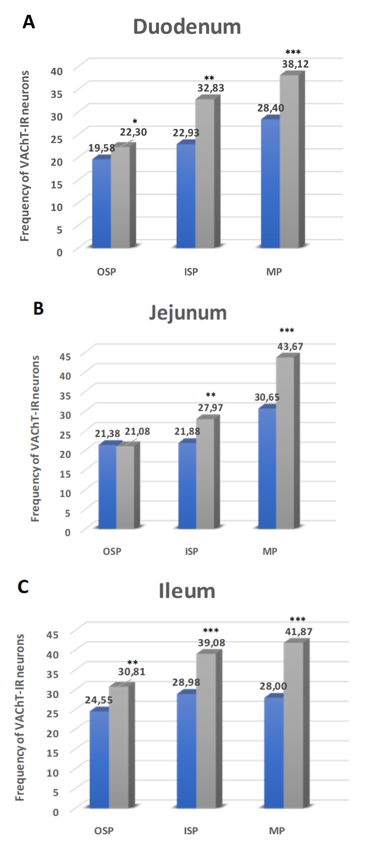
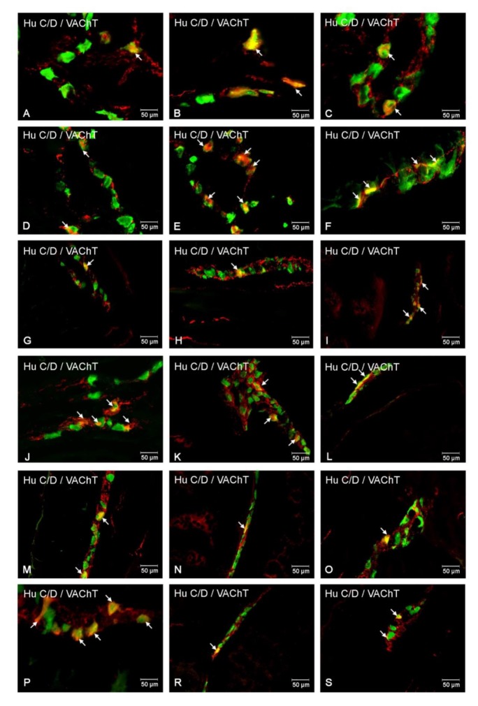
Hyperglycemia is one of the main causes of diabetes complications. Gastrointestinal (GI) disturbances are one of the most frequent complications during diabetes. The porcine digestive tract possesses physiological and pathological similarities to the human digestive tract. This also applies to the innervation of the gastrointestinal tract. In this study, the influence of experimentally-inducted hyperglycemia was examined on the expression of vesicular acetylcholine transporter (VAChT), cocaine- and amphetamine-regulated transcript (CART), galanin (GAL), vasoactive intestinal polypeptide (VIP), and calcitonin gene-related peptide (CGRP) in the enteric nervous system (ENS) neurons in the small intestine of the pig. During the current study, an increased number of neurons containing CART, VIP, GAL, and CGRP under streptozotocin injection were observed. The augmentation of expression included all enteric plexuses present in the small intestine. The same results were obtained in the case of VAChT; namely, chronic hyperglycemia led to an increase in the number of neurons utilizing VAChT in all investigated plexuses. The obtained results suggested that the function of neuropeptides studied in this experiment depended on their localization in the ENS structures, as well as part of the GI tract. Diabetes led to alterations in the neurochemical phenotype of small intestine enteric neurons.

Keywords: diabetes; enteric nervous system; gastrointestinal complications; neuropeptides; pig.


Conflict of interest statement

The authors declare no conflict of interest.

Figures

Figure 1
Figure 1
Diagram of the numbers of perikarya immunoreactive to Cocaine- and amphetamine-regulated transcript (CART) (A–C) of the control (blue bars) and experimental group (grey bars) in the particular parts of the small intestine. OSP – outer submucosal plexus, ISP—inner submucosal plexus, MP—myenteric plexus. * p < 0.05, ** p < 0.01, *** p < 0.001—indicate differences between all groups for the same neuronal populations.
Figure 2
Figure 2
Immunofluorescent staining presenting CART immunoreactivity in cell bodies in the particular intramural plexuses in the small intestine in both control and experimental groups. All photographs have been created by the digital superimposition of two color channels; Hu C/D-positive—used here as a pan-neuronal marker (green), and CART-positive (red). The arrow shows perikaryon containing both examined substances. Myenteric plexus of the duodenum (A,D), jejunum (B,E), and ileum (C,F) under physiological condition (A–C) and after streptozotocin administration (D–F). Outer submucosal plexus of the duodenum (G,J), jejunum (H,K), and ileum (I,L) under physiological condition (G–I) and after streptozotocin administration (J–L). Inner submucosal plexus of the duodenum (M,P), jejunum (N,R), and ileum (O,S) under physiological condition (M–O) and after streptozotocin administration (P–S).
Figure 3
Figure 3
Schematic diagram of the numbers of perikarya immunoreactive to galanin (GAL) (A–C) of the control (blue bars) and experimental group (grey bars) in the particular parts of the small intestine. OSP – outer submucosal plexus, ISP—inner submucosal plexus, MP—myenteric plexus. * p < 0.05, ** p < 0.01, *** p < 0.001 – indicate differences between all groups for the same neuronal populations.
Figure 4
Figure 4
Immunofluorescent staining presenting GAL immunoreactivity in cell bodies in the particular intramural plexuses in the small intestine in both control and experimental groups. All photographs have been created by the digital superimposition of two color channels; Hu C/D-positive—used here as a pan-neuronal marker (green), and GAL-positive (red). The arrow shows perikaryon containing both examined substances. Myenteric plexus of the duodenum (A,D), jejunum (B,E), and ileum (C,F) under physiological condition (A–C) and after streptozotocin administration (D–F).Outer submucosal plexus of the duodenum (G,J), jejunum (H,K), and ileum (I,L) under physiological condition (G–I) and after streptozotocin administration (J–L). Inner submucosal plexus of the duodenum (M,P), jejunum (N,R), and ileum (O,S) under physiological condition (M–O) and after streptozotocin administration (P–S).
Figure 5
Figure 5
Schematic diagram of the numbers of perikarya immunoreactive to a vasoactive intestinal polypeptide (VIP) (A–C) of the control (blue bars) and experimental group (grey bars) in the particular parts of the small intestine. OSP – outer submucosal plexus, ISP—inner submucosal plexus, MP—myenteric plexus. * p < 0.05, ** p < 0.01, *** p < 0.001—indicate differences between all groups for the same neuronal populations.
Figure 6
Figure 6
Immunofluorescent staining presenting VIP immunoreactivity in cell bodies in the particular intramural plexuses in the small intestine in both control and experimental groups. All photographs have been created by the digital superimposition of two color channels; Hu C/D-positive—used here as a pan-neuronal marker (green), and VIP-positive (red). The arrow shows perikaryon containing both examined substances. Myenteric plexus of the duodenum (A,D), jejunum (B,E), and ileum (C,F) under physiological condition (A–C) and after streptozotocin administration (D–F). Outer submucosal plexus of the duodenum (G,J), jejunum (H,K), and ileum (I,L) under physiological condition (G–I) and after streptozotocin administration (J–L). Inner submucosal plexus of the duodenum (M,P), jejunum (N,R), and ileum (O,S) under physiological condition (M–O) and after streptozotocin administration (P–S).
Figure 7
Figure 7
Schematic diagram of the numbers of perikarya immunoreactive to calcitonin gene-related peptide (CGRP) (A–C) of the control (blue bars) and experimental group (grey bars) in the particular parts of the small intestine. OSP – outer submucosal plexus, ISP – inner submucosal plexus, MP – myenteric plexus. * p < 0.05, ** p < 0.01, *** p < 0.001 – indicate differences between all groups for the same neuronal populations.
Figure 8
Figure 8
Immunofluorescent staining presenting CGRP immunoreactivity in cell bodies in the particular intramural plexuses in the small intestine in both control and experimental groups. All photographs have been created by the digital superimposition of two color channels; Hu C/D-positive—used here as a pan-neuronal marker (green), and CGRP-positive (red). The arrow shows perikaryon containing both examined substances. Myenteric plexus of the duodenum (A,D), jejunum (B,E), and ileum (C,F) under physiological condition (A–C) and after streptozotocin administration (D–F). Outer submucosal plexus of the duodenum (G,J), jejunum (H,K), and ileum (I,L) under physiological condition (G-I) and after streptozotocin administration (J–L). Inner submucosal plexus of the duodenum (M,P), jejunum (N,R), and ileum (O,S) under physiological condition (M–O) and after streptozotocin administration (P–S).
Figure 9
Figure 9
Schematic diagram of the numbers of perikarya immunoreactive to vesicular acetylcholine transporter (VAChT) (A–C) of the control (blue bars) and experimental group (grey bars) in the particular parts of the small intestine. OSP – outer submucosal plexus, ISP – inner submucosal plexus, MP – myenteric plexus. * p < 0.05, ** p < 0.01, *** p < 0.001 – indicate differences between all groups for the same neuronal populations.
Figure 10
Figure 10
Immunofluorescent staining presenting VAChT immunoreactivity in cell bodies in the particular intramural plexuses in the small intestine in both control and experimental groups. All photographs have been created by the digital superimposition of two color channels; Hu C/D-positive—used here as a pan-neuronal marker (green), and VAChT-positive (red). The arrow shows perikaryon containing both examined substances. Myenteric plexus of the duodenum (A,D), jejunum (B,E), and ileum (C,F) under physiological condition (A–C) and after streptozotocin administration (D–F). Outer submucosal plexus of the duodenum (G,J), jejunum (H,K), and ileum (I,L) under physiological condition (G-I) and after streptozotocin administration (J–L). Inner submucosal plexus of the duodenum (M,P), jejunum (N,R), and ileum (O,S) under physiological condition (M–O) and after streptozotocin administration (P–S).

Similar articles

Cited by

References

    1. Furness J.B., Callaghan B.P., Rivera L.R., Cho H.J. The enteric nervous system and gastrointestinal innervation: Integrated local and central control. Adv. Exp. Med. Biol. 2014;817:39–71. - PubMed
    1. Furness J.B. The enteric nervous system and neurogastroenterology. Nat. Rev. Gastroenterol. Hepatol. 2012;9:286–294. doi: 10.1038/nrgastro.2012.32. - DOI - PubMed
    1. Furness J.B. The enteric nervous system: Normal functions and enteric neuropathies. Neurogastroenterol. Motil. 2008;20(Suppl. 1):32–38. - PubMed
    1. Schemann M., Neunlist M. The human enteric nervous system. Neurogastroenterol. Motil. 2004;16(Suppl. 1):55–59. - PubMed
    1. Furness J.B. The organisation of the autonomic nervous system: Peripheral connections. Auton Neurosci. 2006;130:1–5. doi: 10.1016/j.autneu.2006.05.003. - DOI - PubMed

MeSH terms