. 2020 Mar 17;10(3):163.
doi: 10.3390/diagnostics10030163.

Microbiome Profile of Deep Endometriosis Patients: Comparison of Vaginal Fluid, Endometrium and Lesion

Affiliations

Microbiome Profile of Deep Endometriosis Patients: Comparison of Vaginal Fluid, Endometrium and Lesion

Camila Hernandes et al. Diagnostics (Basel). .

Abstract

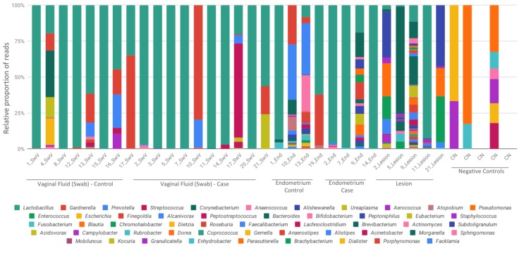
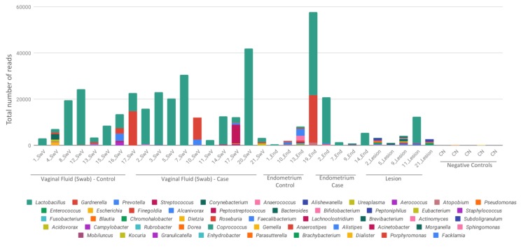
This work aimed to identify and compare the bacterial patterns present in endometriotic lesions, eutopic endometrium and vaginal fluid from endometriosis patients with those found in the vaginal fluid and eutopic endometrium of control patients. Vaginal fluid, eutopic endometrium and endometriotic lesions were collected. DNA was extracted and the samples were analyzed to identify microbiome by high-throughput DNA sequencing of the 16S rRNA marker gene. Amplicon sequencing from vaginal fluid, eutopic endometrium and endometriotic lesion resulted in similar profiles of microorganisms, composed most abundantly by the genus Lactobacillus, Gardnerella, Streptococcus and Prevotella. No significant differences were found in the diversity analysis of microbiome profiles between control and endometriotic patients; however deep endometriotic lesions seems to present different bacterial composition, less predominant of Lactobacillus and with more abundant Alishewanella, Enterococcus and Pseudomonas.

Keywords: 16S rRNA; endometriosis; microbiome; next generation sequencing (NGS); pathogenesis; vaginal fluid.


Conflict of interest statement

The authors declare no conflicts of interest.

Figures

Figure 1
Figure 1
Relative abundance of bacterial profiles. Bacterial composition of each sample is reported by color bars relative to a 100% scale. Results are presented by the taxonomic rank of genus. SwV—vaginal fluid (Swab), End—endometrium, CN—negative controls.
Figure 2
Figure 2
Total reads sequenced for each sample. Bacterial composition of each sample is reported by different color bars and scaled as the total number of reads for each sample. Results are presented by the taxonomic rank of genus. SwV—vaginal fluid (Swab), End—endometrium, CN—negative controls.
Figure 3
Figure 3
Alpha and beta-diversity analysis. (A) Principal coordinates analysis (PCoA) made with weighted UniFrac distances and Bray‐Curtis dissimilarity. Sample groups are represented by the letters: A (dark green)-control swab from vaginal fluid; B (orange)-case swab from vaginal fluid; C (purple)-control endometrial tissue; D (pink)-case endometrial tissue; and E (light green)-lesion. (B) Alpha diversity indexes of Shannon and Simpson are sowed in the boxplots for each collection site group of samples (A, B, C, D and E). Kruskall‐Wallis and Wilcoxon tests are showed for group and paired groups comparison, respectively (represented by solid lined above boxplots, only significative comparisons were shown and marked with an *). Black solid points represent outlier samples.
Figure 4
Figure 4
Relative and differential abundances. (A) Boxplots represent the most abundant genera detected and their distribution in the samples. Swab_ctrl—control swab from vaginal fluid; Swab—case swab from vaginal fluid; Endo_ctrl—control endometrial tissue; Endo—case endometrial tissue; lesion—samples from endometriosis lesion. Black solid points represent outlier samples for each group. (B) Differential abundance heatmap comparing all the collection sites from case and control samples. Results are showed as the log2FC for the differentially detected genera. Most abundant genera are showed as red values considering the first sample category in the bottom legend.

Similar articles

Cited by

References

    1. De Paula Andres M., Lopes L.A., Baracat E.C., Podgaec S. Dienogest in the treatment of endometriosis: Systematic review. Arch. Gynecol. Obstet. 2015;292:523–529. doi: 10.1007/s00404-015-3681-6. - DOI - PubMed
    1. Parasar P., Ozcan P., Terry K.L. Endometriosis: Epidemiology, Diagnosis and Clinical Management. Curr. Obstet. Gynecol. Rep. 2017;6:34–41. doi: 10.1007/s13669-017-0187-1. - DOI - PMC - PubMed
    1. Nisolle M., Donnez J. Peritoneal endometriosis, ovarian endometriosis, and adenomyotic nodules of the rectovaginal septum are three different entities. Fertil. Steril. 1997;68:585–596. doi: 10.1016/S0015-0282(97)00191-X. - DOI - PubMed
    1. Bassi M.A., Arias V., D’Amico N., Gueuvoghlanian-Silva B.Y., Abrao M.S., Podgaec S. Deep Invasive Endometriosis Lesions of the Rectosigmoid May Be Related to Alterations in Cell Kinetics. Reprod Sci. 2015;22:1122–1128. doi: 10.1177/1933719115574341. - DOI - PubMed
    1. Santulli P., Tran C., Gayet V., Bourdon M., Maignien L., Pocate-Cheriet K., Chapron C., de Ziegler D. Oligo-anovulation is not a rarer feature in women with documented endometriosis. Fertil. Steril. 2018;110:941–948. doi: 10.1016/j.fertnstert.2018.06.012. - DOI - PubMed

LinkOut - more resources