. 2020 Mar 20;9(1):651-663.
doi: 10.1080/22221751.2020.1741326. eCollection 2020.

Tropism, pathology, and transmission of equine parvovirus-hepatitis

Affiliations

Tropism, pathology, and transmission of equine parvovirus-hepatitis

Joy Ellen Tomlinson et al. Emerg Microbes Infect. .

Abstract

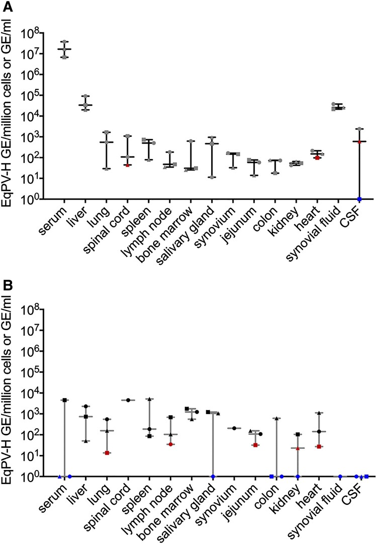
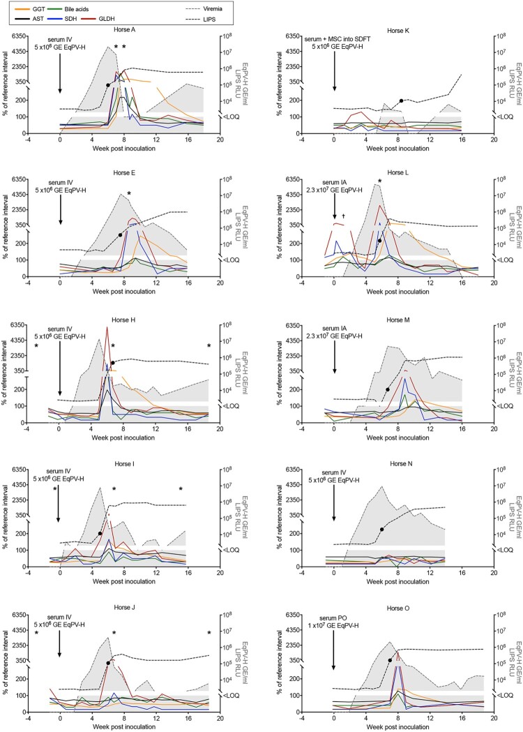
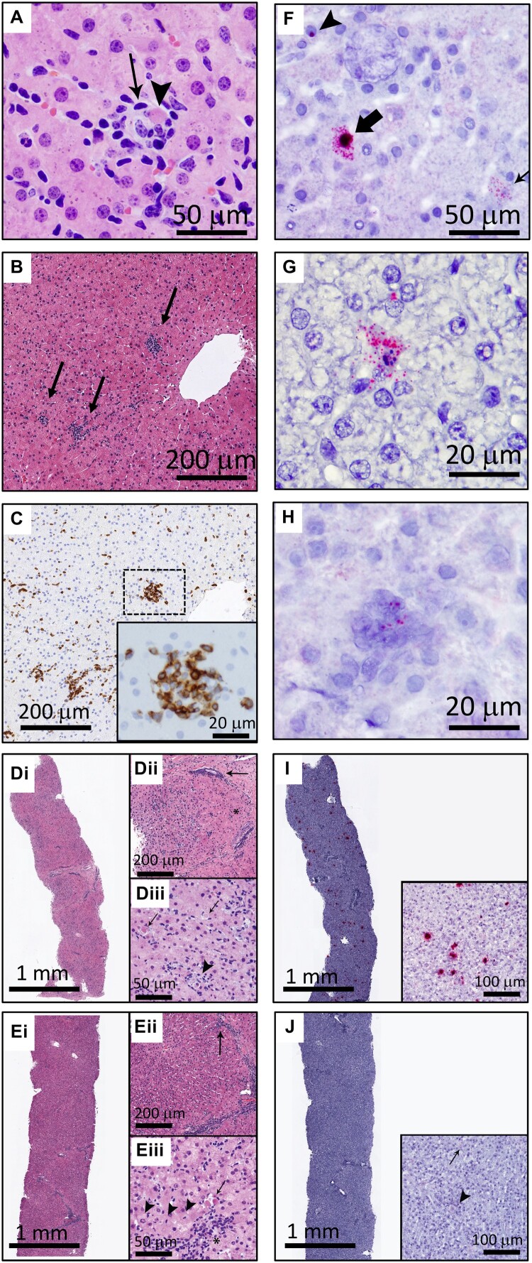
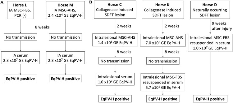
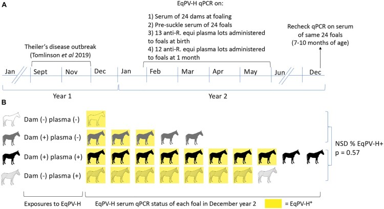
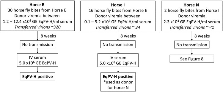
Equine parvovirus-hepatitis (EqPV-H) has recently been associated with cases of Theiler's disease, a form of fulminant hepatic necrosis in horses. To assess whether EqPV-H is the cause of Theiler's disease, we first demonstrated hepatotropism by PCR on tissues from acutely infected horses. We then experimentally inoculated horses with EqPV-H and 8 of 10 horses developed hepatitis. One horse showed clinical signs of liver failure. The onset of hepatitis was temporally associated with seroconversion and a decline in viremia. Liver histology and in situ hybridization showed lymphocytic infiltrates and necrotic EqPV-H-infected hepatocytes. We next investigated potential modes of transmission. Iatrogenic transmission via allogeneic stem cell therapy for orthopedic injuries was previously suggested in a case series of Theiler's disease, and was demonstrated here for the first time. Vertical transmission and mechanical vectoring by horse fly bites could not be demonstrated in this study, potentially due to limited sample size. We found EqPV-H shedding in oral and nasal secretions, and in feces. Importantly, we could demonstrate EqPV-H transmission via oral inoculation with viremic serum. Together, our findings provide additional information that EqPV-H is the likely cause of Theiler's disease and that transmission of EqPV-H occurs via both iatrogenic and natural routes.

Keywords: Theiler’s disease; horse fly; serum hepatitis; stem cells; vertical transmission.


Conflict of interest statement

No potential conflict of interest was reported by the author(s).

Figures

Figure 1.
Figure 1.
EqPV-H is hepatotropic and persists in tissues for months. (A) qPCR of tissues from 3 acutely infected horses at 5 weeks post inoculation and (B) three horses at least 15 weeks after infection. Viral load was significantly associated with tissue type in acutely infected horses (p = 0.028). Viral load was normalized to cell count by B2M qPCR for solid tissues or to volume for liquids. EqPV-H positive samples below the limit of quantitation are indicated in red, and samples below the limit of detection are indicated in blue.
Figure 2.
Figure 2.
Serum biochemical, serologic, and virologic profiles of 10 horses experimentally infected with EqPV-H. Inoculation routes and doses varied and are indicated on each panel. *Times when liver biopsies were obtained. •First seropositive sample (positive cut-off 104 RLU). tHorse L was Equine hepacivirus (EqHV) negative when screened before inclusion in the study, however, she was naturally infected during the study and was found to be EqHV serum qPCR positive starting week −1 of IA EqPV-H+ serum inoculation. This was cleared by week 3, which was associated with a rise in liver enzymes, as is typical with EqHV infection. Liver markers were normalized to the maximum of the reference interval. Reference intervals: AST, 222–489 U/L; SDH, 1–6 U/L; GLDH, 2–10 U/L; GGT, 8–33 U/L; Bile acids, 2–10 μmol/L. GE, genome equivalents; EqPV-H, equine parvovirus-hepatitis; LOQ, limit of quantitation of PCR; RLU, relative light units for LIPS serology.
Figure 3.
Figure 3.
EqPV-H experimentally inoculated horses developed hepatitis at peak viremia. (A) Viral parameters (n = 10). (B) Seroconversion timing (n = 10). (C) Clinical parameters of horses that developed hepatitis (n = 8). Two horses did not develop hepatitis (Horses K and N) and one horse (Horse A) developed clinical signs of hepatitis including icterus, inappetence, and lethargy. (D) Peak serum biochemical markers (n = 10). Values above reference interval (RI) are indicated in red. RI: AST, 222–489 U/L; SDH, 1–6 U/L; GLDH, 2–10 U/L; GGT, 8–33 U/L; Bile acids, 2–10 μmol/L; Direct bilirubin, 0.1–0.3 mg/dL; Triglycerides, 14–65 mg/dL.
Figure 4.
Figure 4.
EqPV-H infects hepatocytes and results in hepatocellular necrosis with lymphocytic infiltrates. (A) An individual, necrotic hepatocyte (arrowhead) surrounded by small lymphocytes (arrow). Horse I, HE. (B) Random multifocal clusters of lymphocytes in the hepatic parenchyma (arrows). Horse J, HE. (C) Cells within the clusters of lymphocytes are CD3 positive. Horse J, CD3 IHC. (D) Horse A had the most severe biochemical and clinical hepatitis, which corresponded with the most severe liver pathology. At the beginning of hepatitis (week 7), there was marked increase in cellularity throughout the parenchyma (Di). Pathology was minimal in periportal regions (asterisk, Dii) and portal tracts contained few inflammatory cells (arrow, Dii). Increased numbers of individual necrotic hepatocytes (arrows, Diii), and clusters of lymphocytes, neutrophils, and macrophages surrounding necrotic hepatocytes (arrowhead, Diii), were found throughout zones 2 and 3 of lobules. (E) Seven days later, Horse A’s parenchymal cellularity was already reduced (Ei, Eii). Inflammatory cells and individual necrotic hepatocytes were reduced overall, although portal tracts still contained increased numbers of mixed inflammatory cells (arrow, Eii). Increased numbers of hepatocytes with mitotic figures (arrowheads, Eiii) and fewer individual necrotic cells (arrow, Eiii) were found throughout the parenchyma, compared to the week 7 biopsy (D). Inflammatory cells in portal tracts narrowly breached the limiting plate (asterisk, Eiii). (F) Three hepatocytes are shown demonstrating the range of in situ hybridization intensity with punctate nuclear (arrowhead), cytoplasmic (thin arrow), and combined nuclear and cytoplasmic (broad arrow) hybridization. Horse J, EqPV-H ISH, antisense probe. (G) An individual necrotic cell with nuclear karyorrhexis and EqPV-H hybridization within the cytoplasm and extending into the surrounding parenchyma. Horse B, EqPV-H ISH, antisense probe. (H) A focal aggregate of cells in the parenchyma with positive hybridization. Horse I, EqPV-H ISH, sense probe. (I) The biopsy with the most severe pathology had large numbers of hepatocytes with mild to strong positive nuclear and cytoplasmic hybridization (Horse A week 7, same sample as D, EqPV-H ISH, antisense probe). (J) One week later, EqPV-H hybridization was rare (Horse A week 8, same sample as E, EqPV-H ISH, antisense probe) with only mild nuclear (arrowhead) and/or cytoplasmic (arrow) hybridization.
Figure 5.
Figure 5.
EqPV-H can be transmitted by intra-articular (A) or intra-tendinous (B) injection. Allogeneic bone marrow-derived MSC were collected from a highly EqPV-H viremic horse. Cells were cultured in media containing either foetal bovine serum (MSC-FBS) or EqPV-H+ autologous horse serum (MSC-AHS), and washed twice in PBS before re-suspension in 1 ml PBS or EqPV-H+ AHS for inoculation. If horses did not become EqPV-H+ by 8 weeks, an additional inoculation with a higher viral load was administered, as indicated. SDFT, superficial digital flexor tendon; MSC, mesenchymal stromal cells; FBS, foetal bovine serum; PBS, phosphate-buffered saline; AHS, autologous horse serum; GE, genome equivalents.
Figure 6.
Figure 6.
EqPV-H was not transmitted vertically, but was efficiently transmitted to foals by 7–10 months of age. (A) Following an outbreak of Theiler’s disease in September–November of year 1, all foals born on the farm were monitored for EqPV-H infection. EqPV-H qPCR was performed on dam and foal serum collected at birth (ranged from January-May) and foal serum from December. All foals received hyperimmune plasma at birth and at one month of age and these plasma lots were tested by qPCR. (B) Foals were exposed to EqPV-H positive dams and/or plasma and no clear association between these exposures and foal viremia at 7–10 month of age was discernable. Fifteen foals were born to EqPV-H+ dams and 9 to EqPV-H dams, but no in utero transmission was observed, as all foals were EqPV-H serum qPCR negative at birth. After birth, foals were exposed to EqPV-H+ dams, EqPV-H+ hyperimmune plasma, or both (indicated by each row). In December of their birth year, 79% (19/24) of foals were EqPV-H serum qPCR positive (indicated by yellow colour). There was no statistical difference in proportion of infected foals between those that had received EqPV-H or EqPV-H+ plasma (p = 0.57). R. equi, Rhodococcus equi; NSD, no significant difference.
Figure 7.
Figure 7.
EqPV-H transmission via horse flies could not be demonstrated. Horse flies were fed on EqPV-H viremic horses, dependent on horse fly capture and feeding behaviour. Flies that fed on EqPV-H+ donors were immediately transferred to EqPV-H recipient horses, where they fed to repletion. The estimated number of virions transmitted to each horse was calculated based on donor viremia (GE/ml serum) at the time of each fly bite, 2 nl of blood transfer per bite, and an estimated 40% packed cell volume of the donor (60% serum volume). If horses did not become EqPV-H+ by 8 weeks after fly feeding, an additional inoculation was administered, as indicated, to demonstrate susceptibility to infection. GE, genome equivalents.
Figure 8.
Figure 8.
EqPV-H oral transmission was demonstrated. Two horses were first inoculated intranasally (IN) with horse serum containing 1 × 106 GE/ml EqPV-H, but did not become EqPV-H+ by 8 weeks. An additional inoculation was performed orally (PO) with horse serum containing 1 × 107 GE/ml EqPV-H. Horse N did not become EqPV-H+ by 8 weeks after PO inoculation, therefore an additional inoculation was administered, as indicated. GE, genome equivalents.

References

    1. Divers TJ, Tennant BC, Kumar A, et al. A new parvovirus associated with serum hepatitis in horses following inoculation of a common equine biological. Emerg Infect Dis. 2018;24:303–310. doi: 10.3201/eid2402.171031 - DOI - PMC - PubMed
    1. Tomlinson JE, Tennant B, Struzyna A, et al. Viral testing of 10 cases of Theiler’s disease and 37 in-contact horses in the absence of equine biologic product administration: a prospective study (2014-2018). J Vet Intern Med. 2019;33:258–265. doi: 10.1111/jvim.15362 - DOI - PMC - PubMed
    1. Ramsay JD, Evanoff R, Mealey RH, et al. The prevalence of elevated gamma-glutamyltransferase and sorbitol dehydrogenase activity in racing Thoroughbreds and their associations with viral infection. Equine Vet J. 2019;51:738–742. doi: 10.1111/evj.13092 - DOI - PubMed
    1. Lu G, Sun L, Ou J, et al. Identification and genetic characterization of a novel parvovirus associated with serum hepatitis in horses in China. Emerg Microbes Infect [Internet]. 2018;7:170, Available from: http://www.nature.com/articles/s41426-018-0174-2. - PMC - PubMed
    1. Meister TL, Tegtmeyer B, Brüggemann Y, et al. Characterization of equine parvovirus in thoroughbred breeding horses from Germany. Viruses. 2019;11:965. doi: 10.3390/v11100965 - DOI - PMC - PubMed

MeSH terms