. 2020 Mar 19;17(1):89.
doi: 10.1186/s12974-020-01761-0.

Astrocyte-derived exosomes enriched with miR-873a-5p inhibit neuroinflammation via microglia phenotype modulation after traumatic brain injury

Affiliations

Astrocyte-derived exosomes enriched with miR-873a-5p inhibit neuroinflammation via microglia phenotype modulation after traumatic brain injury

Xiaobing Long et al. J Neuroinflammation. .

Abstract

Background: The interaction between astrocytes and microglia plays a vital role in the damage and repair of brain lesions due to traumatic brain injury (TBI). Recent studies have shown that exosomes act as potent mediators involved in intercellular communication.

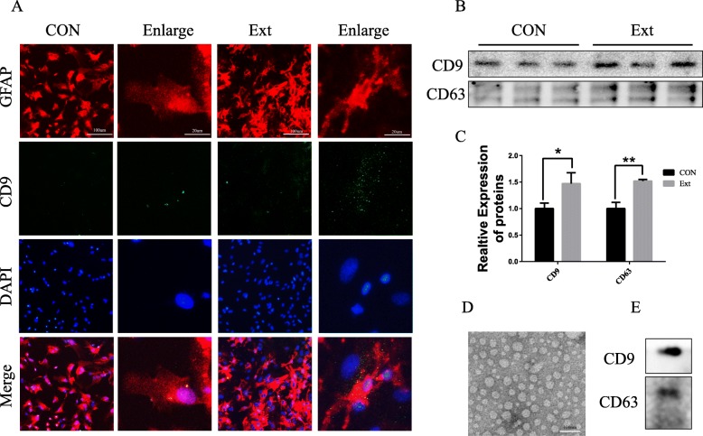
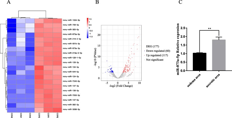
Methods: In the current study, the expression of inflammatory factors and miR-873a-5p in the lesion area and oedema area was evaluated in 15 patients with traumatic brain injury. Exosomes secreted by astrocytes were detected by immunofluorescence, Western blot and electron microscopy. A mouse model of TBI and an in vitro model of LPS-induced primary microglia were established to study the protective mechanism of exosomes from miR-873a-5p overexpressing in TBI-induced nerve injury.

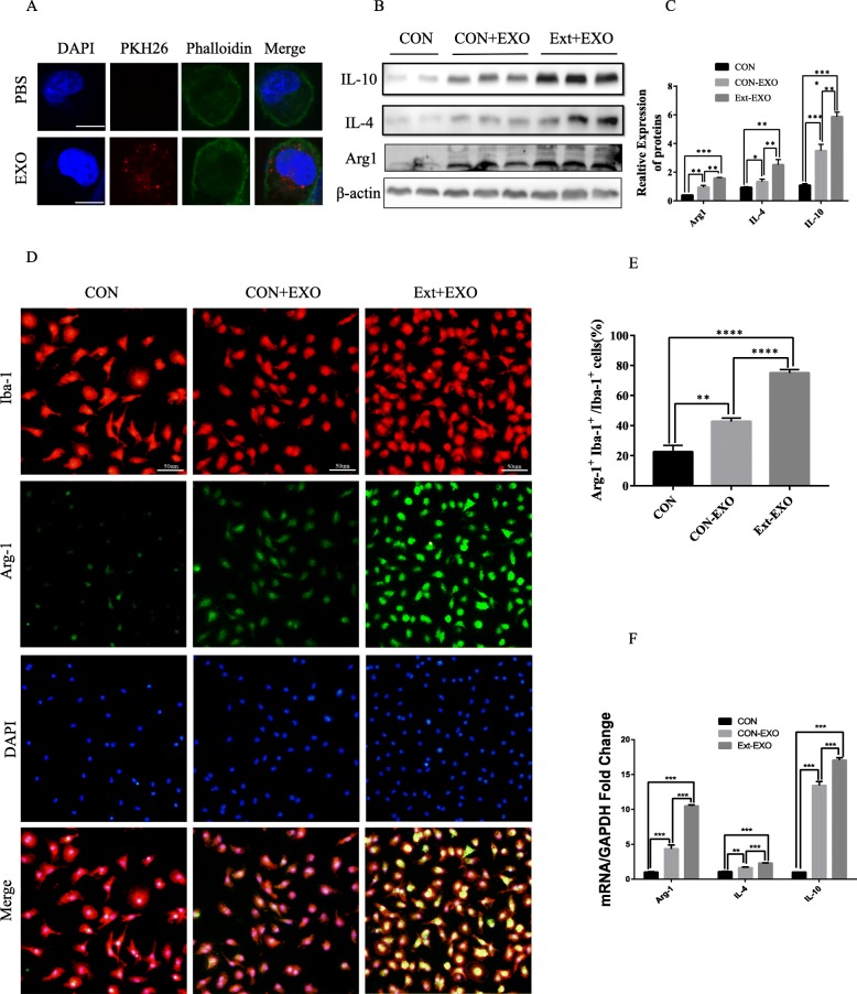
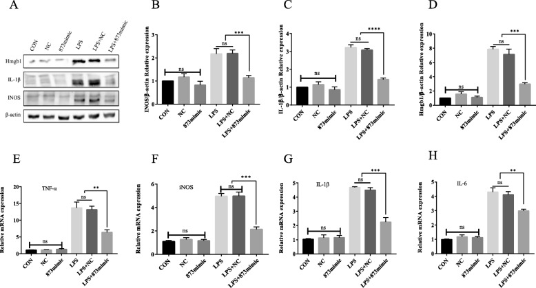
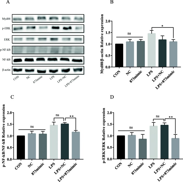
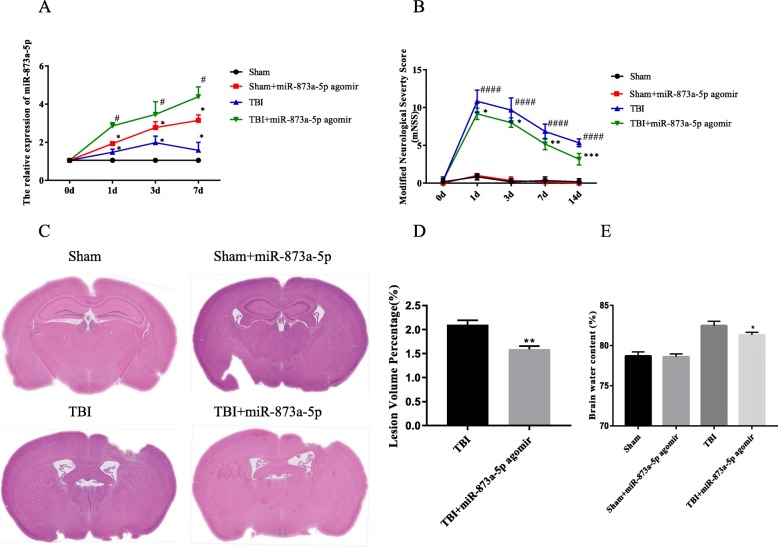
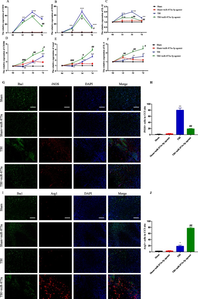
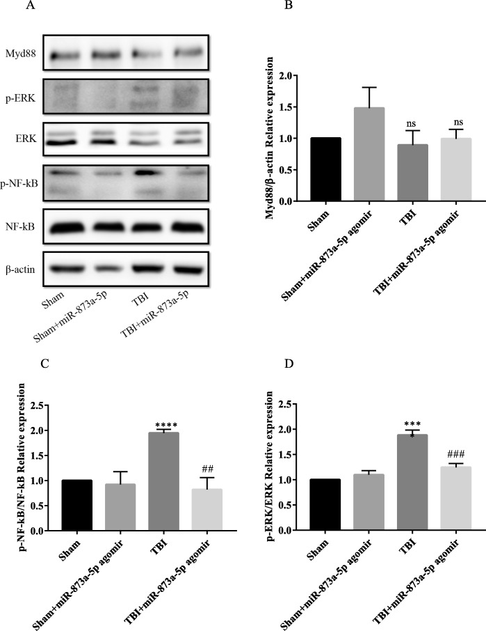
Results: We discovered that exosomes derived from activated astrocytes promote microglial M2 phenotype transformation following TBI. More than 100 miRNAs were detected in these astrocyte-derived exosomes. miR-873a-5p is a major component that was highly expressed in human traumatic brain tissue. Moreover, miR-873a-5p significantly inhibited LPS-induced microglial M1 phenotype transformation and the subsequent inflammation through decreased phosphorylation of ERK and NF-κB p65. This effect also greatly improved the modified neurological severity score (mNSS) and attenuated brain injury in a strictly controlled cortical impact mouse model.

Conclusions: Taken together, our research indicates that miRNAs in the exosomes derived from activated astrocytes play a key role in the astrocyte-microglia interaction. miR-873a-5p, as one of the main components of these astrocyte-derived exosomes, attenuated microglia-mediated neuroinflammation and improved neurological deficits following TBI by inhibiting the NF-κB signalling pathway. These findings suggest a potential role for miR-873a-5p in treating traumatic brain injury.

Keywords: Astrocyte; Exosome; M1/M2; Microglia; Traumatic brain injury; miR-873a-5p.


Conflict of interest statement

The authors declare that they have no competing interests.

Figures

Fig. 1
Fig. 1
“Brain extracts” stimulated the synthesis and release of exosomes from astrocytes. a Exosomes (marked by CD9 (green)) derived from astrocytes were significantly increased under “brain extract” stimulation. b, c After 24 h of treatment, the medium of primary cultured astrocytes was harvested to detect the markers CD9 and CD63 by Western blotting (data were presented as mean ± SD, *p < 0.05, **p < 0.01, n = 5, t test). d A representative transmission electron microscopy image of purified exosomes from the culture medium of stimulated astrocytes after 24 h of treatment. Scale bar 100 nm. e The astrocyte-derived exosomes were identified by the markers CD9 and CD63 by Western blotting
Fig. 2
Fig. 2
Astrocyte-derived exosomes are taken up by primary microglia and promote microglial M2 phenotype transformation. a PKH26 staining and exosome uptake. It was shown by confocal microscopy that exosomes (red) were taken up by the cells and existed in the cytoplasm and around the cell nucleus. Immunofluorescence of the primary microglia showing DAPI (blue), exosomes (red) and F-actin (green). No red fluorescent signal was detected in the PBS control group. PBS: microglial staining with PBS. Exo: the exosomes labelled with pkh26 were incubated with microglia for 24 h. Bar = 10 μm. b, c Primary cultured microglia were divided into three groups: con, con+Exo and Ext+Exo. Exo: The microglia were incubated with astrocyte-derived exosomes for 4 h. Ext+Exo: primary cultured microglia were stimulated with “brain extracts” for 24 h and washed before incubation with exosomes. These M2 markers were also detected by Western blot analysis. d, e Fluorescence confocal microscopic images showing both Iba-1 (microglia marker) and Arg-1 (M2 marker) expression increased after astrocyte-derived exosome treatment. Bar = 50 μm. Quantification of the percentage of Arg-1+Iba-1+ cells among total Iba-1+ cells. This effect was more significant after “brain extract” stimulation. f The mRNA expression of microglia M2 markers (Arg-1, IL-4, IL-10) was detected by RT-PCR (data were presented as mean ± SD, *p < 0.05, **p < 0.01, ***p < 0.001, n = 5, one-way ANOVA)
Fig. 3
Fig. 3
MicroRNA microarray analysis of exosomes released from Ext-stimulated astrocytes. a CON: exosomes secreted by astrocytes under physiological conditions. COR: exosomes secreted by astrocytes under simulated trauma. The miRNA component of the exosomes released from stimulated astrocytes was studied by microRNA microarray analysis. The heat map shows the change of the first 20 miRNAs. b The miRNA distribution diagram. The transverse axis represents multiple miRNAs in the Ext group compared to those in the CON group, and the longitudinal axis represents the Log10 of the p value. c The expression of miR-873a-5p in necrotic brain tissue and oedema brain tissue in traumatic brain injury was measured by RT-PCR (data were presented as mean ± SD, compared with the oedema area group, **p < 0.01, n = 15, t test)
Fig. 4
Fig. 4
The effects of miR-873a-5p on LPS-activated primary cultured microglia. ad Western blotting and statistical analysis of the pro-inflammatory cytokines Hmgb1, IL-1β and iNOS in LPS- and miR-873a-5p-treated cells. eh RT-qPCR analyses of changes in the pro-inflammatory cytokines TNF-α, iNOS, IL-1β and IL-6 at the mRNA level (data were presented as mean ± SD, *p < 0.05, **p < 0.01, ***p < 0.001, n = 5, one-way ANOVA)
Fig. 5
Fig. 5
The effects of miR-873a-5p on NF-κB activation in primary cultured microglia. ad Western blotting and statistical analysis of Myd88 and phosphorylated NF-κB and ERK (data were presented as mean ± SD, *p < 0.05, **p < 0.01, n = 5, one-way ANOVA)
Fig. 6
Fig. 6
miR-873a-5p attenuated brain defect area, cerebral oedema content and neurological deficit in mice after TBI. a miR-873a-5p treatment significantly increased the expression of miR-873a-5p in the cortex at 1, 3 and 7 days after TBI. b The nerve function of mice was assessed by mNSS (data were presented as mean ± SD, compared with the sham group *p < 0.05, compared with the TBI group #p < 0.05, n = 5, one-way ANOVA). c, d The area of the mice brain defect was determined by the proportional method (data were presented as mean ± SD, compared with the TBI group **p < 0.01, n = 5, one-way ANOVA). e The water content of brain tissue was measured by the dry-wet method (data were presented as mean ± SD, compared with the TBI group *p < 0.05, n = 3, one-way ANOVA)
Fig. 7
Fig. 7
miR-873a-5p treatment inhibits the inflammatory response by promoting microglia polarization to M2 after TBI. ac miR-873a-5p treatment significantly reduced the mRNA expression of CD32, INOS and IL-1β in the cortex at 1, 3 and 7 days after TBI. df miR-873a-5p increased the expression of the M2 microglia signature genes CD206, IL4 and Arg1 in the cortex at 1, 3 and 7 days after TBI. g, h Representative images of double immunofluorescent staining in the injured cortex with the microglial marker Iba1 (green) and the M1 marker iNOS (red). i, j Representative images of double immunofluorescent staining in the injured cortex with the microglia marker Iba1 (green) and the M2 marker Arg1 (red). Bar = 50 μm (data were presented as mean ± SD, compared with the sham group *p < 0.05, **p < 0.01, ***p < 0.001; compared with the TBI group #p < 0.05, ##p < 0.01, ###p < 0.001, n = 5, one-way ANOVA)
Fig. 8
Fig. 8
The in vivo effects of miR-873a-5p on NF-κB activation in mice TBI model. ad Brain tissue of the in vivo mice TBI model was collected at the seventh day post-TBI. Western blot demonstrating that miR-873a-5p agomir administration suppressed Myd88 signal activation and reversed TBI-induced phosphorylation of NF-κB and ERK (data were presented as mean ± SD, compared with the sham group ****p < 0.0001, compared with the TBI group ##p < 0.01, ###p < 0.001, n = 5, one-way ANOVA)

Similar articles

Cited by

References

    1. Andelic N. The epidemiology of traumatic brain injury. Lancet Neurol. 2013;12:28–29. doi: 10.1016/S1474-4422(12)70294-6. - DOI - PubMed
    1. Faden AI, Wu J, Stoica BA, Loane DJ. Progressive inflammation-mediated neurodegeneration after traumatic brain or spinal cord injury. Br J Pharmacol. 2016;173:681–691. doi: 10.1111/bph.13179. - DOI - PMC - PubMed
    1. Loane DJ, Kumar A. Microglia in the TBI brain: the good, the bad, and the dysregulated. Exp Neurol. 2016;275(Pt 3):316–327. doi: 10.1016/j.expneurol.2015.08.018. - DOI - PMC - PubMed
    1. Xia CY, Zhang S, Gao Y, Wang ZZ, Chen NH. Selective modulation of microglia polarization to M2 phenotype for stroke treatment. Int Immunopharmacol. 2015;25:377–382. doi: 10.1016/j.intimp.2015.02.019. - DOI - PubMed
    1. Chen Y, Swanson RA. Astrocytes and brain injury. J Cereb Blood Flow Metab. 2003;23:137–149. doi: 10.1097/01.WCB.0000044631.80210.3C. - DOI - PubMed