. 2020 Mar 27;23(3):100961.
doi: 10.1016/j.isci.2020.100961. Epub 2020 Mar 4.

Serpina3c Regulates Adipogenesis by Modulating Insulin Growth Factor 1 and Integrin Signaling

Affiliations

Serpina3c Regulates Adipogenesis by Modulating Insulin Growth Factor 1 and Integrin Signaling

Yoonjeong Choi et al. iScience. .

Abstract

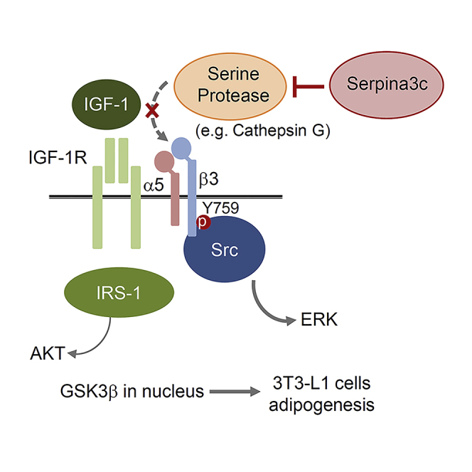
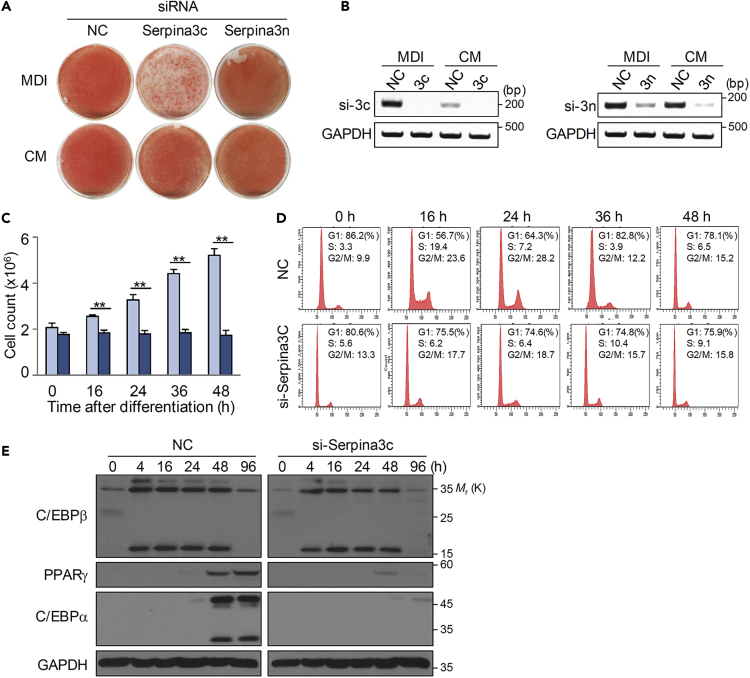
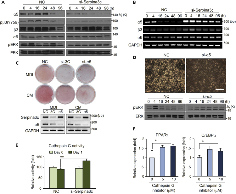
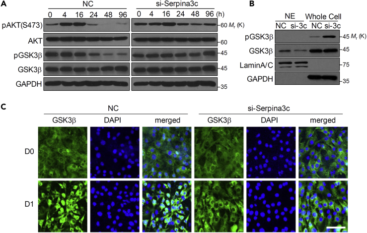
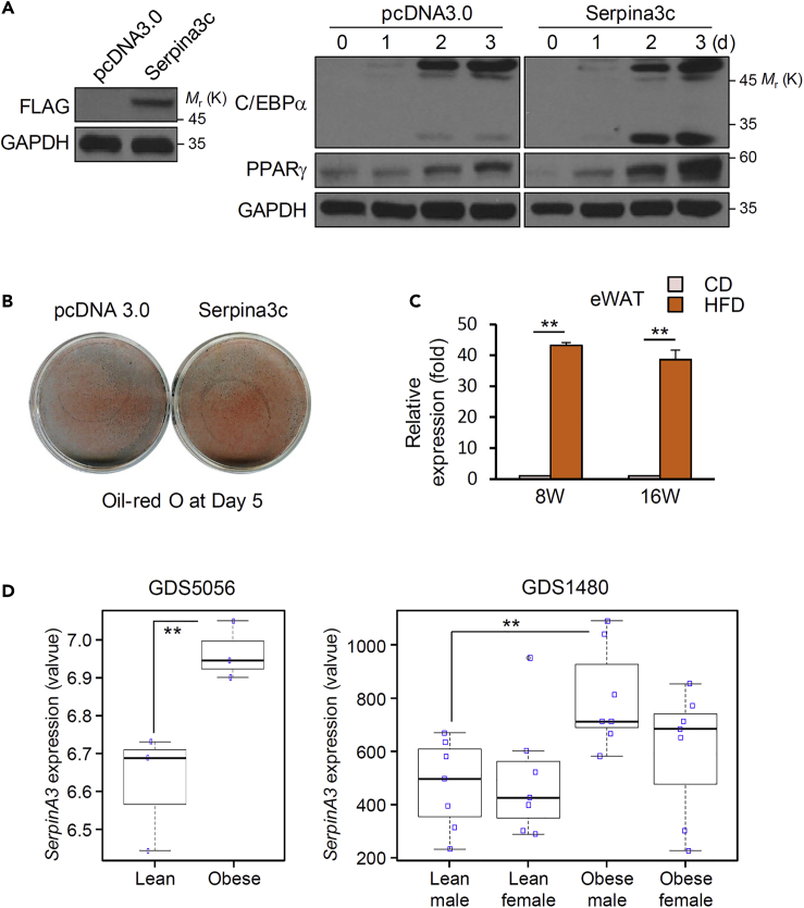
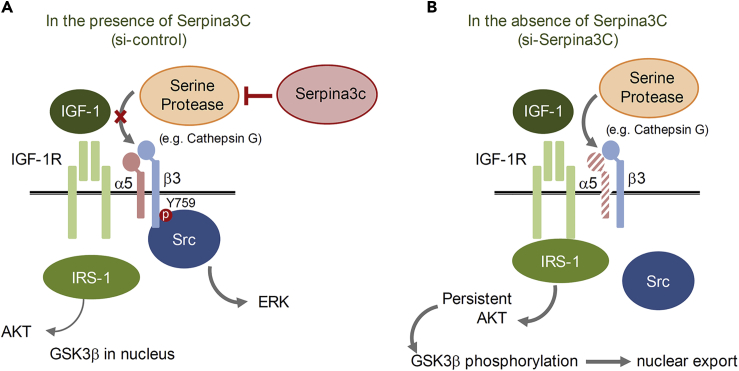
Preadipocyte differentiation can be induced upon a hormonal treatment, and various factors secreted by the cells may contribute to adipogenesis. In this study, RNA-seq revealed Serpina3c as a critical factor regulating the signaling network during adipogenesis. Serpina3c is a secretory protein and is highly expressed in fat tissues. Knockdown of Serpina3c decreased adipogenesis by attenuating the mitotic clonal expansion of 3T3-L1 cells. These cells exhibited decreases in integrin α5, which abolished the phosphorylation of integrin β3. We found that Serpina3c inhibits a serine protease that regulates integrin α5 degradation. Knockdown of Serpina3c disrupted integrin-mediated insulin growth factor 1 (IGF-1) signaling and ERK activation. Serpina3c-mediated regulation of integrin-IGF-1 signaling is also associated with AKT activation, which affects the nuclear translocation of GSK3β. Altogether, our results indicate that Serpina3c secreted from differentiating adipocytes inhibits serine proteases to modulate integrin/IGF-1-mediated ERK and AKT signaling and thus is a critical factor contributing to adipogenesis.

Keywords: Developmental Biology; Molecular Biology; Transcriptomics.


Conflict of interest statement

Declaration of Interest The authors declare no conflict of interest.

Figures

None
Graphical abstract
Figure 1
Figure 1
Gene Expression Profile in Differentiating 3T3-L1 Cells Treated with CM (A) A schematic model of CM preparation using a 3T3-L1 preadipocyte differentiation system. Confluent growth-arrested 3T3-L1 preadipocytes were induced to differentiate with MDI and FBS. After 2 days, cell-exposed medium was collected to treat other preadipocytes. Differentiating cells were harvested after 0, 4, and 24 h for RNA sequencing. MDI + FBS-treated cells were used as a control group. (B) CM accelerated 3T3-L1 adipocyte differentiation as shown by Oil Red O staining at day 5 of induction. (C) Strategy for the identification of CM-containing factors via RNA sequencing data. (D) Heatmap showing relative expression of 20 genes identified from RNA sequencing data. Red, high expression; blue, low expression. Fold changes of the total genes are presented in Data S1. These RNA sequencing data using MDI and CM are publicly available from Gene Expression Omnibus (GEO: GSE144130).
Figure 2
Figure 2
General Characterization of Serpina3c in Adipocytes (A–C) Serpina3c expression patterns induced by MDI or CM treatment in 3T3-L1 adipocytes. Cell lysate were prepared at various times, (A) 0∼6 h or (B) 0∼120 h after treatment for RT-PCR analysis of Serpina3cSerpina3n, and Gapdh. (C) Also, the expression of adipokines, Leptin and AdipoQ, was compared with those of Serpina3c and Serpina3n. (D) Tissue distribution of Serpina3c by RT-PCR in C57BL/6 male mice. (E) Amino acid sequence of Serpina3c was analyzed by SignalP-4.1 prediction (www.cbs.dtu.dk/services/SignalP-4.1/) showing the N-terminal signal peptide for secretory proteins. (F) Overexpression vector was generated by inserting mouse Serpina3c (or Serpina3n) encoding a C-terminal FLAG tag into pcDNA3.0. HEK293T cells were transfected with this construct, and 2 days later, the medium was collected, precipitated by cold acetone, and subjected to western blot analysis to detect the FLAG-tagged protein secreted from cells. pcDNA3.0 was used as a negative control. (G) Serpina3c expression and secretion patterns induced by MDI in 3T3 L1 adipocytes. RT-PCR was performed to detect expression of Serpina3c mRNA after treatment. Western blot analysis was performed to investigate expression and secretion of Serpina3c protein in precipitated media or cell lysate after treatment.
Figure 3
Figure 3
Serpina3c Is Required for Mitotic Clonal Expansion during Adipogenesis (A and B) 3T3-L1 cells were transfected with negative control (NC), Serpina3c, or Serpina3n siRNAs. Differentiation of transfected cells was induced by MDI or CM. Serpina3c depletion abolishes 3T3-L1 differentiation in MDI-treated cells. Oil Red O staining was conducted on day 8 (A) and knockdown of Serpina3c was assessed by RT-PCR (B). (C) Cell proliferation determined by cell counting at 0, 16, 24, 36, and 48 h after MDI treatment. Data are represented as mean ± SE. p Values less than 0.05 were considered significant, with ∗p < 0.05, ∗∗p < 0.01 as determined by Student's t test. (D) Cell-cycle analysis by flow cytometry at the indicated time points. (E) Western blot analysis was performed for differentiation markers of adipocytes in control cells and those with Serpina3c knockdown (si-Serpina3c).
Figure 4
Figure 4
Knockdown of Serpina3c Results in Integrin α5-mediated ERK Inactivation (A) 3T3-L1 cells were transfected with either negative control siRNA (NC) or si-Serpina3c and then differentiated using MDI. Integrin α5, β3, and α6 and phosphorylated integrin β3 and ERK were detected by western blotting at the indicated time points. (B) RT-PCR was performed to assess the expression of integrin α5, β3, and α6. 3T3-L1 cells were transfected with NC or Serpina3c siRNA, and then differentiation was induced by MDI. (C) Cells treated with NC, si-Serpina3c (si-3C), or si-Integrin α5 (si-α5) were stained with Oil Red O on day 6, and depletion of Serpina3c or integrin α5 was assessed by RT-PCR. (D) Microscopy images of cells 2 days after transfections with NC and si-α5. Cell structure was not changed. Western blot analysis was performed for ERK and phosphorylated ERK. Scale bar, 50 μm. (E) Cathepsin G activity was measured in 3T3-L1 cells transfected with NC or si-Serpina3c on day 0 or day 1 after differentiation. Data are represented as mean ± SD. p Values less than 0.05 were considered significant, with ∗p < 0.05, ∗∗p < 0.01 as determined by Student's t test. (F) Gene expression changes of PPARγ and C/EBPα during differentiation after treatment with cathepsin G inhibitor. Data are represented as mean ± SD. p Values less than 0.05 were considered significant, with ∗p < 0.05, ∗∗p < 0.01 as determined by Student's t test.
Figure 5
Figure 5
Serpina3c knockdown Induces Prolonged AKT Phosphorylation Resulting in Inhibited GSK3β Translocation 3T3-L1 cells were transfected with NC or si-Serpina3c and then differentiated using MDI. (A) Protein levels in transfected cells were measured by western blotting. (B) Nuclear extracts (NE) and whole-cell lysates were prepared 24 h after MDI treatment and phosphorylated (p) GSK3β and GSK3β levels were measured by western blotting. LaminA/C and GAPDH were used as nuclear and cytosol protein marker, respectively. (C) Cells transfected with NC or si-Serpina3c were induced to differentiate with MDI and fixed for immunocytochemistry against GSKβ 24 h later. Scale bar, 50 μm.
Figure 6
Figure 6
Serpina3c Overexpression Increases Adipocyte Differentiation 3T3-L1 cells were transfected with a control (pcDNA 3.0) or Serpina3c overexpression vector and induced to differentiate. (A) Overexpressed Serpina3c was confirmed 2 days after transfection by detection of the FLAG tag from whole-cell lysates. Proteins collected at the indicated times after induction were used to assay the levels of C/EBPα and PPARγ by western blot (GAPDH was used as loading control). (B) Oil Red O staining on day 5. (C) Serpina3c expression by quantitative real-time PCR in epididymal WAT tissues from C57BL/6 male mice fed a chow diet (CD) or a high-fat diet (HFD) for 8 or 16 weeks. WAT, white adipose tissue. Data are represented as mean ± SD. p Values less than 0.05 were considered significant, with ∗p < 0.05, ∗∗p < 0.01 as determined by Student's t test. (D) SERPINA3 gene expression levels in adipose stem cells or preadipocytes from obese and lean individuals, using publicly available repository data (Gene Expression Omnibus accessions GDS5056 and GDS1480). Data are represented as mean ± SD. p Values less than 0.05 were considered significant, with ∗p < 0.05, ∗∗p < 0.01 as determined by Student's t test.
Figure 7
Figure 7
Schematic Illustration of How Serpina3c Regulates Adipogenesis (A) IGF-1 receptors form a complex with the integrin α5-β3 heterodimer that is required to activate ERK. With ligand binding, integrin β3 is phosphorylated at Tyr759, leading to Src binding and ERK activation. In this situation, Serpina3c blocks any serine proteases, which can degrade integrin α5. Insulin receptor substrate 1 (IRS-1) may transiently induce AKT signaling. (B) In the absence of Serpina3c, serine proteases degrade integrin α5, and the integrin-IGF-1 receptor complex is not formed, resulting in persistent activation of AKT by IRS-1 and phosphorylation of GSK3β, thereby attenuating adipose differentiation.

Similar articles

Cited by

References

    1. Barrett A.J., Rawlings N.D., O'Brien E.A. The MEROPS database as a protease information system. J. Struct. Biol. 2001;134:95–102. - PubMed
    1. Choi H., Lee H., Kim T.H., Kim H.J., Lee Y.J., Lee S.J., Yu J.H., Kim D., Kim K.S., Park S.W. G0/G1 switch gene 2 has a critical role in adipocyte differentiation. Cell Death Differ. 2014;21:1071–1080. - PMC - PubMed
    1. Cowan K.J., Law D.A., Phillips D.R. Identification of Shc as the primary protein binding to the tyrosine-phosphorylated beta(3) subunit of alpha(IIb)beta(3) during outside-in integrin platelet signaling. J. Biol. Chem. 2000;275:36423–36429. - PubMed
    1. El Ouaamari A., Dirice E., Gedeon N., Hu J., Zhou J.Y., Shirakawa J., Hou L., Goodman J., Karampelias C., Qiang G. SerpinB1 promotes pancreatic beta cell proliferation. Cell Metab. 2016;23:194–205. - PMC - PubMed
    1. Fagerholm S.C., Hilden T.J., Gahmberg C.G. P marks the spot: site-specific integrin phosphorylation regulates molecular interactions. Trends Biochem. Sci. 2004;29:504–512. - PubMed