. 2020 Apr 7;117(14):8001-8012.
doi: 10.1073/pnas.1912617117. Epub 2020 Mar 19.

Targeting the cyclin-dependent kinase 5 in metastatic melanoma

Affiliations

Targeting the cyclin-dependent kinase 5 in metastatic melanoma

Samanta Sharma et al. Proc Natl Acad Sci U S A. .

Abstract

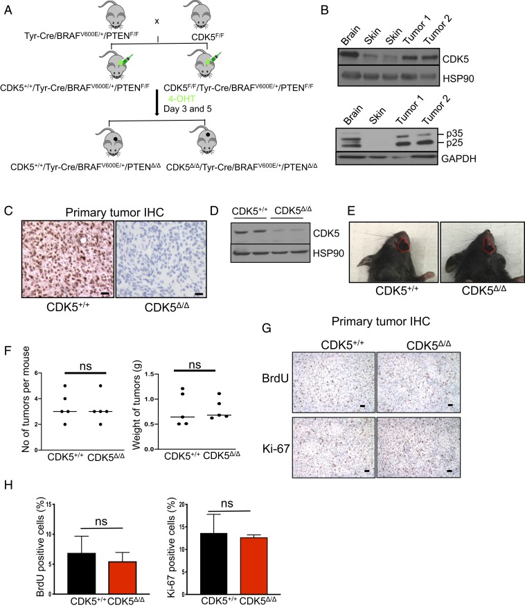
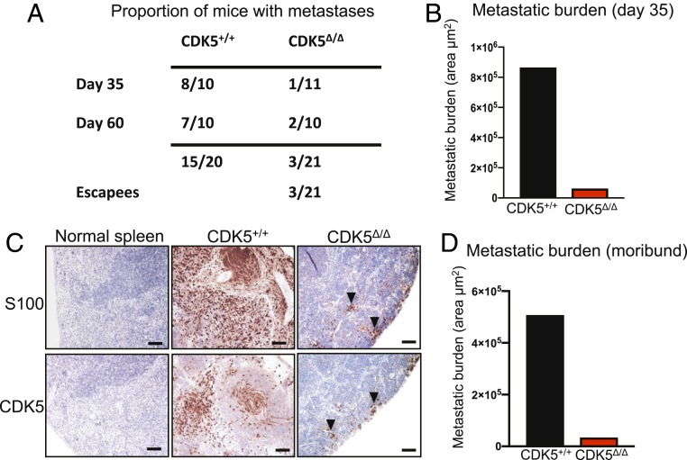
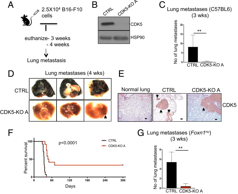
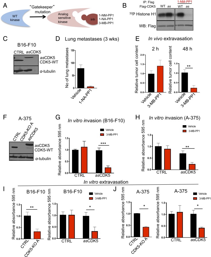
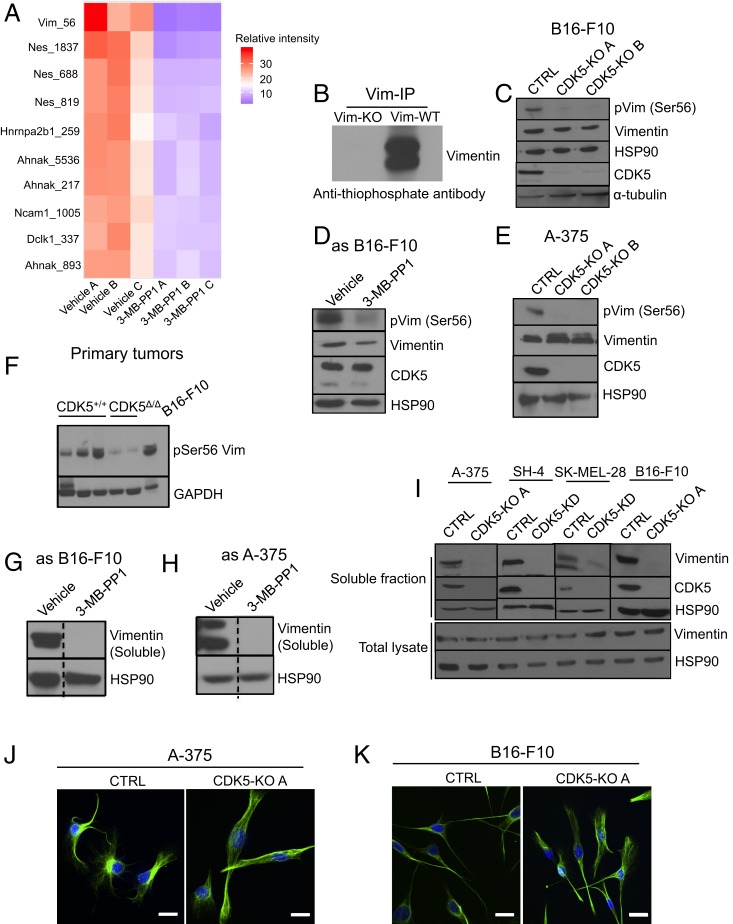
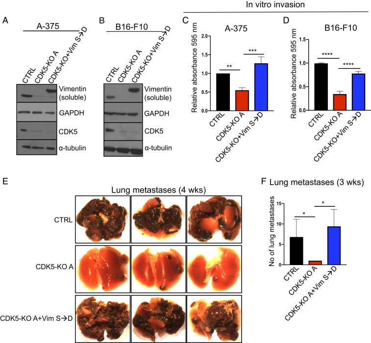
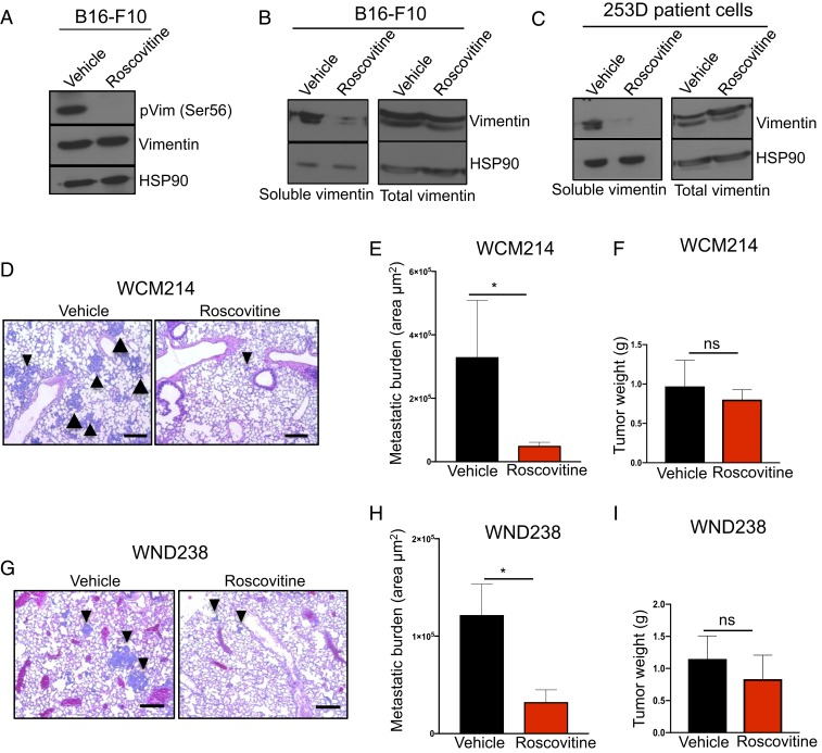
The cyclin-dependent kinase 5 (CDK5), originally described as a neuronal-specific kinase, is also frequently activated in human cancers. Using conditional CDK5 knockout mice and a mouse model of highly metastatic melanoma, we found that CDK5 is dispensable for the growth of primary tumors. However, we observed that ablation of CDK5 completely abrogated the metastasis, revealing that CDK5 is essential for the metastatic spread. In mouse and human melanoma cells CDK5 promotes cell invasiveness by directly phosphorylating an intermediate filament protein, vimentin, thereby inhibiting assembly of vimentin filaments. Chemical inhibition of CDK5 blocks the metastatic spread of patient-derived melanomas in patient-derived xenograft (PDX) mouse models. Hence, inhibition of CDK5 might represent a very potent therapeutic strategy to impede the metastatic dissemination of malignant cells.

Keywords: CDK5; cyclin-dependent kinases; metastasis; mouse cancer models.


Conflict of interest statement

Competing interest statement: P.S. has been a consultant at Novartis, Genovis, Guidepoint, The Planning Shop, ORIC Pharmaceuticals, and Exo Therapeutics; his laboratory receives research funding from Novartis. W.M. is currently an employee of Cedilla Therapeutics.

Figures

Fig. 1.
Fig. 1.
Analyses of mouse melanoma tumors. (A) Schematic representation of the crosses to generate mice bearing CDK5+/+/Tyr-Cre/BRAFV600E/+/PTENΔ/Δ and CDK5Δ/Δ/Tyr-Cre/BRAFV600E/+/PTENΔ/Δ tumors. (B) Immunoblotting of tumor lysates for CDK5 (Upper) and p35 (Lower). p25 denotes proteolytically cleaved p35 species. Adjacent normal skin and brain (the latter as a positive control) were also analyzed. Immunoblotting for HSP90 and GAPDH was used as a loading control (n = 2). (C) Immunohistochemistry (IHC) staining of tumor sections with an anti-CDK5 antibody. (Scale bars, 50 μm.) (D) Immunoblotting of tumor lysates for CDK5 and HSP90 (loading control) (n = 3). (E) Examples of skin tumors (denoted by red contours) arising in CDK5+/+ and CDK5F/F mice. (F) Quantification of the number of tumors per mouse and total tumor weight per mouse in CDK5+/+ and CDK5F/F animals (n = 5 mice/group). ns, not significant; unpaired t test. (G) IHC staining of tumors for BrdU (Upper), or Ki-67 (Lower) to mark proliferating cells. (H) Quantification of the fractions of BrdU- or Ki-67–positive cells in CDK5+/+ and CDK5Δ/Δ tumors (n = 5 mice/group). Shown are mean values ± SD (Right, unpaired t test; Left, unpaired t test with Welch’s correction).
Fig. 2.
Fig. 2.
CDK5 is essential for the metastatic spread. (A) Table showing the proportion of CDK5+/+/Tyr-Cre/BRAFV600E/+/PTENF/F (CDK5+/+) and CDK5F/F/Tyr-Cre/BRAFV600E/+/PTENF/F (CDK5Δ/Δ) animals displaying macroscopic or microscopic metastases in distal organs. P < 0.05 (Fisher’s exact test). (B) Quantification of the total metastatic burden in the internal organs of CDK5+/+/Tyr-Cre/BRAFV600E/+/PTENF/F (CDK5+/+) and CDK5F/F/Tyr-Cre/BRAFV600E/+/PTENF/F (CDK5Δ/Δ) animals, euthanized on day 35. To estimate total metastatic burden in a given animal, we calculated the total area occupied by metastases in step sections of internal organs (SI Appendix, Materials and Methods). (C) Immunostaining of spleen sections from a control mouse (normal spleen), or spleens with metastases from CDK5+/+/Tyr-Cre/BRAFV600E/+/PTENF/F (CDK5+/+) or CDK5F/F/Tyr-Cre/BRAFV600E/+/PTENF/F (CDK5Δ/Δ) mice. Sections were immunostained for S100 (a marker of melanocytes and melanoma cells) or CDK5, and counterstained with hematoxylin. Note that small clusters of metastatic cells in a CDK5F/F/Tyr-Cre/BRAFV600E/+/PTENF/F mouse were CDK5-positive (arrowheads), indicating that they arose from cells that escaped CDK5 deletion (SI Appendix, Fig. S2). (Scale bars, 50 μm.) (D) Quantification of the total metastatic burden in the internal organs of CDK5+/+/Tyr-Cre/BRAFV600E/+/PTENF/F (CDK5+/+) and CDK5F/F/Tyr-Cre/BRAFV600E/+/PTENF/F (CDK5Δ/Δ) animals, euthanized when the tumor sizes reached allowable limit, or the animals became moribund or paralyzed (by day 40 in CDK5+/+ and day 60 in CDK5F/F mice). Total metastatic burden was estimated as in B.
Fig. 3.
Fig. 3.
In vivo metastasis assays. (A) Schematic representation of the experimental approach to generate lung metastases. (B) Immunoblotting of CDK5+/+ (CTRL) and CDK5-knockout (CDK5-KO A) mouse melanoma B16-F10 cells for CDK5 and HSP90 (loading control) (n = 3). (C) The numbers of lung metastases in C57BL/6 mice injected with CDK5+/+ (CTRL) or CDK5-KO melanoma cells, quantified after 3 wk (n = 5 mice/group). Shown are mean values ± SD **P < 0.01 (Mann–Whitney U test). (D) The appearance of lungs from animals as above, 4 wk after injection of melanoma cells (n = 5 mice/group). An arrowhead points to a metastatic lesion in a mouse injected with CDK5-KO cells. (E) Sections of lungs from a healthy normal animal (normal lung), or from animals injected with CDK5+/+ (CTRL), or CDK5-KO melanoma cells were immunostained for CDK5 (n = 5 mice/group). Note that a metastatic lesion found in a mouse injected with CDK5-KO cells was CDK5-positive, indicating that it arose from cells that escaped CDK5 deletion. Metastatic tumors are marked by arrowheads. (Scale bars, 50 μm.) (F) Survival curves of mice injected with CDK5+/+ (CTRL) or CDK5-KO melanoma cells (n = 10 mice/group). P < 0.0001 (Log-rank test). (G) The numbers of lung metastases in immunocompromised Foxn1nu mice injected with CDK5+/+ (CTRL) or CDK5-KO melanoma cells, quantified after 3 wk (n = 5 mice/group). Shown are mean values ± SD **P < 0.01 (Mann–Whitney U test).
Fig. 4.
Fig. 4.
The kinase activity of CDK5 is required for tumor cells extravasation. (A) Schematic representation of the analog-sensitive approach. (B) Flag-tagged wild-type (WT) or analog-sensitive (as) CDK5 were expressed in 293T cells, immunoprecipitated with an anti-Flag antibody and subjected to in vitro kinase reactions using histone H1 as a substrate (a canonical substrate of CDKs), in the presence of radioactive [γ32P]-ATP and in the presence or absence of 5 μM 1-NM-PP1 (inhibitor of as kinases). Radiolabeled histone H1 was detected by autoradiography. Note that in the absence of 1-NM-PP1, wild-type and as CDK5 display comparable kinase activities. Addition of 1-NM-PP1 blocks the activity of as CDK5 without affecting wild-type CDK5. (C) Immunoblotting for CDK5 in mouse melanoma B16-F10 cells (CTRL), or B16-F10 cells in which we knocked out the endogenous CDK5 using CRISPR/Cas9 and expressed Flag-tagged as CDK5 (asCDK5). CDK5-WT, endogenous wild-type Cdk5. α-Tubulin was used as a loading control (n = 2). (D) The numbers of lung metastases in C57BL/6 mice injected with asCDK5 B16-F10 melanoma cells, quantified after 3 wk. Mice were treated with vehicle or with an inhibitor of as kinases 1-NA-PP1, from day 0 until the end of the experiment (n = 5 mice/group) (SI Appendix, Materials and Methods). Shown are mean values ± SD ***P < 0.001 (unpaired t test with Welch’s correction). (E) Fluorescently labeled asCDK5 B16-F10 cells were injected into tail veins of Foxn1nu. Recipient mice were treated with vehicle or with an inhibitor of as kinases 3-MB-PP1 (n = 3 mice/group). Lungs were imaged after 2 (Left) and 48 h (Right). Shown are mean values ± SD **P < 0.01 (unpaired t test). (F) Immunoblotting for CDK5 in human melanoma A-375 cells (CTRL), A-375 cells in which we knocked out the endogenous CDK5 using CRISPR/Cas9 (CDK5-KO A), and CDK5-KO cells in which we expressed Flag-tagged asCDK5 (asCDK5). α-Tubulin was used as a loading control (n = 2). (G) In vitro invasion assays. Parental mouse melanoma B16-F10 cells (CTRL) or B16-F10 cells engineered to express asCDK5 in place of wild-type CDK5 (asCDK5) were subjected to invasion assays in the presence of vehicle or 3-MB-PP1 (n = 3; in triplicate). Shown are mean values ± SD ***P < 0.001 (two-way ANOVA, Bonferroni’s multiple comparisons test). (H) Similar assays as in G, using parental human melanoma A-375 cells (CTRL) or A-375 cells expressing asCDK5 (n = 3; in triplicate). Shown are mean values ± SD. **P < 0.01 (two-way ANOVA, Bonferroni’s multiple comparisons test). (I) In vitro transendothelial migration (in vitro extravasation) assays using parental (CTRL) and CDK5-KO mouse melanoma B16-F10 cells (Left) (n = 3; in triplicate), or parental B16-F10 (CTRL) and asCDK5 B16-F10 cells (asCDK5) assayed in the presence of vehicle or 3-MB-PP1 (Right) (n = 3; in triplicate). (J) Similar assays as in I, using human melanoma A-375 cells (n = 3; in triplicate). In I and J data are shown as mean values ± SD **P < 0.01, *P < 0.05 (unpaired t test).
Fig. 5.
Fig. 5.
CDK5 phosphorylates vimentin in melanoma cells and regulates its polymerization. (A) Heat map depicting 10 phosphosites (only S/T-P sequences are shown) which displayed the most strongly decreased phosphorylation upon CDK5 inhibition. Three independent cultures of vehicle-treated (vehicle A, B, and C) or 3-MB-PP1-treated (3-MB-PP1 A, B, and C) asCDK5 B16-F10 cells were analyzed. For each phosphopeptide, the intensities (abundance) across all six samples were scaled to 100, and the top 10 phosphorylated peptides displaying the highest fold decrease in phosphorylation levels in CDK5-inhibited samples were used to make the heat map. The relative abundance in each sample was color coded, as indicated. (B) In cell kinase reactions. In vitro cultured asCDK5 B16-F10 cells (Vim-WT) or, for control, vimentin knockout asCDK5 B16-F10 cells (Vim-KO) were permeabilized by treatment with 30 μg/mL digitonin and supplemented with N6-substituted bulky ATP analog, N6-furfuryl-ATPγS for 30 min, resulting in thio-phosphorylation of direct CDK5 substrates. Proteins were then alkylated with p-nitrobenzyl mesylate to generate epitopes for an anti-thiophosphate ester antibody. Endogenous vimentin was immunoprecipitated with an anti-vimentin antibody, and immunoblots were probed with an anti-thiophosphate ester antibody to evaluate thio-phosphorylation of vimentin. Please note the absence of signal for thio-phosphorylated vimentin in vimentin knockout asCDK5 B16-F10 cells (Vim-KO). (C) Protein lysates from CDK5+/+ (CTRL) and CDK5-knockout (CDK5-KO A, CDK5-KO B; A and B denote different sgRNAs, see SI Appendix, Materials and Methods) B16-F10 cells were immunoblotted with antibodies against phospho-serine56-vimentin, total vimentin, or CDK5. HSP90 and α-tubulin were used as loading controls (n = 3). (D) Similar analysis as in C, using asCDK5 B16-F10 cells cultured for 4 h in the presence of vehicle or 3-MB-PP1 (to inhibit CDK5) (n = 2). (E) Similar analysis as in C, using parental (CTRL) and CDK5-KO human melanoma A-375 cells (n = 2). (F) Similar analysis as in C, using tumors arising in CDK5+/+/Tyr-Cre/BRAFV600E/+/PTENF/F (CDK5+/+) or CDK5F/F/Tyr-Cre/BRAFV600E/+/PTENF/F (CDK5Δ/Δ) mice. B16-F10 cells were used as a positive control. Immunoblotting for GAPDH was used as a loading control (n = 2). (G) asCDK5 mouse melanoma B16-F10 cells were cultured for 4 h in the presence of vehicle or 3-MB-PP1. Cells were lysed, the soluble fraction was obtained as described in SI Appendix, Materials and Methods, and immunoblotted with an anti-vimentin antibody. HSP90 served as a loading control (n = 2). The lines were spliced together from the same blot (indicated by dashed lines). (H) Similar analysis as in G, using asCDK5 human melanoma A-375 cells (n = 2). The lines were spliced together from the same blot (indicated by dashed lines). (I) Upper (soluble fraction) human (A-375, SH-4, SK-MEL-28), and mouse (B16-F10) melanoma cells: parental (CTRL), CDK5-KO, or cells in which we depleted CDK5 using anti-CDK5 shRNA (CDK5-KD), were lysed and the soluble fractions obtained as described in SI Appendix, Materials and Methods. Immunoblots were probed for vimentin, CDK5, and HSP90 (loading control). Extract from each melanoma cell line was run on a separate blot (indicated by solid vertical lines). Lower (total lysate) total cell lysates (containing soluble and insoluble proteins) were immunoblotted for vimentin and HSP90. (J) In vitro cultured parental (CTRL) and CDK5-KO human A-375 melanoma cells were immunostained using an anti-vimentin antibody and analyzed by confocal microscopy. Hoechst staining was used to visualize nuclei. (K) Similar analysis as in J with cultured parental (CTRL) and CDK5-KO mouse B16-F10 melanoma cells. (Scale bars, 50 μm.)
Fig. 6.
Fig. 6.
Rescue of the invasion defect in CDK5-null cells by phosphomimicking vimentin. (A) Parental (CTRL), CDK5-KO, and CDK5-KO cells expressing Flag-tagged vimentin phosphomimicking mutant containing serine 56 to aspartic acid substitution (Vim S→D) human melanoma A-375 cells were lysed to obtain soluble protein fractions. Immunoblots were probed for vimentin and CDK5. GAPDH and α-tubulin were used as loading controls (n = 2). (B) Similar analysis as in A using mouse melanoma B16-F10 cells (n = 2). (C) In vitro invasion assays using parental human melanoma A-375 cells (CTRL), A-375 CDK5-KO cells, or CDK5-KO cells expressing S56D phosphomimicking vimentin (CDK5-KO+VimS→D, from A) (n = 3; in triplicate). Shown are mean values ± SD **P < 0.01, ***P < 0.001 (one-way ANOVA with Tukey’s multiple comparisons test). (D) Similar assays as in C, using parental mouse B16-F10 melanoma cells (CTRL), or B16-F10 CDK5-KO cells, or CDK5-KO cells expressing S56D phosphomimicking vimentin (CDK5-KO+VimS→D, from B) (n = 3; in triplicate). Shown are mean values ± SD ****P < 0.0001 (one-way ANOVA with Tukey’s multiple comparisons test). (E) Parental mouse melanoma B16-F10 cells (CTRL), or CDK5 knockout B16-F10 cells (CDK5-KO), or CDK5-KO cells expressing phosphomimicking S56D vimentin (CDK5-KO+VimS→D) were injected into tail veins of C57BL/6 mice (n = 5 mice/group). Animals were euthanized and lungs photographed after 4 wk. (F) Similar experiment as in E, the numbers of lung metastases were quantified after 3 wk (n = 5 mice/group). Shown are mean values ± SD *P < 0.05 (Kruskal-Wallis test with Dunn’s multiple comparisons test).
Fig. 7.
Fig. 7.
CDK5 inhibition in melanoma cells and patient-derived xenografts. (A) Mouse B16-F10 cells were cultured in the presence of vehicle or 5 μM roscovitine for 4 h, lysed, and immunoblotted with antibodies against phospho-serine56-vimentin and total vimentin. HSP90 was used as a loading control (n = 2). (B) B16-F10 cells were cultured as above. Left (soluble vimentin), cells were lysed and the soluble fraction obtained as described in SI Appendix, Materials and Methods. Immunoblots were probed for vimentin and HSP90 (loading control). Right (total vimentin), total lysates (containing soluble and insoluble fractions) were also immunoblotted as above. (C) Patient-derived melanoma cells (253D) were cultured in the presence of vehicle or 5 μM roscovitine for 4 h, and analyzed as in B for the presence of soluble and total vimentin. (D) Histological sections of lungs from mice implanted with patient-derived melanoma tumors (model WCM214). Mice were treated with roscovitine (daily i.p. injection, 150 mg/kg) or with vehicle for 28 d (n = 10 mice/group; see SI Appendix, Materials and Methods). Arrowheads point to examples of metastases. (Scale bars, 50 μm.) (E) Total metastatic burden in mice implanted with patient-derived melanoma tumors (model WCM214). Mice were treated as above with vehicle or roscovitine, euthanized, and their internal organs subjected to histopathological analyses for the presence of metastases. Only lung metastases were detected. To estimate total metastatic burden in a given animal, the total area occupied by metastases was calculated in step sections of lungs (n = 5 mice/group; see SI Appendix, Materials and Methods). (F) The total weight of primary tumors in mice implanted with patient-derived melanomas (model WCM214) at the end of treatment. Mice were treated as above with vehicle or roscovitine, euthanized after 28 d, and tumors were weighted (n = 10 mice/group). (G) Similar analysis as in D in mice bearing xenografts of patient-melanoma tumors (model WND238). Mice were treated with vehicle or roscovitine for 49 d (n = 10 mice/group). (Scale bars, 50 μm.) (H) Similar analysis as in E in mice bearing xenografts of patient-melanoma tumors (model WND238) (n = 10 mice/group). Mice were treated with vehicle or roscovitine for 49 d. (I) Similar analysis as in F in mice bearing xenografts of patient-melanoma tumors (model WND238). Shown in E, F, H, and I are mean values ± SE *P < 0.05 (Mann–Whitney U test). ns, not significant.

References

    1. Malumbres M., Cyclin-dependent kinases. Genome Biol. 15, 122 (2014). - PMC - PubMed
    1. Lim S., Kaldis P., Cdks, cyclins and CKIs: Roles beyond cell cycle regulation. Development 140, 3079–3093 (2013). - PubMed
    1. Malumbres M., Barbacid M., Cell cycle, CDKs and cancer: A changing paradigm. Nat. Rev. Cancer 9, 153–166 (2009). - PubMed
    1. Sherr C. J., Beach D., Shapiro G. I., Targeting CDK4 and CDK6: From discovery to therapy. Cancer Discov. 6, 353–367 (2016) - PMC - PubMed
    1. Otto T., Sicinski P., Cell cycle proteins as promising targets in cancer therapy. Nat. Rev. Cancer 17, 93–115 (2017). - PMC - PubMed

Publication types

MeSH terms