. 2020 Apr 7;117(14):7917-7928.
doi: 10.1073/pnas.1918659117. Epub 2020 Mar 19.

Centromere scission drives chromosome shuffling and reproductive isolation

Affiliations

Centromere scission drives chromosome shuffling and reproductive isolation

Vikas Yadav et al. Proc Natl Acad Sci U S A. .

Abstract

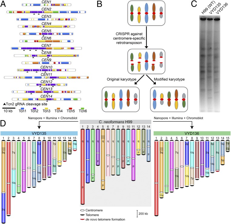
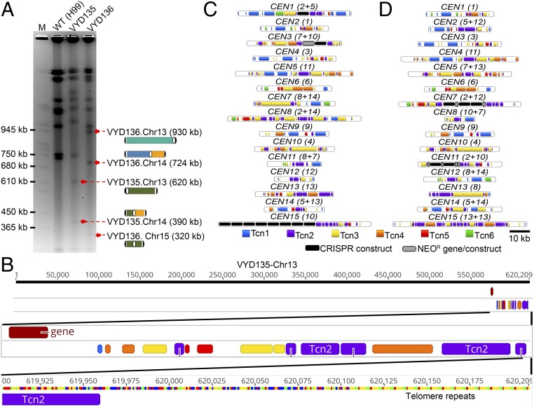
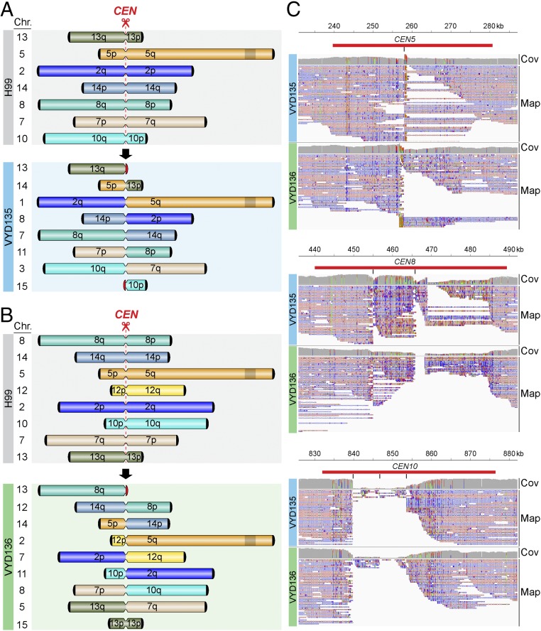
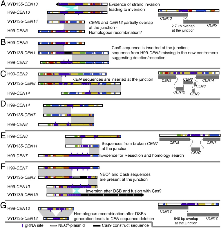
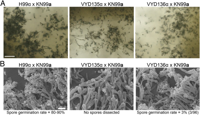
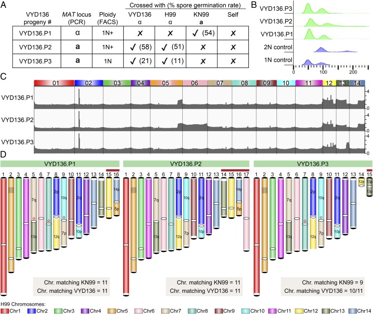
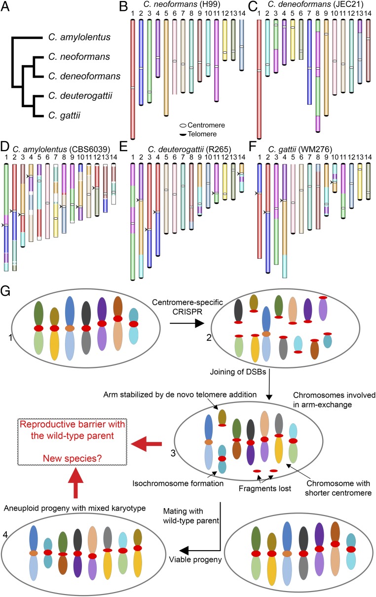
A fundamental characteristic of eukaryotic organisms is the generation of genetic variation via sexual reproduction. Conversely, significant large-scale genome structure variations could hamper sexual reproduction, causing reproductive isolation and promoting speciation. The underlying processes behind large-scale genome rearrangements are not well understood and include chromosome translocations involving centromeres. Recent genomic studies in the Cryptococcus species complex revealed that chromosome translocations generated via centromere recombination have reshaped the genomes of different species. In this study, multiple DNA double-strand breaks (DSBs) were generated via the CRISPR/Cas9 system at centromere-specific retrotransposons in the human fungal pathogen Cryptococcus neoformans The resulting DSBs were repaired in a complex manner, leading to the formation of multiple interchromosomal rearrangements and new telomeres, similar to chromothripsis-like events. The newly generated strains harboring chromosome translocations exhibited normal vegetative growth but failed to undergo successful sexual reproduction with the parental wild-type strain. One of these strains failed to produce any spores, while another produced ∼3% viable progeny. The germinated progeny exhibited aneuploidy for multiple chromosomes and showed improved fertility with both parents. All chromosome translocation events were accompanied without any detectable change in gene sequences and thus suggest that chromosomal translocations alone may play an underappreciated role in the onset of reproductive isolation and speciation.

Keywords: Cryptococcus neoformans; DSB repair; chromosome translocation; karyotype evolution; retrotransposons.


Conflict of interest statement

The authors declare no competing interest.

Figures

Fig. 1.
Fig. 1.
Centromere-specific DSBs mediated by CRISPR led to chromosome rearrangements. (A) Centromere maps showing the distribution of retrotransposons (Tcn1–Tcn6) in the centromeres of wild-type strain H99 of C. neoformans. (B) An outline depicting the model for achieving multiple chromosome translocations in C. neoformans. (C) PFGE revealed many differences in the karyotype of VYD135 and VYD136 compared with wild type, H99. (D) Chromosome maps for VYD135 and VYD136 compared to the H99 genome revealed multiple chromosome translocations in these strains. Chromosomes are colored with H99 chromosomes as reference. “q” represents the longer arm, while “p” represents the shorter arm according to the wild-type chromosome configuration.
Fig. 2.
Fig. 2.
Sequence analysis of new centromeres and telomeres. (A) PFGE ethidium bromide (EtBr) staining showing bands for newly generated short chromosomes and new telocentric chromosomes. M represents S. cerevisiae chromosomes. (B) Chromosome map of one of the newly formed telomeres showing the presence of telomere sequence repeats next to Tcn2 elements present in the centromere. (C and D) Maps showing the distribution of retrotransposons, along with the integration of foreign sequences from CRISPR/Cas9 and the neomycin resistance gene (NEOR), in the centromeres of VYD135 (C) and VYD136 (D). Numbers in brackets next to CEN numbers represent the wild-type CEN numbers that rearranged to form new centromeres.
Fig. 3.
Fig. 3.
Chromosome rearrangements are mediated via the gRNA cleavage site. (A and B) Simplified outline maps depicting the chromosomes that underwent translocation in VYD135 (A) and VYD136 (B). Black semicircles, telomeres; red semicircles, de novo telomeres; narrow bands, centromeres; shaded box on chromosome 5, MAT locus. (C) Nanopore reads mapping to the wild-type H99 genome revealed the sites of DSB formation or repair junctions at centromeres. The reads either converge on a single site (CEN5, CEN8) or exhibit sequence gaps between sites (CEN10) marking the location of junctions. Red bars indicate the centromeres, whereas the black vertical lines mark the site of gRNA cleavage. Cov, coverage of nanopore reads; Map, mapping of nanopore reads.
Fig. 4.
Fig. 4.
Synteny analysis of rearranged centromeres with wild-type H99 centromeres reveals complex rearrangements. (AG) A pairwise comparison of newly generated and wild-type centromeres revealed that translocations are mediated by double-strand breaks (DSBs) generated via CRISPR. Centromere-specific events are described in the individual panels. Gray shades represent direct synteny, while the cyan shade represents inversion events. In the cases that are shown in detail, the cross represents the evidence for HR, whereas the connecting gray lines represent joining events marking NHEJ. VYD135-CEN7 was generated after artificial fusion of two contigs and hence was not analyzed in detail.
Fig. 5.
Fig. 5.
Chromosome-shuffled isolates exhibit defects in sexual reproduction. (A) Light microscopy images showing hyphae, basidia, and spore chains in crosses between MATa wild-type strain KN99a and MATα wild-type (H99) and rearranged strains (VYD135 and VYD136). (Scale bar, 100 µm.) (B) Scanning electron microscope (SEM) images depicting a complete or partial sporulation defect when wild-type KN99a was crossed with VYD135 and VYD136, respectively. (Scale bar, 10 µm.)
Fig. 6.
Fig. 6.
VYD136 progeny are aneuploid and exhibit mixed karyotypes. (A) Analysis of the mating-type locus and the mating efficiency of three progeny of VYD136 with either parent. The numbers in brackets represent spore germination rates of respective crosses. (B) Flow cytometry profiles of wild-type haploid, diploid, and three progeny of VYD136. (C) Illumina sequencing data mapped to the wild-type H99 genome revealed aneuploidy for multiple chromosomes in the three VYD136 progeny. (D) Karyotypes for three progeny showing synteny compared to the wild-type H99 genome. The red stars represent breaks that were fused later based on synteny and ploidy. The chromosomes shown with red bar on top were not assembled de novo but represent possible chromosomes configuration based on Illumina and nanopore sequencing analysis. Contigs 14 in VYD136.P3 could not be resolved into their chromosome configuration. “q” represents longer arm, while “p” represents shorter arm according to the wild-type chromosome configuration.
Fig. 7.
Fig. 7.
A model proposing the evolution of reproductive isolation induced by centromere breaks. (A) A schematic representing phylogeny showing the relationships of five Cryptococcus species, for which chromosome-level genome assemblies are currently available and published. (BF) Chromosome maps for five species: C. neoformans, C. deneoformans, C. amylolentus, C. deuterogattii, and C. gattii. The synteny maps for all species were generated with C. neoformans as the reference and are colored accordingly. Chromosomal translocations involving centromeres are marked with arrowheads. (G) A model showing the events observed in the study. DSBs generated using CRISPR at centromeres (step 2) reshapes the karyotype following complex repair events. These complex events include the loss of centromere DNA, isochromosome formation, and de novo telomere formation (step 3), similar to what is observed during the process of chromothripsis. The new karyotype can generate a reproductive barrier with the parental isolate and lead to speciation. On the other hand, the strain with the rearranged karyotype could mate with a wild-type isolate, albeit at low frequency, leading to aneuploid progeny (step 4), which can independently establish itself as a new species.

References

    1. Hunter N., Meiotic recombination: The essence of heredity. Cold Spring Harb. Perspect. Biol. 7, a016618 (2015). - PMC - PubMed
    1. Andersen S. L., Sekelsky J., Meiotic versus mitotic recombination: Two different routes for double-strand break repair: The different functions of meiotic versus mitotic DSB repair are reflected in different pathway usage and different outcomes. BioEssays 32, 1058–1066 (2010). - PMC - PubMed
    1. LaFave M. C., Sekelsky J., Mitotic recombination: Why? when? how? where? PLoS Genet. 5, e1000411 (2009). - PMC - PubMed
    1. Harewood L., Fraser P., The impact of chromosomal rearrangements on regulation of gene expression. Hum. Mol. Genet. 23, R76–R82 (2014). - PubMed
    1. Hasty P., Montagna C., Chromosomal rearrangements in cancer: Detection and potential causal mechanisms. Mol. Cell. Oncol. 1, e29904 (2014). - PMC - PubMed

Publication types

LinkOut - more resources