. 2020 May 1;34(9-10):688-700.
doi: 10.1101/gad.335570.119. Epub 2020 Mar 19.

Autophagy promotes mammalian survival by suppressing oxidative stress and p53

Affiliations

Autophagy promotes mammalian survival by suppressing oxidative stress and p53

Yang Yang et al. Genes Dev. .

Abstract

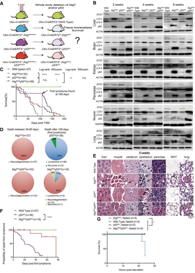
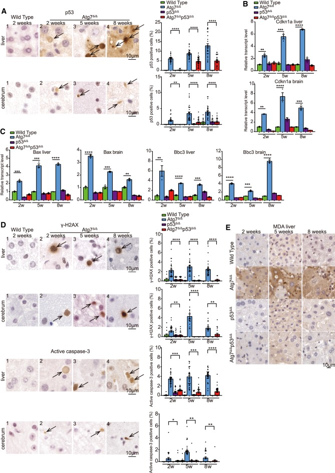
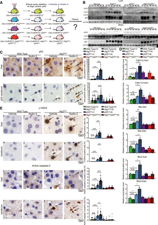
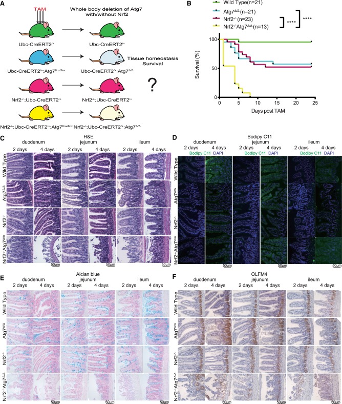
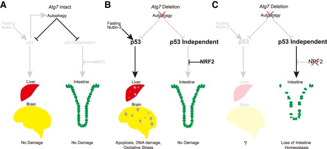
Autophagy captures intracellular components and delivers them to lysosomes for degradation and recycling. Conditional autophagy deficiency in adult mice causes liver damage, shortens life span to 3 mo due to neurodegeneration, and is lethal upon fasting. As autophagy deficiency causes p53 induction and cell death in neurons, we sought to test whether p53 mediates the lethal consequences of autophagy deficiency. Here, we conditionally deleted Trp53 (p53 hereafter) and/or the essential autophagy gene Atg7 throughout adult mice. Compared with Atg7Δ/Δ mice, the life span of Atg7Δ/Δp53Δ/Δ mice was extended due to delayed neurodegeneration and resistance to death upon fasting. Atg7 also suppressed apoptosis induced by p53 activator Nutlin-3, suggesting that autophagy inhibited p53 activation. To test whether increased oxidative stress in Atg7Δ/Δ mice was responsible for p53 activation, Atg7 was deleted in the presence or absence of the master regulator of antioxidant defense nuclear factor erythroid 2-related factor 2 (Nrf2). Nrf2-/-Atg7Δ/Δ mice died rapidly due to small intestine damage, which was not rescued by p53 codeletion. Thus, Atg7 limits p53 activation and p53-mediated neurodegeneration. In turn, NRF2 mitigates lethal intestine degeneration upon autophagy loss. These findings illustrate the tissue-specific roles for autophagy and functional dependencies on the p53 and NRF2 stress response mechanisms.

Keywords: ATG7; DNA damage; NRF2; apoptosis; autophagy; oxidative stress; p53.


Figures

Figure 1.
Figure 1.
Atg7Δ/Δ, p53Δ/Δ mice have extended life span, delayed tissue damage and neurodegeneration compared with Atg7Δ/Δ mice. (A) Experimental design for generation of Atg7Δ/Δ mice, p53Δ/Δ mice, and Atg7Δ/Δ p53Δ/Δ mice. Ubc-CreERT2/+, Ubc-CreERT2/+; Atg7flox/flox mice, Ubc-CreERT2/+; p53flox/flox, Ubc-CreERT2/+; p53flox/flox; Atg7flox/flox mice were treated with TAM at 8–10 wk of age and analyzed at certain time points afterward. (B) Western blot for ATG7, p62, and LC3 at the indicated times of the indicated tissues from wild-type mice, Atg7Δ/Δ mice, p53Δ/Δ mice, and Atg7Δ/Δp53Δ/Δ mice. β-Actin was used as a loading control. (C) Kaplan-Meier survival curve of wild-type mice, Atg7Δ/Δ mice, p53Δ/Δ mice, and Atg7Δ/Δp53Δ/Δ mice. Dotted line indicates 109 d, when the first lymphoma was identified in p53Δ/Δ mice. (n.s,) Not significant; (*) P < 0.05; (**) P < 0.01; (****) P < 0.0001 (log-rank test and Gehan-Breslow-Wilcoxon test as indicated). (D) Percentage distribution for the cause of death of Atg7Δ/Δ, p53Δ/Δ, and Atg7Δ/Δp53Δ/Δ mice. The cause of death was analyzed at 30–90 d after TAM and 109–180 d after TAM. (E) Representative histology of liver, muscle, cerebrum, cerebellum, pancreas, white adipose tissue (WAT), and lung by hematoxylin and eosin stain (H&E) from wild-type, Atg7Δ/Δ, p53Δ/Δ, and Atg7Δ/Δp53Δ/Δ mice at the 8-wk time point. Black arrows indicate the damage site for these tissues. (F) Kaplan-Meier survival curve of wild-type mice, p53Δ/Δ mice, and Atg7Δ/Δp53Δ/Δ mice that died after 109 d. Black dots on the survival curve indicate the censoring times that mice died of no tumor development. (****) P < 0.0001 (log-rank test). (G) Kaplan-Meier survival curve of wild-type mice, Atg7Δ/Δ mice, p53Δ/Δ mice, and Atg7Δ/Δp53Δ/Δ mice during starvation at 10 d after TAM. (*) P < 0.05 (log-rank test). See also Supplemental Figure S1.
Figure 2.
Figure 2.
Autophagy is required to protect liver and brain from p53 accumulation, DNA damage response activation, and apoptosis. (A) Representative liver and cerebrum IHC staining of p53 and quantification at the indicated times from wild-type and Atg7Δ/Δ mice. Black arrows indicate p53-positive cells. (2w) 2-wk time point; (5w) 5-wk time point; (8w) 8-wk time point. (B,C) Quantitative real-time PCR of Cdkn1a, Bax, and Bbc3 for liver and brain tissues from wild-type, Atg7Δ/Δ, p53Δ/Δ, and Atg7Δ/Δp53Δ/Δ mice at the indicated times. (**) P < 0.01; (***) P < 0.001; (****) P < 0.0001 (unpaired t-test). (D) Representative liver and cerebrum IHC staining for γ-H2AX and active caspase-3 with quantification at the indicated times from wild-type and Atg7Δ/Δ mice. Black arrows indicate γ-H2AX or active caspase-3-positive cells. (2w) 2 wk; (5w) 5 wk; (8w) 8 wk. (*) P < 0.05; (**) P < 0.01; (***) P < 0.001 (****) P < 0.0001(unpaired t-test). (E) Representative liver IHC staining for MDA at the indicated times from wild-type, Atg7Δ/Δ, p53Δ/Δ, and Atg7Δ/Δp53Δ/Δ mice. See also Supplemental Figure S2.
Figure 3.
Figure 3.
Activation of p53 by MDM2 antagonist Nutlin-3a in Atg7Δ/Δ mice leads to further increased DNA damage response and apoptosis in the liver and brain. (A) Experimental design for generation of Atg7Δ/Δ, p53Δ/Δ, and Atg7Δ/Δp53Δ/Δ mice and Nutlin-3 administration. Nutlin-3 was administered to mice by oral gavage 2 wk after TAM administration at a dosage of 200 mg/kg for 1 wk. (B) Western blot for ATG7, p62, and LC3 for liver and brain tissues from wild-type, Atg7Δ/Δ, p53Δ/Δ, and Atg7Δ/Δp53Δ/Δ mice treated with vehicle or Nutlin-3. β-Actin was used as a loading control. (V) Treated with vehicle; (N) treated with Nutlin-3. (C) Representative liver and cerebrum IHC staining for p53 and quantification from wild-type and Atg7Δ/Δ mice treated with vehicle or Nutlin-3. Black arrows indicate p53-positive cells. (V) Vehicle; (N) Nutlin-3. (*) P < 0.05; (****) P < 0.0001; (n.s.) not significant (unpaired t-test). (D) Quantitative real-time PCR of Cdkn1a, Bax, and Bbc3 for liver and brain tissues from wild-type, Atg7Δ/Δ, p53Δ/Δ, and Atg7Δ/Δp53Δ/Δ mice treated with vehicle or Nutlin-3. (V) Vehicle; (N) Nutlin-3. (*) P < 0.05; (**) P < 0.01; (***) P < 0.001; (n.s.) not significant (unpaired t-test). (E) Representative liver and cerebrum IHC staining for γ-H2AX and active caspase-3 with quantification from wild-type and Atg7Δ/Δ mice treated with vehicle or Nutlin-3. Black arrows indicate γ-H2AX or active caspase-3-positive cells. (V) Vehicle; (N) Nutlin-3. (*) P < 0.05; (***) P < 0.001; (****) P < 0.0001; (n.s.) not significant (unpaired t-test).
Figure 4.
Figure 4.
Atg7 deficiency is synthetically lethal in the absence of Nrf2. (A) Experimental design for generation of Atg7Δ/Δ mice, Nrf2−/− mice, and Nrf2−/−Atg7Δ/Δ mice. (B) Kaplan-Meier survival curve of wild-type, Atg7Δ/Δ, Nrf2−/−, and Nrf2−/−Atg7Δ/Δ mice. (****) P < 0.0001(log-rank [Mantel-Cox] test). (C) Representative histology of duodenum, jejunum, and ileum by H&E at the indicated times from wild-type, Atg7Δ/Δ, Nrf2−/−, and Nrf2−/−Atg7Δ/Δ mice. (D) Representative Bodipy C11 stain of duodenum, jejunum, and ileum at the indicated times from wild-type, Atg7Δ/Δ, Nrf2−/−, and Nrf2−/−Atg7Δ/Δ mice. (E) Representative Alcian blue stain of duodenum, jejunum, and ileum at the indicated times from wild-type, Atg7Δ/Δ, Nrf2−/−, and Nrf2−/−Atg7Δ/Δ mice. (F) Representative duodenum, jejunum, and ileum IHC stain of OLFM4 at the indicated times from wild-type, Atg7Δ/Δ, Nrf2−/−, and Nrf2−/−Atg7Δ/Δ mice.
Figure 5.
Figure 5.
Mechanism by which autophagy interacts with the p53 and NRF2 stress response mechanisms to protect tissues in a tissue specific manner. See the text for details.

References

    1. Asano J, Sato T, Ichinose S, Kajita M, Onai N, Shimizu S, Ohteki T. 2017. Intrinsic autophagy is required for the maintenance of intestinal stem cells and for irradiation-induced intestinal regeneration. Cell Rep 20: 1050–1060. 10.1016/j.celrep.2017.07.019 - DOI - PubMed
    1. Bode AM, Dong Z. 2004. Post-translational modification of p53 in tumorigenesis. Nat Rev Cancer 4: 793–805. 10.1038/nrc1455 - DOI - PubMed
    1. Cadwell K, Liu JY, Brown SL, Miyoshi H, Loh J, Lennerz JK, Kishi C, Kc W, Carrero JA, Hunt S, et al. 2008. A key role for autophagy and the autophagy gene Atg16l1 in mouse and human intestinal Paneth cells. Nature 456: 259–263. 10.1038/nature07416 - DOI - PMC - PubMed
    1. Chan K, Lu R, Chang JC, Kan YW. 1996. NRF2, a member of the NFE2 family of transcription factors, is not essential for murine erythropoiesis, growth, and development. Proc Natl Acad Sci 93: 13943–13948. 10.1073/pnas.93.24.13943 - DOI - PMC - PubMed
    1. Cox DR, Oakes D. 1984. Analysis of survival data. Chapman and Hall, London; New York.

Publication types

MeSH terms