. 2020 Mar 19;11(1):1455.
doi: 10.1038/s41467-020-15257-1.

Ca2+-Daptomycin targets cell wall biosynthesis by forming a tripartite complex with undecaprenyl-coupled intermediates and membrane lipids

Affiliations

Ca2+-Daptomycin targets cell wall biosynthesis by forming a tripartite complex with undecaprenyl-coupled intermediates and membrane lipids

Fabian Grein et al. Nat Commun. .

Abstract

The lipopeptide daptomycin is used as an antibiotic to treat severe infections with gram-positive pathogens, such as methicillin resistant Staphylococcus aureus (MRSA) and drug-resistant enterococci. Its precise mechanism of action is incompletely understood, and a specific molecular target has not been identified. Here we show that Ca2+-daptomycin specifically interacts with undecaprenyl-coupled cell envelope precursors in the presence of the anionic phospholipid phosphatidylglycerol, forming a tripartite complex. We use microbiological and biochemical assays, in combination with fluorescence and optical sectioning microscopy of intact staphylococcal cells and model membrane systems. Binding primarily occurs at the staphylococcal septum and interrupts cell wall biosynthesis. This is followed by delocalisation of components of the peptidoglycan biosynthesis machinery and massive membrane rearrangements, which may account for the pleiotropic cellular events previously reported. The identification of carrier-bound cell wall precursors as specific targets explains the specificity of daptomycin for bacterial cells. Our work reconciles apparently inconsistent previous results, and supports a concise model for the mode of action of daptomycin.


Conflict of interest statement

The authors declare no competing interests.

Figures

Fig. 1
Fig. 1. DAP specifically induces the LiaRS stress response in the presence of Ca2+ and binds to the cell division site.
a PliaI-lux induction in B. subtilis was monitored upon addition of DAP (0.5 µg ml−1) in the absence (black line) or presence (blue line) of 1.25 mM Ca2+ and teixobactin (0.5 µg ml−1) (green line). Luciferase activity is presented as relative luminescence units (RLU). Representative graph of three independent experiments. b, c DAP localises to the septum of S. aureus. Cells were grown to mid-exponential phase (OD600 = 0.5) followed by the addition of Ca2+ and a mixture of labelled and unlabelled DAP (7 µg ml−1 DAP; 0.8 µg ml−1 DAP-FL) b or unlabelled DAP (7 µg ml−1) c. Cells were washed and imaged by fluorescence microscopy; phase, phase contrast. Cell outlines in c are indicated by dashed lines. Scale bar: 1 µm. Representative images from five independent experiments are shown. Source data are provided as a Source Data file.
Fig. 2
Fig. 2. DAP binds to S. aureus in a biphasic manner.
a DAP-FL binding to S. aureus monitored over time (0–60 min). S. aureus HG003 was grown to mid-exponential phase (OD600 = 0.5) followed by the addition of Ca2+ and a mixture of labelled and unlabelled DAP (7 µg ml−1 DAP; 0.8 µg ml−1 DAP-FL). At different time points, samples were taken, washed and imaged by fluorescence microscopy. Representative pictures are shown from three independent experiments. Phase, phase contrast. Scale bar 1 µm. b Survival of cells from the experiment described in a and schematic depiction of DAP-FL-binding behaviour in two phases. Source data are provided as a Source Data file.
Fig. 3
Fig. 3. Pre-incubation with lipid II-binding antibiotics prevents DAP binding to S. aureus and delays DAP-induced killing.
a Binding of DAP-FL to S. aureus cells (OD600 = 0.5) pre-incubated with cell wall targeting antibiotics (oxacillin (OXA), teixobactin (TEIX) and oritavancin (ORI) (4-fold MIC)). After pre-incubation for 2 min, cells were washed and incubated with DAP-FL in the presence of Ca2+ for 10 min followed by washing and the addition of nile red membrane stain. Cells were washed again and subjected to fluorescence microscopy. In a control (CTRL) pre-incubuation with antibiotics was omitted. Scale bar 1 µm. Representative images are shown. b Quantification of DAP-FL binding measured in the experiment as described in a. Binding is expressed as mean grey value per cell. Box plots represent the interquartile range of the data. The black bar represents the mean and whiskers represent minimal and maximal values, respectively. At least n = 350 cells were evaluated for each condition from three biologically independent experiments. Significance was determined by unpaired Student’s t-test with a 95% confidence interval. ****p < 0.0001, n.s., not significant (p = 0.0835). c Survival of S. aureus challenged with DAP (10 µg ml−1) without (red line) or with (blue line) pre-incubation with TEIX as described in a. Bactericidal effects were not observed in cells only pre-treated with TEIX (black line). Data presented are mean values from n = 3 biologically independent experiments. Error bars represent the standard deviation (SD). Source data are provided as a Source Data file.
Fig. 4
Fig. 4. DAP binding to supported bilayers doped with cell wall lipid intermediates.
a Binding of DAP to supported bilayers is drastically increased in presence of bactoprenyl-coupled lipid intermediates and PG. Supported planar bilayers were prepared on coverslips using neutral DOPC lipids. Negatively charged PG lipids and bactoprenyl lipids were added, either 0.1 mol% bactoprenyl-coupled lipid intermediates or mixtures with PG (0.1 mol% each, 0.2 mol% PG served as a control). DAP was applied as a mixture of 1 µM native DAP and 50 nM DAP-FL. Movies with 100 frames were recorded at a frame rate of 60 Hz within 3 min after addition of DAP. The movies allowed to discern single binding events of DAP-FL. Exemplary fluorescence images are shown for each mixture. The field of view corresponds to (32.5 µm)2. b Inhibition of DAP-FL binding to supported planar bilayers (PC + 0.1% PG) by antibiotics that target specific bactoprenyl-coupled lipid intermediates. 300 nM of each antibiotic (FRIU, friulimicin; BACI, bacitracin and ORI, oritavancin) were incubated with the corresponding bactoprenyl-coupled lipid intermediate containing membranes for 5 min (antibiotic:lipid intermediates ratio 100:1). Excess of antibiotics was removed by buffer exchange followed by the addition of DAP as described in a. Exemplary movies are shown in Supporting Movies 2 and 3. Data were obtained from at least 20 movies for each experiment. We determined the mean values in a field of 160 µm2 in the first image of each movie. Data in a and b are plotted as averages of these means and error bars represent the SD of all movies of a specific experiment. Significance was determined by unpaired Student’s t-test with a 95% confidence interval, ****p < 0.0001. All experiments were independently repeated three times and yielded comparable results. Source data are provided as a Source Data file.
Fig. 5
Fig. 5. DAP forms a tripartite complex with lipid II and PG and inhibits cell wall biosynthesis in vitro.
a DAP was incubated with purified lipid II at increasing molar ratio in the presence (left TLC) and absence (right TLC) of PG. Reaction mixtures were extracted with BuOH and the upper solvent phase was spotted to TLC. Free PG, DAP and lipid II migrate to defined positions on the TLC, while components that are locked in complex are retained in the lower aqueous phase after extraction and as a result are diminished on the TLC. Representative images from five independent experiments are shown. b Impact of DAP on the MraY-catalysed synthesis of lipid I and c on the transglycosylation reaction catalysed by PBP2. DAP inhibits both reactions in a dose-dependent manner and almost completely blocks enzymatic activity when added in 10-fold molar excess. The enzymatic activity is expressed as synthesised lipid I (MraY) or converted lipid II (PBP2). The control reactions in the absence of antibiotics were set to 100%. DAP was added at molar ratios of 1:1 to 10:1 with respect to the lipid substrates as indicated. Data presented are means from three independent experiments and error bars represent the SD. Source data are provided as a Source Data file.
Fig. 6
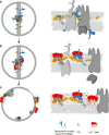
Fig. 6. Proposed model for the mechanism of action of DAP.
a Orchestration of the cell wall biosynthesis machinery at the division septum of S. aureus in the absence of DAP. b Ca2+-DAP oligomerises and preferentially localises at the division septum enriched in anionic lipids, primarily PG, and C55P-coupled cell wall precursors. The formation of a tripartite complex of Ca2+-DAP with PG and bactoprenyl-coupled lipid intermediates blocks cell wall synthesis and triggers the delocalisation of the cell wall biosynthetic machinery. c Prolonged treatment results in a progressive dispersion of DAP throughout the entire cytoplasmic membrane, followed by disintegration of the membrane bilayer finally resulting in membrane leakage and cell death.

References

    1. Debono M, et al. A21978C, a complex of new acidic peptide antibiotics: isolation, chemistry, and mass spectral structure elucidation. J. Antibiot. 1987;40:761–777. doi: 10.7164/antibiotics.40.761. - DOI - PubMed
    1. Debono M, et al. Enzymatic and chemical modifications of lipopeptide antibiotic A21978C: the synthesis and evaluation of daptomycin (LY146032) J. Antibiot. 1988;41:1093–1105. doi: 10.7164/antibiotics.41.1093. - DOI - PubMed
    1. Wood TM, Martin NI. The calcium-dependent lipopeptide antibiotics: structure, mechanism, & medicinal chemistry. MedChemComm. 2019;10:634–646. doi: 10.1039/C9MD00126C. - DOI - PMC - PubMed
    1. Strieker M, Marahiel MA. The structural diversity of acidic lipopeptide antibiotics. Chembiochem. 2009;10:607–616. doi: 10.1002/cbic.200800546. - DOI - PubMed
    1. Straus SK, Hancock REW. Mode of action of the new antibiotic for Gram-positive pathogens daptomycin: comparison with cationic antimicrobial peptides and lipopeptides. Biochim. Biophys. Acta. 2006;1758:1215–1223. doi: 10.1016/j.bbamem.2006.02.009. - DOI - PubMed

Publication types

MeSH terms