. 2020 Mar 19;10(1):5001.
doi: 10.1038/s41598-020-61767-9.

Nuclear receptor CAR-ERα signaling regulates the estrogen sulfotransferase gene in the liver

Affiliations

Nuclear receptor CAR-ERα signaling regulates the estrogen sulfotransferase gene in the liver

MyeongJin Yi et al. Sci Rep. .

Abstract

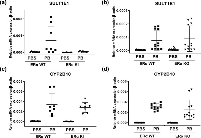
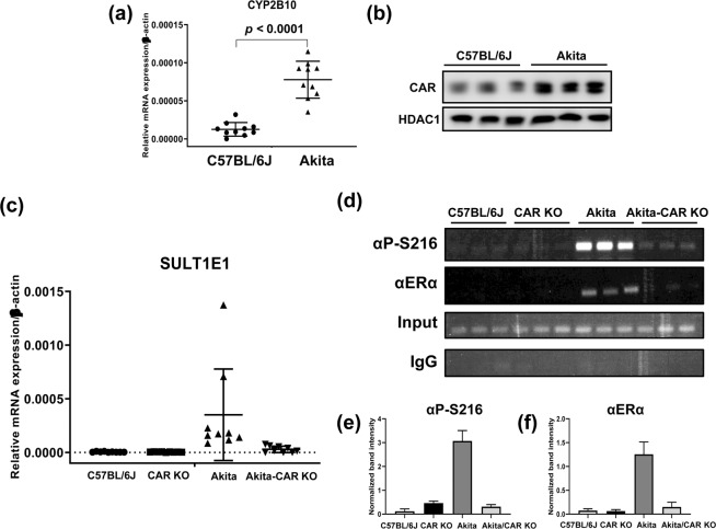
Estrogen sulfotransferase (SULT1E1) inactivates estrogen and regulates its metabolic homeostats. Whereas SULT1E1 is expressed low in the liver of adult mice, it is induced by phenobarbital (PB) treatment or spontaneously in diabetic livers via nuclear receptors. Utilizing constitutive active/androstane receptor (CAR) KO, estrogen receptor α (ERα KO, phosphorylation-blocked ERα S216A KI mice, it is now demonstrated that, after being activated by PB, CAR binds and recruits ERα onto the Sulte1 promoter for subsequent phosphorylation at Ser216. This phosphorylation tightens CAR interacting with ERα and to activates the promoter. Hepatic SULT1E1 mRNA levels are constitutively up-regulated in type 1 diabetic Akita mice; CAR spontaneously accumulates in the nucleus and activates the Sult1e1 promoter by recruiting phosphorylated ERα in the liver as observed with PB-induced livers. Thus, this CAR-phosphorylated ERα signaling enables these two nuclear receptors to communicate, activating the Sult1e1 gene in response to either PB or diabetes in mice. ERα phosphorylation may integrate CAR into estrogen actions, providing insights into understanding drug-hormone interactions in clinical therapy.


Conflict of interest statement

The authors declare no competing interests.

Figures

Figure 1
Figure 1
PB-induced expression of SULT1E1 in CAR WT and CAR KO males. (a) Cytosolic extracts were prepared from C57BL/6J male livers which were treated with PB or PBS. SULT1E1 was induced by PB-treatment, and a polyclonal antibody for α-tubulin was used as a loading control. (b) PB induction of SULT1E1 mRNA in CAR wild-type and KO mouse livers treated with PBS or PB (N = 8). P-value derived from ANOVA is 0.0013. (c) Hepatic RNAs were prepared from CAR WT (N = 8) and CAR KO (N = 8) males treated with PB or PBS for 24 h. The same RNAs used for SULT1E1 were utilized to measure the levels of CYP2B10 mRNA. All p-values for four different groups are lower than 0.0001. All data are presented as means ± S.D. of values of individual mice, and one-way ANOVA was used as a statistical analysis for multiple groups.
Figure 2
Figure 2
PB-induced expression of SULT1E1 mRNA in ERα S216A KI and ERα KO males. Hepatic RNAs were prepared from (a) ERα WT (N = 8), and ERα S216A KI (N = 8), males or (b) ERα WT (N = 12) and ERα KO (N = 12) males treated with PB or PBS for 24 h, to measure each SULT1E1 mRNA. Each p-value of both data set is derived from as 0.0059, and <0.0001, respectively. The same RNA samples from (c) ERα WT (N = 8), and ERα S216A KI (N = 8), males or (d) ERα WT (N = 12) and ERα KO (N = 12) males were used to measure CYP2B10 mRNA. The both p-values of data are estimated as lower than 0.0001. All data are presented as means ± S.D. of values of individual mice. One-way ANOVA was used as a statistical analysis for data set of ERα S216A KI, and Kruskal-Wallis test was used to analyze the data set of ERα KO.
Figure 3
Figure 3
PB-induced binding of ERα phosphorylated at Ser216 to the Sult1e1 promoter in male mice. ChIP assays were employed to show ERα bindings to a proximal Sult1e1 promoter. (a) Schematic representation of a region that was amplified and primers that were used for PCR amplification. (b) Chromatins were prepared from livers of CAR WT and KO males treated with PB or PBS for 6 h. And those were subjected to ChIP assays with either an ERα antibody or a P-S216 peptide antibody. Both of (c,d) show the quantification, which was normalized by input intensity, for P-ERα and ERα respectively. The densitometry was performed by ImageJ. Kruskal-Wallis test and one-way ANOVA were used as statistical analyses, and p-values are 0.0004 and <0.0001 respectively. (e) Chromatins were prepared from livers of ERα and KI males treated with PB or PBS for 6 h. And those chromatins were subjected to ChIP assays with either an ERα antibody or a P-S216 peptide antibody. Rabbit IgG was used as a negative control. Both of (f,g) present the quantifying data from ERα and KI males, which was normalized by input intensity, for P-ERα and ERα respectively. The densitometry was performed by ImageJ. Kruskal-Wallis test and one-way ANOVA were used as statistical analyses, and p-values are 0.0064 and <0.0001 respectively. All data are presented as means ± S.D. of values of individual mice.
Figure 4
Figure 4
Interactions between CAR and phosphorylated ERα. (a) The DR4 sequence was used as a probe for gel shift assays. (b) 32P-labeled double stranded probe was mixed with nuclear extracts prepared from mouse livers as indicated in the figure. For super shifts, CAR or phosphorylated ERα antibody was added to a mixture of probe and nuclear extract as indicated in the figure. (c) FLAG-tagged CAR was co-expressed with either EYFP-tagged ERα WT, ERα S216A, and ERα S216D in Huh7 cells in the presence of 10 nM 17β-estradiol for 24 h. Whole cell lysates were isolated and precipitated with αGFP-resin, eluted proteins from which were loaded on a SDS-PAGE gel and subjected to staining with an αFLAG antibody. Full-length gel images are described as Supplement Fig. 3. (d) 3D model of the hypothetical heterotetramer between an ERα homodimer (monomers in brown and magenta) and the CAR/RXRα heterodimer (CAR in yellow and RXRα in ash-rose). In this model one surface of CAR interacts with RXRα, while another surface interacts with an ERα subunit. Ligands (17β-estradiol (ERα), 9-cis-retinoic acid (RXRα), 3,5-dichloro-2-(4-[(3,5-dichloropyridin-2-yl)oxy]phenoxy)pyridine (CAR)) are shown with atoms in spheres (oxygen in red, nitrogen in blue and chlorine in green, carbon atoms are colored as protein the ligands are bound to). This nuclear receptor tetramer may comprise a complex which regulates the expression of Sult1e1 gene. A description of how the model tetramer was created can be found in the Methods.
Figure 5
Figure 5
Hepatic SULT1E1 expression in Akita-CAR KO males. (a) Hepatic RNAs were prepared from C57BL/6J (N = 10) and Akita (N = 10) males. CYP2B10 mRNA, the classic CAR target showed the higher expression in Akita compared to C57BL/6J. Data set was analyzed by a Student’s t-test, and p-value is lower than 0.0001. (b) Nuclear extracts were separately prepared form livers of C57BL/6J (N = 3) and Akita (N = 3). The expression of CAR in nucleus was higher than that of C57BL/6J. (c) Hepatic RNAs were prepared from CAR WT (N = 9), CAR KO (N = 8), Akita (N = 9) and Akita-CAR KO (N = 6). Data set was analyzed by Kruskal-Wallis test, and p-value is 0.0001. (d) Chromatins were separately prepared from three livers of each of the above-mentioned groups for subsequent ChIP assays. Both of (e,f) presented the quantifying data, which were normalized by input intensity, for P-ERα and ERα respectively. The densitometry was performed by ImageJ. All data are presented as means ± S.D. of values of individual mice and one-way ANOVA was used as a statistical analysis. Each p-value of both groups is lower than 0.0001.
Figure 6
Figure 6
Non-phosphorylated ERα is not bound to the Sult1e1 promoter in male mice with P-RORα. ChIP assays were applied to show RORα bindings to a proximal Sult1e1 promoter. (a) Chromatins were prepared from livers of ERα WT and KI males which were subjected to ChIP assays with either an RORα antibody or a P-S100 peptide antibody. Both of (b,c) indicate the data quantification, which was normalized by input intensity, for P-RORα and RORα respectively. The assay was examined by ImageJ, and one-way ANOVA were used as statistical analyses, and p-values are 0.0247 and 0.1080 respectively. (d) V5-CAR WT (no tag) was co-expressed with FLAG-tagged RORα WT, RORα S100D, EYFP-tagged ERα WT, and ERα S216D in Huh7 cells. Whole cell lysates were isolated and precipitated with αGFP-resin, and eluted proteins were loaded on a SDS-PAGE gel and subjected to staining with an αGFP- or αFLAG antibody.
Figure 7
Figure 7
Schematic depiction of the CAR-ERα communication mechanism. CAR phosphorylated at Thr38 forms a homodimer through surface A and remains inactive in the cytoplasm,. In response to PB, Thr38 is dephosphorylated and non-phosphorylated CAR homodimer dissociates, allowing the interaction between the DBD and LBD to translocate into the nucleus,. In the nucleus, CAR utilizes its surface B to form an RXRα-CAR heterodimer. ERα dimerizes through B surfaces upon estrogen binding. Subsequently, RXRα-CAR heterodimer recruits ERα homodimer to the Sulte1 promoter by between the heterodimer-bound CAR and homodimer-bound ERα via their A surfaces. While phosphorylated ERα is unable to directly bind DNA, its homodimer sandwiches RXRα-CAR heterodimer and phosphorylated RORα, resulting in an activation of the promoter.

Similar articles

Cited by

References

    1. Paterni I, Granchi C, Katzenellenbogen JA, Minutolo F. Estrogen receptors alpha (ERalpha) and beta (ERbeta): subtype-selective ligands and clinical potential. Steroids. 2014;90:13–29. doi: 10.1016/j.steroids.2014.06.012. - DOI - PMC - PubMed
    1. Yasar P, Ayaz G, User SD, Gupur G, Muyan M. Molecular mechanism of estrogen-estrogen receptor signaling. Reprod. Med. Biol. 2017;16:4–20. doi: 10.1002/rmb2.12006. - DOI - PMC - PubMed
    1. Svenson JL, EuDaly J, Ruiz P, Korach KS, Gilkeson GS. Impact of estrogen receptor deficiency on disease expression in the NZM2410 lupus prone mouse. Clin. Immunol. 2008;128:259–268. doi: 10.1016/j.clim.2008.03.508. - DOI - PMC - PubMed
    1. Song WC, Qian Y, Sun X, Negishi M. Cellular localization and regulation of expression of testicular estrogen sulfotransferase. Endocrinol. 1997;138:5006–5012. doi: 10.1210/endo.138.11.5512. - DOI - PubMed
    1. Kakuta Y, et al. Mouse steroid sulfotransferases: substrate specificity and preliminary X-ray crystallographic analysis. Biochem. Pharmacol. 1998;55:313–317. doi: 10.1016/s0006-2952(97)00465-6. - DOI - PubMed

Publication types