. 2020 Mar 19;10(1):4987.
doi: 10.1038/s41598-020-61921-3.

Participation of Intussusceptive Angiogenesis in the Morphogenesis of Lobular Capillary Hemangioma

Affiliations

Participation of Intussusceptive Angiogenesis in the Morphogenesis of Lobular Capillary Hemangioma

Lucio Díaz-Flores et al. Sci Rep. .

Abstract

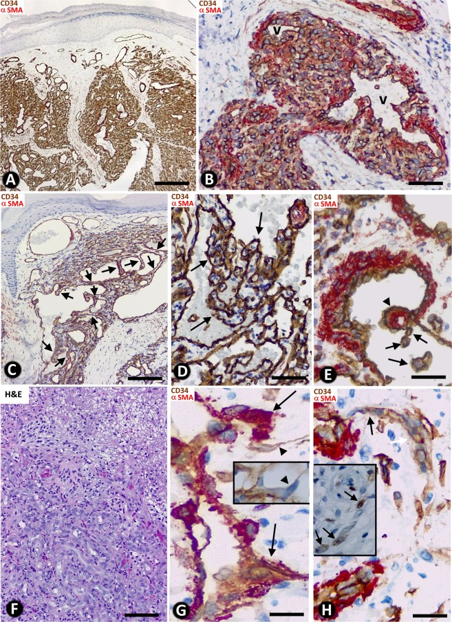
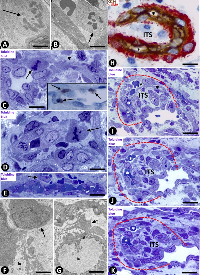
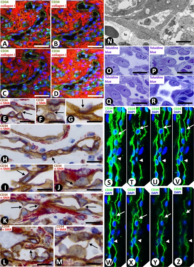
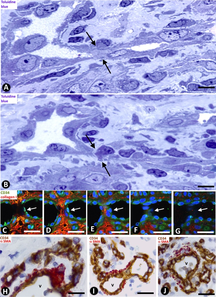
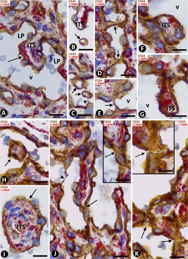
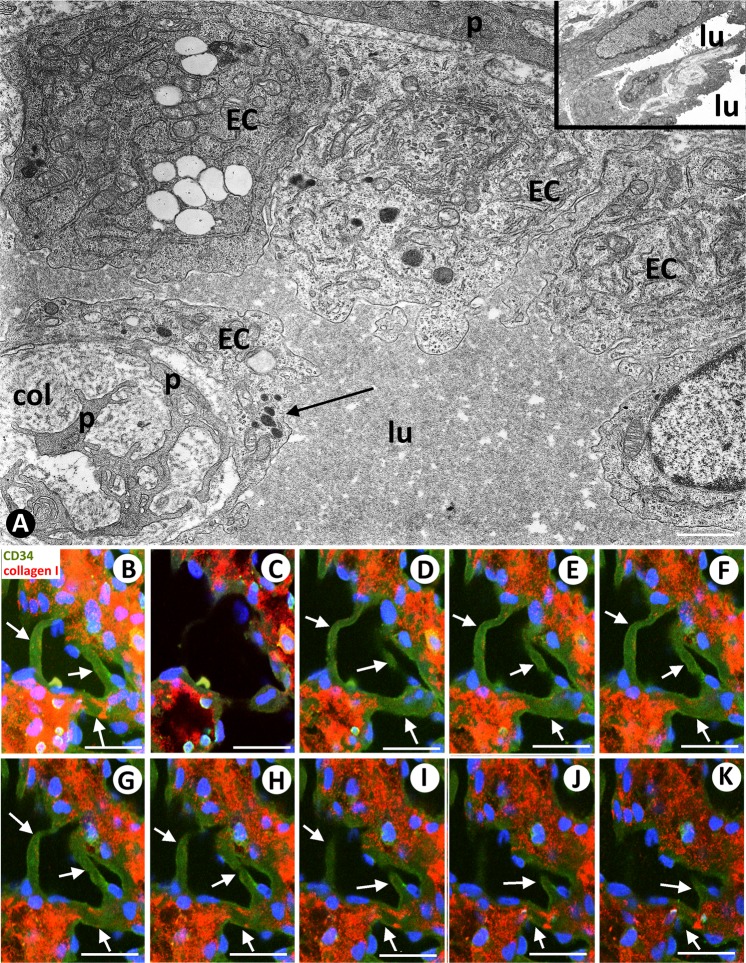
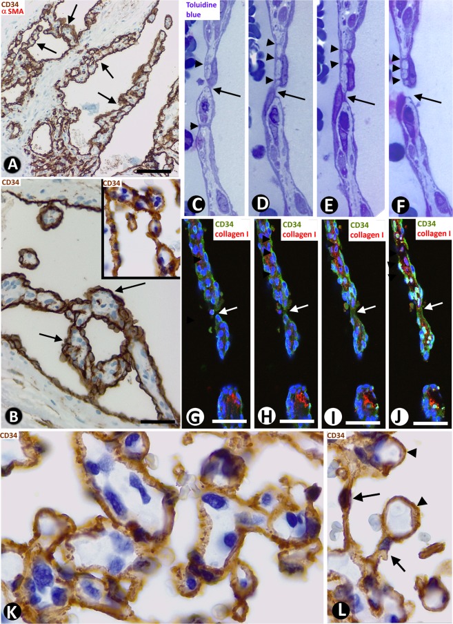
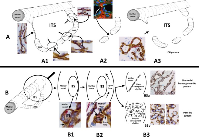
In lobular capillary hemangioma (LCH), misnamed pyogenic granuloma, only sprouting angiogenesis (SA) has been considered. We assess the occurrence of intussusceptive angiogenesis (IA) in LCH and whether IA determines the specific and other focal patterns in the lesion. For this purpose, we study specimens of 120 cases of LCH, using semithin sections (in 10), immunohistochemistry, and confocal microscopy (in 20). In addition to SA, the results in LCH showed (1) intussusceptive phenomena, including pillars/folds and associated vessel loops, which encircled interstitial tissue structures (ITSs). (2) Two types of evolved loops depending on interendothelial contacts from opposite walls: (a) numerous interendothelial contacts, alternating with capillary-sized lumens (main capillary pattern of the lesion) and (b) few interendothelial contacts, wide open lumens, and intravascular transport of pillars/folds, which were arranged linearly, forming septa (focal sinusoidal-like pattern) or were irregularly grouped (focal intravascular papillary endothelial hyperplasia, IPEH-like pattern). In conclusion, we demonstrate that IA participates in synergistic interaction with SA in LCH development and that the prevalence of specific intussusceptive phenomena determines the predominant capillary pattern and associated sinusoidal hemangioma-like and IPEH-like patterns in the lesion, which suggest a role of IA as conditioner of vessel tumour/pseudo-tumour morphology.


Conflict of interest statement

The authors declare no competing interests.

Figures

Figure 1
Figure 1
General characteristics of lobular capillary hemangioma (LCH). (A) Several lobules are observed in the lesion (lobular pattern). (B) In a lobule, presence of a branched venule (v) and numerous capillary-sized vessels, showing ECs (brown) and pericytes (red). (C) A zone with sinusoidal hemangioma-like morphology is observed, presenting wide vascular spaces with pillars, some of which are arranged in a linear fashion, forming incomplete septa (arrows). (D) An IPEH-like zone with numerous pillars irregularly arranged(arrows) in a wide vascular space. (E) CD34+ ECs (brown) and αSMA+ smooth muscle cells (red) are observed in the vessel walls. A large (arrowhead) and small pillars (arrows) are present. (F) Zone of ulceration with capillary-sized vessels, fibrin deposits and inflammatory infiltrate. (G) Sprouts are observed from a mother vessel (outward growth - arrows). In one of them, note a tip EC growing toward the interstitium (arrowhead). In the insert, the collagen IV stained basal membrane disappears in a tip EC (arrowhead). (H) Stalk ECs from a mother vessel are observed initiating a loop (arrows). In the insert, stalk ECs expressing ki67 in a loop are observed (arrows). (A to E,G,H) Double-staining with anti-CD34 (brown) and anti-αSMA (red). (F) H&E staining. Bar: (A,C) 160 µm; (B,F) 25 µm; (D,E,G,H) 20 µm.
Figure 2
Figure 2
(AD) Mitoses are observed in a stalk cell of a loop (A, arrow), and in endothelial cells (B and C, arrows) and in a pericyte (D, arrow) of mother vessels. In the insert of C, note cells expressing ki67 (arrows) in the wall of a mother vessel. In C, note a sprout emerging from a mother vessel (arrowhead). (EG) Incorporation of pericytes or their processes (arrows) around ECs and formation of new vessel lumens (lu). (HK) Vessel loops surrounding interstitial tissue structures (ITSs) are observed in double-staining (H) and in serial semi-thin sections (a dashed line delimits the loop, (IK). In H, the loop encircling an ITS shows CD34+ ECs (brown) and αSMA+ pericytes (red). Perforated and unperforated interendothelial contacts from the opposite walls of the loops are seen alternating with zones in which the loop lumen (asterisk) is open (HK). Note the presence of red blood cells in the open spaces of the loop (IK). (A,B,F,G) Ultrathin sections. Uranyl acetate & Lead citrate. (CE,IK) Semithin sections stained with Toluidine Blue (IK) serial sections). (H) Double-stained section with anti-CD34 (brown) and anti-αSMA (red). Bar: (A,B,F,G) 4 µm; (C,D) 10 µm; (H) 20 µm; (EK) 25 µm.
Figure 3
Figure 3
(AD) Images showing the spatial path of a loop zone, in single (AC) and whole-mount (D) views in confocal microscopy (frontal view, 6 µm section, immunofluorescence label with anti-CD34, green, and anti-collagen I, red, DAPI: blue). (EM) Contacts between ECs from opposite walls of the loops (arrows). Note that the contacting ECs may have triangular (E), ovoid (F) or flattened (G) morphology in the histological sections and that planar contact may be extensive (H). Contacts can be symmetric (E) or asymmetric (I,J), in proximity (K) or not, and can acquire a peg-like aspect when ECs surround connective tissue (L). (M) Occasionally, smaller transcapillary cytoplasmic projections (antenna-like or filipodia-like) were seen. (N) Ultrastructural image of an endothelial projection (arrow) separating two luminal spaces (lu) of a loop. (OR) Serial semithin sections showing an EC projecting intraluminally (in Figs. 4A,B, in which a perforated contact is shown, the region in O-R also appears in the image with a portion of the projecting cell -Fig. 4A- and with no presence of this projecting cell -Fig. 4B-). (SZ) Single (TZ) and whole-mounted (S) views in confocal microscopy (frontal view, 10 µm section and CD34 staining, DAPI: blue) showing the appearance and disappearance of endothelial contacts (arrowhead), including a peg-like structure (arrow). (EM) Double-staining with anti-CD34 (brown) and anti-αSMA (red). (N) Ultrathin section. Uranyl acetate and Lead citrate. (OR) Semithin sections stained with Toluidine Blue. Bar: A-D, 30 µm; (EM,O–Z) 10 µm; (N) 8 µm.
Figure 4
Figure 4
Perforations of interendothelial contacts and loop fragmentation into capillary-sized vessels. (A,B) An interendothelial contact (A, arrows) and its perforation (B, arrows) are shown in two sections obtained from serial semithin sections. Note the presence of neighbouring interstitial cells in the perforated zone. (CG) Images in confocal microscopy with double immunofluorescent label (CD34 and collagen I, DAPI: blue) demonstrating the appearance and disappearance of a pillar (arrows), with an endothelial cover (green) and a core with collagen I (red). (HJ) Images showing loop fragmentation in several capillary-sized vessels after perforation of interendothelial contacts. Note that the loops arise from venules (v) and that the capillary-sized vessels are arranged in a linear arciform path reminiscent of that of loops. (A,B) Semithin section stained with Toluidine Blue. (CG) Immunofluorescent label with CD34, collagen I and DAPI. (HJ) Sections double-stained with anti-CD34 (brown) and anti-αSMA (red). Bar: (A,B) 8 µm; (CG) 30 µm; (HJ) 35 µm.
Figure 5
Figure 5
Vessel loops with scarce interendothelial contacts, open lumens, and intravascular pillars. Pillars (arrows) are covered by CD34+ ECs (corresponding to the inner layer of the loop) and show a core whose connective content (collagen, pericytes and interstitial cells) depends on pillar size. The loop open lumen (LP) is connected to that of the mother vessel (v) and surrounds an ITS (A,B). Nascent pillars are also observed (C and J, arrowhead). Note that pillars/ITSs are isolated in the vessel lumen (H and I, arrows) or adhered to the vessel wall, with a planar (F) or meso-like morphology (G), or to other pillars/ITSs, arranged linearly or grouped irregularly (J,K). The zones of adherence are shown at higher magnification in inserts of (J,K) (arrows). Double-staining with anti-CD34 (brown) and αSMA (red). Bar: (AK) 8 µm.
Figure 6
Figure 6
(A) Ultrastructural image of a zone of a vessel with prominent ECs and a transversally sectioned intravascular pillar (arrow), showing a cover formed by ECs and a core with processes of pericytes (p) and collagen material (col). In the insert, a pillar appears longitudinally sectioned. lu: vessel lumen. (BK) The appearance and disappearance and the whole-mounted view (B) of three intravascular pillars (arrows) are shown in confocal microscopy. A and insert: Ultrathin sections. Uranyl acetate and lead citrate. (BK) confocal microscopy, frontal view, 10 µm section, immunofluorescent label with anti-CD34 (green), anti-collagen I (red), DAPI (blue). Bar: (A) 2 µm; (BK) 20 µm.
Figure 7
Figure 7
(AJ) Zones with sinusoidal hemangioma-like morphology are shown in LCH. (AB) Pillars arranged in a linear fashion are observed forming incomplete septa (arrows), covered by CD34+ ECs. (CF) In serial semithin sections, communications between opposite endothelial cells of the septa (arrowheads) and a zone of continuity and discontinuity between pillars are present (arrows). (GJ) Similar aspect of septa showing continuity and discontinuity between pillars (arrows) in confocal microscopy [single (GI) and whole-mount (J) in frontal view (6 µm section)]. (K,L) Zones with IPEH-like morphology. Note the irregular arrangement of pillars (K), and large pillars (L-arrowheads) connected by thin pillars (L-arrows). (A) Section double-stained with anti-CD34 (brown) and anti-αSMA (red). (B,K,L) Sections stained with anti-CD34 (brown). (CF) Serial semithin sections stained with Toluidine Blue. (GJ) Immunofluorescent label with CD34 (green), collagen I (red) and DAPI (blue). Bar: (A) 80 µm; (BJ) 20 µm; (K,L) 10 µm.
Figure 8
Figure 8
Schematic representation of the IA mechanisms that influence vascular lesion morphogenesis, depending on the formation, or not, of multiple interendothelial contacts. (A) Successive steps of capillary-like space formation are shown in A1, A2 and A3, with their corresponding microphotographs. In A1, a loop originating from a mother vessel. The loop shows several examples of interendothelial contacts, alternating with capillary-like spaces. In A2, perforation of an interendothelial contact. In A3, loop segmented in capillary-like structures, acquiring the capillary aspect of LCH. These images only represent one loop. Spatially, however, multiple converging loops occur in the lesion, and the capillary-sized spaces may connect with others in other loops. These findings could explain the future conservation or involution of these spaces. Note that the ITS surrounded by the loop remains outside the mother vessel (extravascular). (B) Several steps of pillar formation in the mother vessel are shown in B1, B2 and B3, with their corresponding microphotographs. The loop originating from the mother vessel (B1) becomes permeable (B2), and the ITS and its surrounding endothelium (internal side of the loop) is transported to the mother vessel lumen (although some connections between the pillar and vessel wall may persist). In B3, several pillars can be arranged linearly or irregularly, originating the sinusoidal-hemangioma-like (B3a) or IPEH-like (B3b) pattern, respectively.

References

    1. Ackermann M, Tsuda A, Secomb TW, Mentzer SJ, Konerding MA. Intussusceptive remodeling of vascular branch angles in chemically-induced murine colitis. Microvasc. Res. 2013;87:75–82. doi: 10.1016/j.mvr.2013.02.002. - DOI - PMC - PubMed
    1. Burri PH, Djonov V. Intussusceptive angiogenesis–the alternative to capillary sprouting. Mol. Aspects Med. 2002;23:S1–27. doi: 10.1016/S0098-2997(02)00096-1. - DOI - PubMed
    1. Burri PH, Hlushchuk R, Djonov V. Intussusceptive angiogenesis: its emergence, its characteristics, and its significance. Dev. Dyn. 2004;231:474–488. doi: 10.1002/dvdy.20184. - DOI - PubMed
    1. De Spiegelaere W, et al. Intussusceptive angiogenesis: a biologically relevant form of angiogenesis. J. Vasc. Res. 2012;49:390–404. doi: 10.1159/000338278. - DOI - PubMed
    1. Díaz-Flores L, et al. Segmentation of dilated hemorrhoidal veins in hemorrhoidal disease. Cells, Tissues, Organs. 2018;205:120–128. doi: 10.1159/000489250. - DOI - PubMed