. 2020 Mar 19;10(1):5050.
doi: 10.1038/s41598-020-61875-6.

Migratory dendritic cells in skin-draining lymph nodes have nickel-binding capabilities

Affiliations

Migratory dendritic cells in skin-draining lymph nodes have nickel-binding capabilities

Toshinobu Kuroishi et al. Sci Rep. .

Abstract

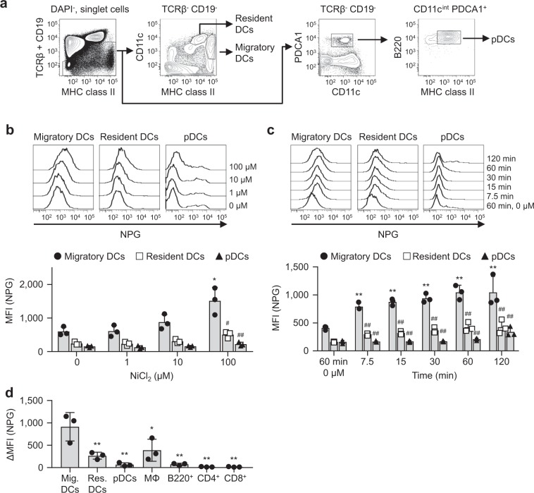
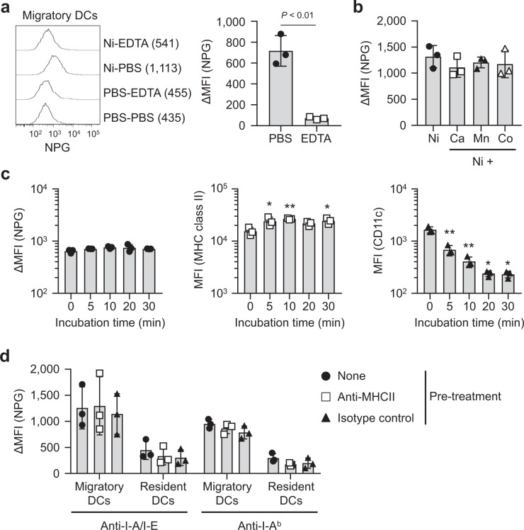
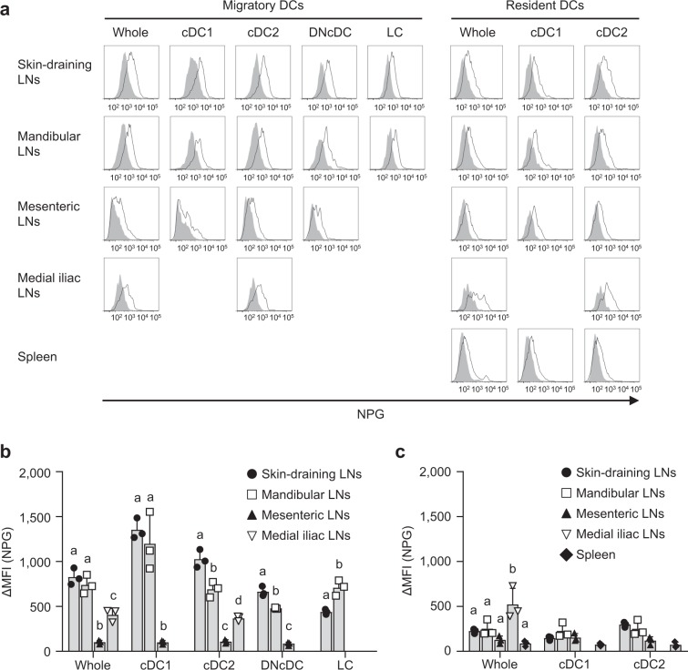
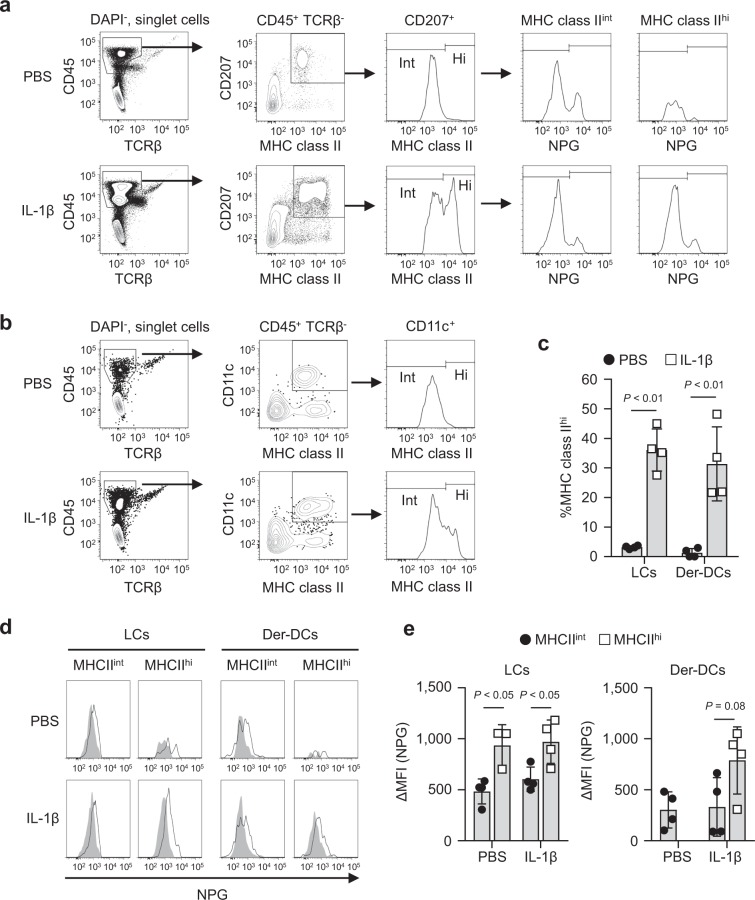
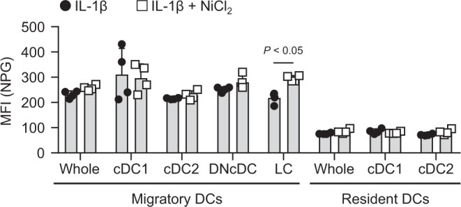
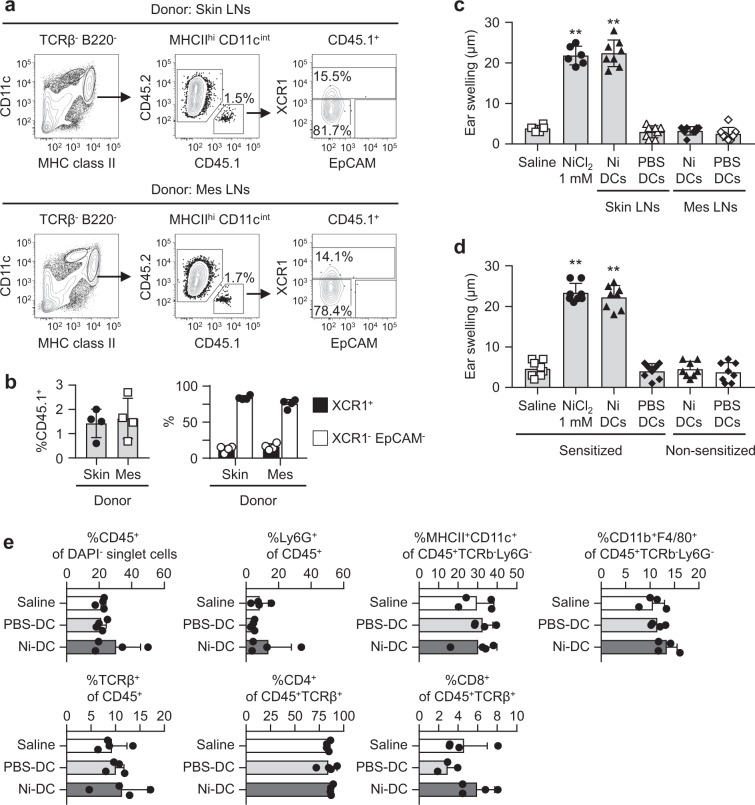
Nickel (Ni) is the most frequent metal allergen and induces Th1-dependent type-IV allergies. In local skin, epidermal Langerhans cells (LCs) and/or dermal dendritic cells (DCs) uptake antigens and migrate to draining lymph nodes (LNs). However, the subsets of antigen-presenting cells that contribute to Ni presentation have not yet been identified. In this study, we analyzed the Ni-binding capabilities of murine DCs using fluorescent metal indicator Newport Green. Elicitation of Ni allergy was assessed after intradermal (i.d.) injection of Ni-treated DCs into ear pinnae of Ni-sensitized mice. The Ni-binding capabilities of MHC class IIhi CD11cint migratory DCs were significantly stronger than those of MHC class IIint CD11chi resident DCs and CD11cint PDCA1+ MHC class IIint B220+ plasmacytoid DCs. Migratory DCs in skin-draining and mandibular LNs showed significantly stronger Ni-binding capabilities than those in mesenteric and medial iliac LNs. An i.d. injection of IL-1β induced the activation of LCs and dermal DCs with strong Ni-binding capabilities. Ni-binding LCs were detected in draining LNs after i.d. challenge with IL-1β and Ni. Moreover, an i.d. injection of Ni-treated DCs purified from skin-draining LNs elicited Ni-allergic inflammation. These results demonstrated that migratory DCs in skin-draining LNs have strong Ni-binding capabilities and elicit Ni allergy.


Conflict of interest statement

The authors declare no competing interests.

Figures

Figure 1
Figure 1
Ni-NPG staining of skin-draining LN cells. (a) The gating strategy used in this analysis. (b) Cells were incubated with the indicated concentration of NiCl2 for 60 min followed by NPG. Histograms represent NPG fluorescence of the indicated DCs. Results are representative of three independent experiments. Graph showing the MFI of NPG. Bars represent the mean ± SD of three independent experiments. Each symbol represents the value from an independent experiment. *P < 0.05, significantly different from the control (0 μM NiCl2). #P < 0.05, ##P < 0.01, significantly different from migratory DCs. (c) Cells were incubated with 100 μM NiCl2 for the indicated times followed by NPG. Histograms represent the NPG fluorescence of the indicated DCs. Results are representative of three independent experiments. Graph showing the MFI of NPG. Bars represent the mean ± SD of three independent experiments. Each symbol represents the value from an independent experiment. **P < 0.01, significantly different from the control (60 min, 0 μM NiCl2). ##P < 0.01, significantly different from migratory DCs. (d) Cells were incubated with 100 μM NiCl2 for 60 min followed by NPG. Graph showing the ΔMFI. Bars represent the mean ± SD of three independent experiments. Each symbol represents the value from an independent experiment. *P < 0.05, **P < 0.01, significantly different from migratory DCs.
Figure 2
Figure 2
Biochemical properties of Ni-binding capabilities. (a) Skin-draining LN cells were incubated with 100 μM of NiCl2 for 60 min followed by 1 mM EDTA for 30 min. Histograms represent the NPG fluorescence of migratory DCs. Numbers shown in parentheses are the MFI of each histogram. Results are representative of three independent experiments. Graph showing ΔMFI. Bars represent the mean ± SD of three independent experiments. Each symbol represents the value from an independent experiment. (b) Skin-draining LN cells were incubated with 100 μM NiCl2 for 60 min with or without divalent cations at 500 μM. Results are shown as ΔMFI. Bars represent the mean ± SD of three independent experiments. Each symbol represents the value from an independent experiment. (c) Skin-draining LN cells were treated with trypsin for the indicated time, then incubated with 100 μM NiCl2 for 60 min followed by NPG. Results are shown as the ΔMFI of NPG, MFI of MHC class II and CD11c. Bars represent the mean ± SD of three independent experiments. Each symbol represents the value from an independent experiment. *P < 0.05, **P < 0.01, significantly different from the control (0 min). (d) Skin-draining LN cells were incubated with anti-MHC class II Abs for 20 min on ice, then incubated with 100 μM NiCl2 for 60 min followed by NPG. Results are shown as the ΔMFI of NPG. Bars represent the mean ± SD of three independent experiments. Each symbol represents the value from an independent experiment.
Figure 3
Figure 3
Ni-NPG staining of DCs from various LNs. (a) Cells were incubated with 100 μM NiCl2 for 60 min followed by NPG. Histograms represent the NPG fluorescence of cells incubated with (black line) or without (gray-shaded) NiCl2. Results are representative of three independent experiments. (b and c) Ni-NPG staining of migratory (b) and resident (c) DCs was analyzed. Results are shown as ΔMFI. Bars represent the mean ± SD of three independent experiments. Each symbol represents the value from an independent experiment. Means without a common letter were significantly different, P < 0.05.
Figure 4
Figure 4
Ni-NPG staining of ear cells i.d. injected with IL-1β. Epidermal and dermal cells were prepared 24 h after injection with 50 ng of IL-1β or PBS. Cells were incubated with 100 μM NiCl2 for 60 min followed by NPG. (a and b) The gating strategies for epidermal (a) and dermal (b) cells are shown. (c) Percentage of MHC class IIhi cells in epidermal LCs and dermal DCs. Results represent the mean ± SD of four independent experiments. Each symbol represents the value from an independent experiment. (d) Histograms represent NPG fluorescence incubated with (black line) or without (gray-shaded) NiCl2. Results are representative of four independent experiments. (e) ΔMFI of epidermal LCs and dermal DCs. Results represent the mean ± SD of four independent experiments. Each symbol represents the value from independent experiment.
Figure 5
Figure 5
NPG staining of DCs in draining LNs after i.d. challenge with IL-1β and Ni. Ear pinnae of naïve mice were i.d. challenged 5 times every 24 h with 50 ng of IL-1β in the presence (left pinna) or absence (right pinna) of 10 mM NiCl2. Twenty-four hours after the last challenge, auricular LNs cells were stained with NPG followed by antibodies. Graph showing the MFI of NPG. Bars represent the mean ± SD of 4 pinnae from two independent experiments. Each symbol represents the value from each pinna.
Figure 6
Figure 6
Induction of IL-1β in pan-DCs stimulated with Ni. (a) The pan-DC fraction from skin-draining or mesenteric LNs was incubated with LPS or NiCl2 for 4 h. The mRNA expression levels of IL-1β were analyzed by quantitative RT-PCR. Results represent the mean ± SD of four independent experiments. Each symbol represents the value from an independent experiment. *P < 0.05 significantly different from the medium. (b) Pan-DCs were incubated with LPS or NiCl2 for 4 h. After washing, cells were incubated further in the absence of any stimulation for 20 h. Intracellular pro-IL-1β was analyzed by flow cytometry. Representative results are shown. (c) The percentage of pro-IL-1β+ in MHC class II+ CD11c+ cells is shown. Bars represent the mean ± SD of three independent experiments. Each symbol represents the value from an independent experiment. *P < 0.05, **P < 0.01, significantly different from the medium.
Figure 7
Figure 7
Elicitation of Ni-allergic inflammation by Ni-DCs. The pan-DC fraction was prepared from skin-draining or mesenteric LNs from CD45.1+ mice inoculated with B16-Flt3L. (a) A flow cytometric analysis of auricular LN cells from CD45.2+ recipient mice 24 h after i.d. injection of CD45.1+ pan-DCs (2 × 105 cells) into the ear pinnae. Dot plots are representative of four mice. (b) Graphs showing %CD45.1+ in MHC class IIhi CD11cint cells (left), %XCR1+, and %XCR1- EpCAM- in CD45.1+ cells (right). Bars represent the mean ± SD of four mice. Each symbol represents the value from each mouse. (c) The ear pinnae of Ni-sensitized mice were i.d. challenged with NiCl2 or DCs (1 × 104 cells) prepared from skin-draining or mesenteric LNs. (d) The ear pinnae of Ni-sensitized and non-sensitized mice were i.d. challenged with NiCl2 or DCs (1 × 104 cells) prepared from skin-draining LNs. Bars show ear swelling 48 h after the challenge. Results represent the mean ± SD of 6–8 pinnae (3–4 mice). Each symbol represents the value from each pinna. **P < 0.01, significantly different from the control (saline). (e) Ear pinnae of Ni-sensitized mice were i.d. challenged with NiCl2 or DCs (1 × 104 cells) prepared from skin-draining LNs. Inflammatory cell recruitments into local ear tissues were analyzed 48 h after the challenge by flow cytometry. Bars represent the mean ± SD of 3–4 independent experiments. Each symbol represents the value from an independent experiment.

References

    1. Kashem SW, Haniffa M, Kaplan DH. Antigen-Presenting Cells in the Skin. Annual review of immunology. 2017;35:469–499. doi: 10.1146/annurev-immunol-051116-052215. - DOI - PubMed
    1. Malissen B, Tamoutounour S, Henri S. The origins and functions of dendritic cells and macrophages in the skin. Nature reviews. Immunology. 2014;14:417–428. doi: 10.1038/nri3683. - DOI - PubMed
    1. Igyarto BZ, et al. Skin-resident murine dendritic cell subsets promote distinct and opposing antigen-specific T helper cell responses. Immunity. 2011;35:260–272. doi: 10.1016/j.immuni.2011.06.005. - DOI - PMC - PubMed
    1. Bedoui S, et al. Cross-presentation of viral and self antigens by skin-derived CD103+ dendritic cells. Nature immunology. 2009;10:488–495. doi: 10.1038/ni.1724. - DOI - PubMed
    1. Gao Y, et al. Control of T helper 2 responses by transcription factor IRF4-dependent dendritic cells. Immunity. 2013;39:722–732. doi: 10.1016/j.immuni.2013.08.028. - DOI - PMC - PubMed

Publication types