Review
. 2020 May;140(5):623-638.
doi: 10.1007/s00402-020-03373-y. Epub 2020 Mar 19.

Arthroscopic assisted treatment of distal radius fractures and concomitant injuries

Affiliations
Review

Arthroscopic assisted treatment of distal radius fractures and concomitant injuries

Tobias Kastenberger et al. Arch Orthop Trauma Surg. 2020 May.

Abstract

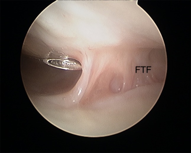
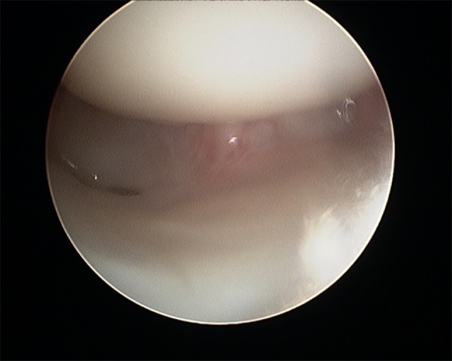
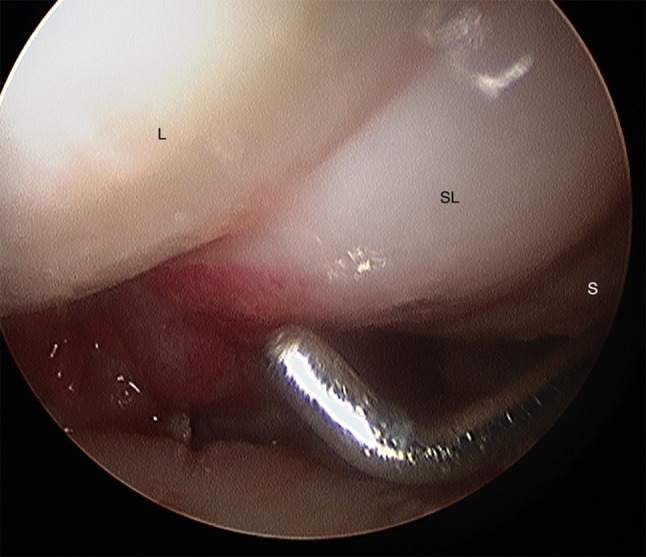
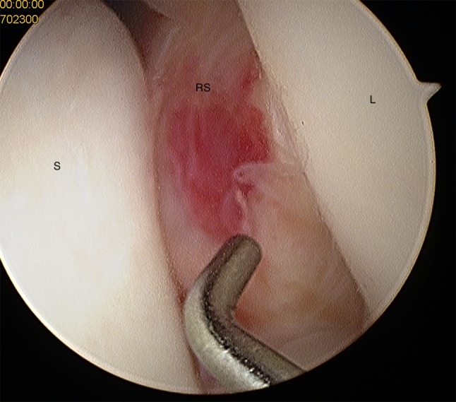
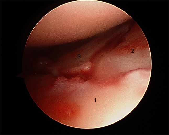
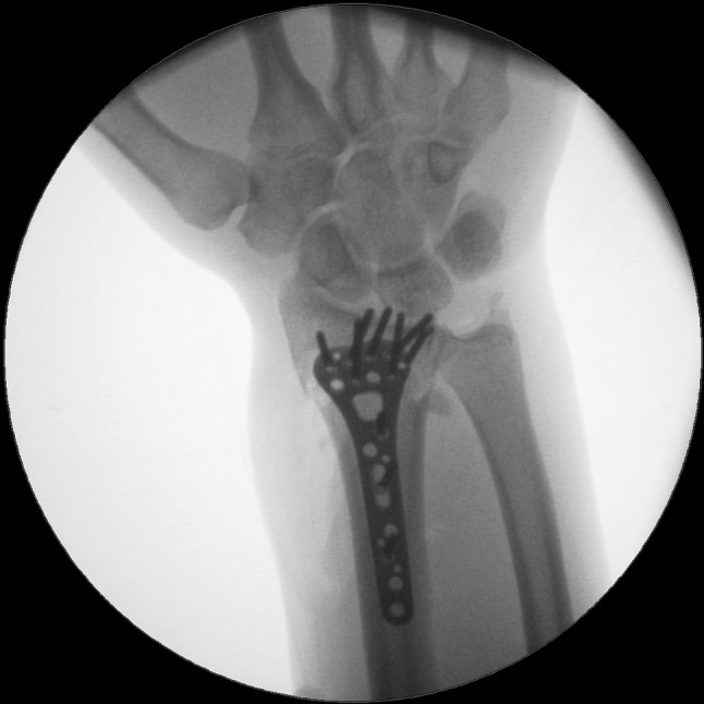
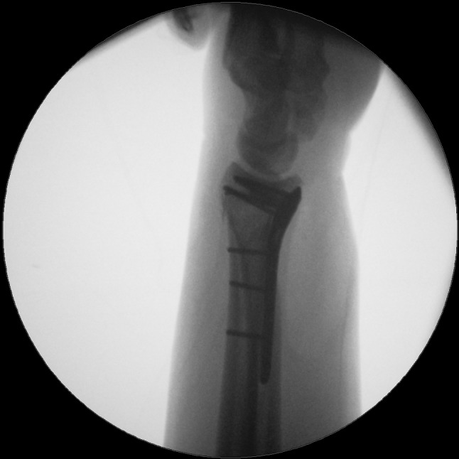
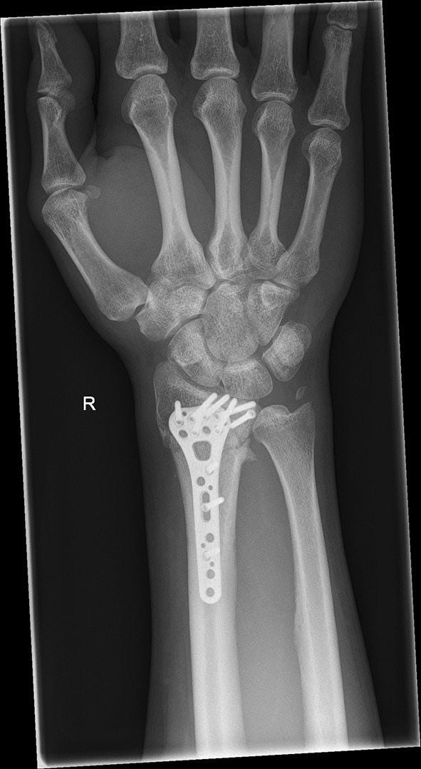
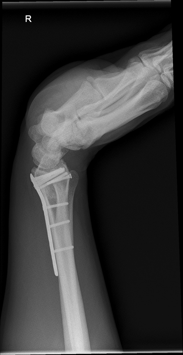
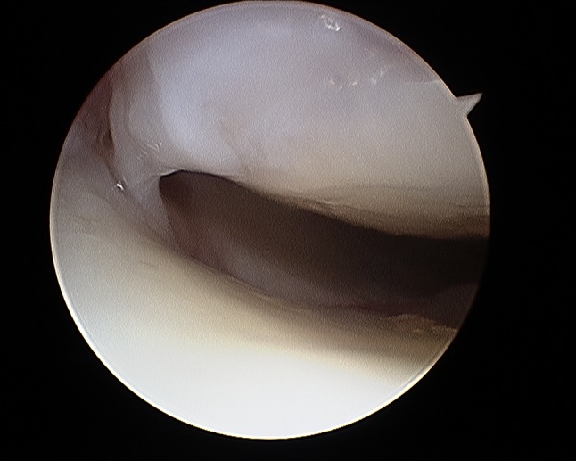
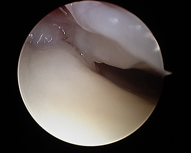
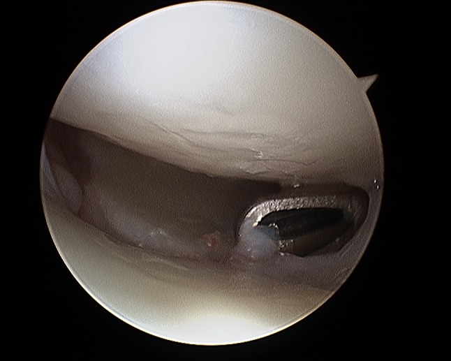
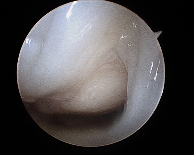
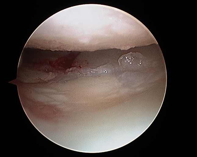
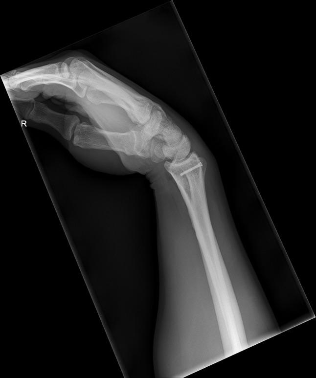
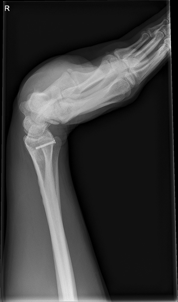
Wrist arthroscopy is mainly used to assist fracture reduction and fixation and to diagnose and treat concomitant injuries mainly to the scapholunate (SL), lunotriquetral (LT) ligament and the triangular fibrocartilage complex (TFCC). Arthroscopy is beneficial in improving anatomical reduction of fracture steps and gaps in intra-articular distal radius fractures (DRFs). Yet, the literature that the functional outcome correlates with the use of arthroscopy, is limited. Non-surgical treatment and immobilization is recommended for Geissler grade I-III Sl-ligament injuries, while open reduction, ligament suture and/or K-wire pinning is mandatory for complete ligament tears according to Geissler grade IV. This manuscript describes the current literature and gives insight into the authors' opinions and practice.

Keywords: Arthroscopic treatment; Concomitant injuries; Distal radius fracture; Injury; Intrinsic ligament.


Conflict of interest statement

No author has a conflict of interest that relates to the content discussed in this manuscript. None of the authors’ professional or financial affiliations that may be perceived as having biased the manuscript. Each author certifies that he or she, or a member of their immediate family, has no commercial associations (i.e., consultancies, stock ownership, equity interest, patent/licensing arrangements, etc.) that might pose a conflict of interest in connection with the submitted article.

Figures

Fig. 1
Fig. 1
Patient lying in a supine position with the upper extremity suspended by the use of finger traps
Fig. 2
Fig. 2
Four kg traction weight applied on the wrist
Fig. 3
Fig. 3
Standard arthroscopy portals 3/4, and 6R with the camera in the 3/4 portal
Fig. 4
Fig. 4
Radiocarpal view a before fracture reduction and b after fracture reduction
Fig. 5
Fig. 5
Intra-articular fibrous tissue formation (FTF)
Fig. 6
Fig. 6
Intra-articular view after debridement of FTF
Fig. 7
Fig. 7
Radiocarpal view: balloon sized scapholunate ligament Geissler grade 1; L lunate, SL scapholunate ligament, S scaphoid
Fig. 8
Fig. 8
Midcarpal view: radius joint surface can be seen with the hook between scaphoid and lunate indicating a scapholunate ligament tear Geissler grade 4; S scaphoid, RS radial joint surface, L lunate
Fig. 9
Fig. 9
Midcarpal view: widening of the lunotriquetral joint with the hook between the triquetrum and lunate indicating a lunotriquetral ligament tear; T triquetrum, L lunate, H hamate
Fig. 10
Fig. 10
Dorsovolar view: initial assessment of the injured wrist with a loss of the radial inclination and radial length
Fig. 11
Fig. 11
Lateral view: initial assessment showing the loss of radio-ulnar and dorso-palmar inclination
Fig. 12
Fig. 12
CT scan of a dorsovolar view after reduction and cast immobilization showing the defect zone of the intermediated column and fracture involvement of the DRUJ
Fig. 13
Fig. 13
CT scan of a lateral view of the intermediate column showing the volar fragment of the intermediate column, a fracture gap and step of more than 2 mm and the volar fragment additionally toppled
Fig. 14
Fig. 14
CT scan of a lateral view of the ulnar column showing the additional ulnar fragment flipped 90°
Fig. 15
Fig. 15
CT scan of an axial view with the numbered intraarticular fragments 1: radial styloid, 2: dorso-ulnar fragment, 3: fragment of the intermediate column 4: volar fragment
Fig. 16
Fig. 16
Initial view of the displaced fracture fragments after inserting the arthroscope through the 3/4 portal
Fig. 17
Fig. 17
Arthroscopic view after debridement using the wrist shaver; all fragments are visualized. 1: radial fragment, 2: volar fragment, 3: intermediate fragment, 4: dorsal fragment
Fig. 18
Fig. 18
Arthroscopic view after arthroscopical fracture reduction showing an anatomical alignment of the involved fragments. 1: radial fragment, 2: volar fragment, 3: intermediate fragment
Fig. 19
Fig. 19
Dorsovolar fluoroscopic view at the end of the surgery showing the main screw fixation directed to the ulnar corner
Fig. 20
Fig. 20
Lateral fluoroscopic view showing the screws in a subchondral position
Fig. 21
Fig. 21
Dorsovolar and lateral x-ray control four weeks after surgery
Fig. 22
Fig. 22
Dorsovolar and lateral X-ray control 4 weeks after surgery
Fig. 23
Fig. 23
Dorsovolar and lateral X-ray control 1 year after surgery
Fig. 24
Fig. 24
Dorsovolar and lateral X-ray control 1 year after surgery
Fig. 25
Fig. 25
Lateral view in a flexed and extended position of the wrist before implant removal
Fig. 26
Fig. 26
Lateral view in a flexed and extended position of the wrist before implant removal
Fig. 27
Fig. 27
Arthroscopic view of the palmar side identifying palmar scar formations
Fig. 28
Fig. 28
Arthroscopic view of the palmar side identifying palmar scar formations
Fig. 29
Fig. 29
Arthroscopic view of the dorsal side identifying palmar scar formations
Fig. 30
Fig. 30
Arthroscopic view of the dorsal side identifying palmar scar formations
Fig. 31
Fig. 31
Arthroscopic view after debridement showing a gap at the volar side
Fig. 32
Fig. 32
Lateral view in a flexed and extended position of the wrist after implant removal and arthroscopic debridement showing an improved range of motion
Fig. 33
Fig. 33
Lateral view in a flexed and extended position of the wrist after implant removal and arthroscopic debridement showing an improved range of motion
Fig. 34
Fig. 34
Clinical photographs in wrist extension and flexion after implant removal
Fig. 35
Fig. 35
Clinical photographs in wrist extension and flexion after implant removal

References

    1. Koval KJ, Harrast JJ, Anglen JO, Weinstein JN. Fractures of the distal part of the radius. The evolution of practice over time. Where's the evidence? J Bone Jt Surg Am. 2008;90(9):1855–1861. - PubMed
    1. Atesok K, Doral MN, Whipple T, Mann G, Mei-Dan O, Atay OA, et al. Arthroscopy-assisted fracture fixation. Knee Surg Sports Traumatol Arthrosc. 2011;19(2):320–329. - PubMed
    1. Dei Giudici L, Di Muzio F, Bottegoni C, Chillemi C, Gigante A. The role of arthroscopy in articular fracture management: the lower limb. Eur J Orthop Surg Traumatol. 2015;25(5):807–813. - PubMed
    1. Smeraglia F, Del Buono A, Maffulli N. Wrist arthroscopy in the management of articular distal radius fractures. Br Med Bull. 2016;119(1):157–165. - PubMed
    1. Ardouin L, Durand A, Gay A, Leroy M. Why do we use arthroscopy for distal radius fractures? Eur J Orthop Surg Traumatol. 2018;28(8):1505–1514. - PubMed

MeSH terms