

Radiomics and Deep Learning: Hepatic Applications
- PMID: 32193887
- PMCID: PMC7082656
- DOI: 10.3348/kjr.2019.0752
Radiomics and Deep Learning: Hepatic Applications
Abstract
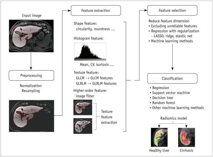
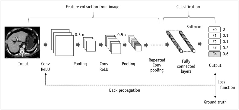
Radiomics and deep learning have recently gained attention in the imaging assessment of various liver diseases. Recent research has demonstrated the potential utility of radiomics and deep learning in staging liver fibroses, detecting portal hypertension, characterizing focal hepatic lesions, prognosticating malignant hepatic tumors, and segmenting the liver and liver tumors. In this review, we outline the basic technical aspects of radiomics and deep learning and summarize recent investigations of the application of these techniques in liver disease.
Keywords: Artificial intelligence; Computer-assisted; Deep learning; Liver; Radiomics.
Copyright © 2020 The Korean Society of Radiology.
Conflict of interest statement
The authors have no potential conflicts of interest to disclose.
Figures
Similar articles
-
Clinical application of deep learning and radiomics in hepatic disease imaging: a systematic scoping review.Br J Radiol. 2022 Aug 1;95(1136):20211136. doi: 10.1259/bjr.20211136. Epub 2022 Jul 14. Br J Radiol. 2022. PMID: 35816550 Free PMC article.
-
Radiomics and deep learning in liver diseases.J Gastroenterol Hepatol. 2021 Mar;36(3):561-568. doi: 10.1111/jgh.15414. J Gastroenterol Hepatol. 2021. PMID: 33709608
-
Conventional and artificial intelligence-based imaging for biomarker discovery in chronic liver disease.Hepatol Int. 2022 Jun;16(3):509-522. doi: 10.1007/s12072-022-10303-0. Epub 2022 Feb 9. Hepatol Int. 2022. PMID: 35138551 Free PMC article. Review.
-
Comparing radiomics models with different inputs for accurate diagnosis of significant fibrosis in chronic liver disease.Eur Radiol. 2021 Nov;31(11):8743-8754. doi: 10.1007/s00330-021-07934-6. Epub 2021 Apr 21. Eur Radiol. 2021. PMID: 33881568
-
Development and validation of a radiomics signature for clinically significant portal hypertension in cirrhosis (CHESS1701): a prospective multicenter study.EBioMedicine. 2018 Oct;36:151-158. doi: 10.1016/j.ebiom.2018.09.023. Epub 2018 Sep 27. EBioMedicine. 2018. PMID: 30268833 Free PMC article.
Cited by
-
Deep Segmentation Feature-Based Radiomics Improves Recurrence Prediction of Hepatocellular Carcinoma.BME Front. 2022 Apr 4;2022:9793716. doi: 10.34133/2022/9793716. eCollection 2022. BME Front. 2022. PMID: 37850181 Free PMC article.
-
Prediction of Ki-67 Expression in HIV-Associated Lung Adenocarcinoma Patients Using Multiple Machine Learning Models Based on CT Imaging Radiomics.Cancer Manag Res. 2025 Apr 25;17:881-892. doi: 10.2147/CMAR.S505390. eCollection 2025. Cancer Manag Res. 2025. PMID: 40303969 Free PMC article.
-
Methodological quality of machine learning-based quantitative imaging analysis studies in esophageal cancer: a systematic review of clinical outcome prediction after concurrent chemoradiotherapy.Eur J Nucl Med Mol Imaging. 2022 Jul;49(8):2462-2481. doi: 10.1007/s00259-021-05658-9. Epub 2021 Dec 23. Eur J Nucl Med Mol Imaging. 2022. PMID: 34939174 Free PMC article.
-
A Clinical-Radiomic Model for Predicting Indocyanine Green Retention Rate at 15 Min in Patients With Hepatocellular Carcinoma.Front Surg. 2022 Mar 24;9:857838. doi: 10.3389/fsurg.2022.857838. eCollection 2022. Front Surg. 2022. PMID: 35402498 Free PMC article.
-
Recent Advances in the Pathogenesis and Clinical Evaluation of Portal Hypertension in Chronic Liver Disease.Gut Liver. 2024 Jan 15;18(1):27-39. doi: 10.5009/gnl230072. Epub 2023 Oct 16. Gut Liver. 2024. PMID: 37842727 Free PMC article. Review.
References
-
- Lee G, Lee HY, Park H, Schiebler ML, van Beek EJR, Ohno Y, et al. Radiomics and its emerging role in lung cancer research, imaging biomarkers and clinical management: state of the art. Eur J Radiol. 2017;86:297–307. - PubMed
-
- Legland D, Kiêu K, Devaux MF. Computation of Minkowski measures on 2D and 3D binary images. Image Anal Stereol. 2007;26:83–92.
-
- Zwanenburg A, Leger S, Vallières M, Löck S. Image biomarker standardisation initiative [updated May 2019]. arXiv:1612.07003 [cs.CV] 2016. [Accessed August 31, 2019]. Available at: https://arxiv.org/abs/1612.07003v9.
Publication types
MeSH terms
LinkOut - more resources
Full Text Sources
Medical
