

Characterization of tissue and functional deficits in a clinically translational pig model of acute ischemic stroke
- PMID: 32194080
- PMCID: PMC10671789
- DOI: 10.1016/j.brainres.2020.146778
Characterization of tissue and functional deficits in a clinically translational pig model of acute ischemic stroke
Abstract
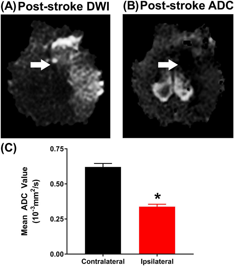
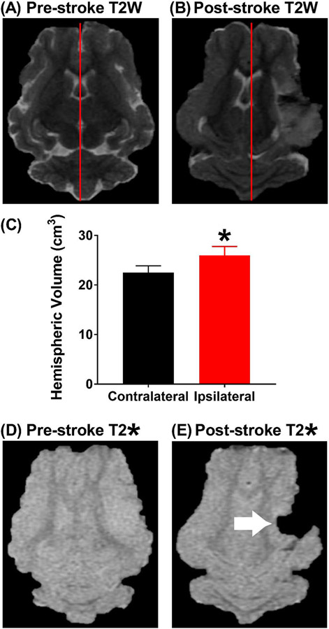
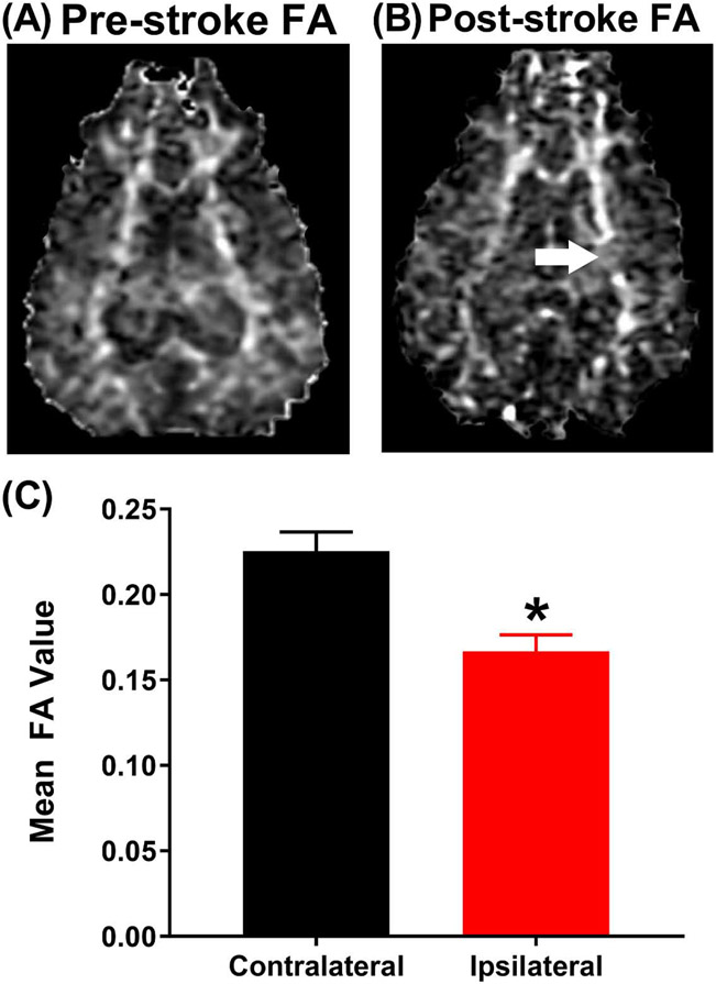
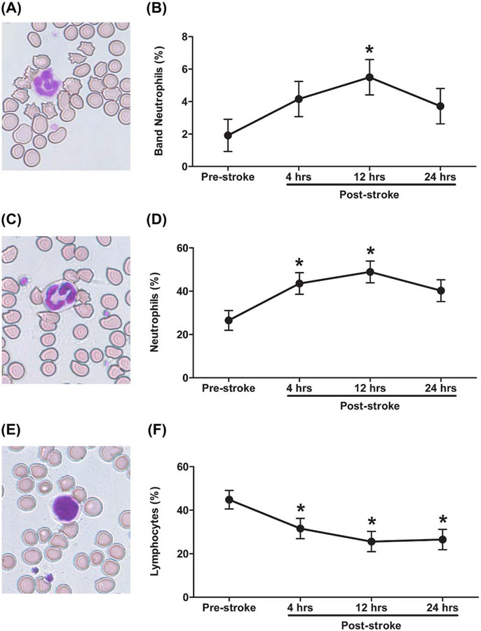
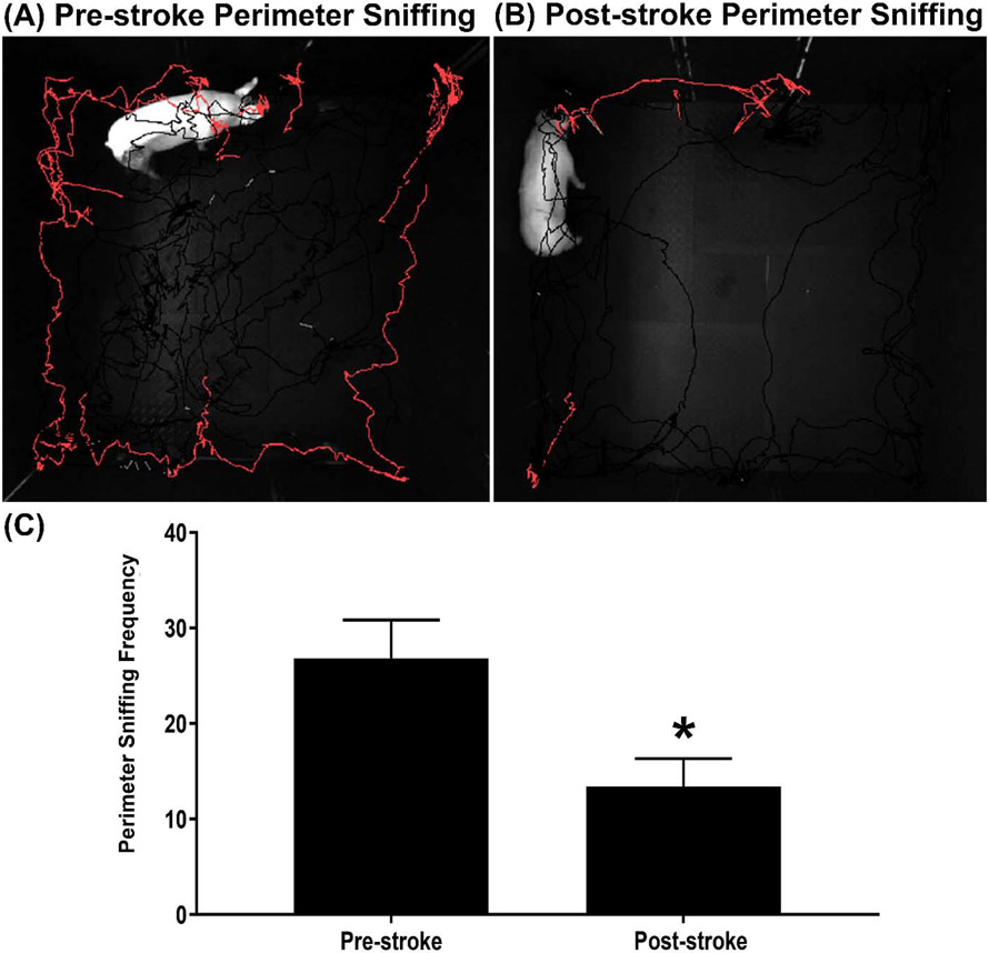
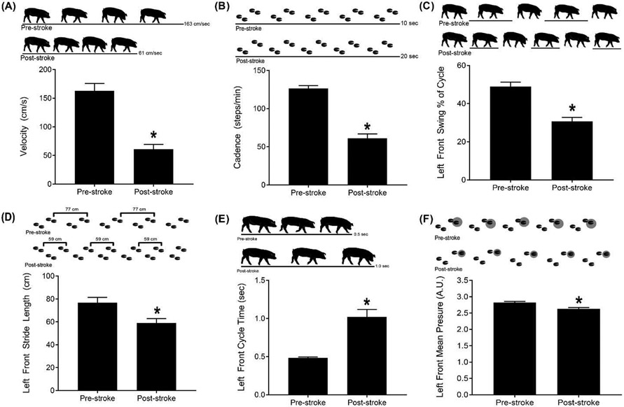
The acute stroke phase is a critical time frame used to evaluate stroke severity, therapeutic options, and prognosis while also serving as a major tool for the development of diagnostics. To further understand stroke pathophysiology and to enhance the development of treatments, our group developed a translational pig ischemic stroke model. In this study, the evolution of acute ischemic tissue damage, immune responses, and functional deficits were further characterized. Stroke was induced by middle cerebral artery occlusion in Landrace pigs. At 24 h post-stroke, magnetic resonance imaging revealed a decrease in ipsilateral diffusivity, an increase in hemispheric swelling resulting in notable midline shift, and intracerebral hemorrhage. Stroke negatively impacted white matter integrity with decreased fractional anisotropy values in the internal capsule. Like patients, pigs showed a reduction in circulating lymphocytes and a surge in neutrophils and band cells. Functional responses corresponded with structural changes through reductions in open field exploration and impairments in spatiotemporal gait parameters. Characterization of acute ischemic stroke in pigs provided important insights into tissue and functional-level assessments that could be used to identify potential biomarkers and improve preclinical testing of novel therapeutics.
Keywords: Acute stroke; Brain ischemia; Gait analysis; Magnetic resonance imaging; Porcine.
Copyright © 2020 The Authors. Published by Elsevier B.V. All rights reserved.
Conflict of interest statement
Declaration of Competing Interest The authors declare that they have no known competing financial interests or personal relationships that could have appeared to influence the work reported in this paper.
Figures
Similar articles
-
Exploring the predictive value of lesion topology on motor function outcomes in a porcine ischemic stroke model.Sci Rep. 2021 Feb 15;11(1):3814. doi: 10.1038/s41598-021-83432-5. Sci Rep. 2021. PMID: 33589720 Free PMC article.
-
Human Neural Stem Cell Extracellular Vesicles Improve Recovery in a Porcine Model of Ischemic Stroke.Stroke. 2018 May;49(5):1248-1256. doi: 10.1161/STROKEAHA.117.020353. Epub 2018 Apr 12. Stroke. 2018. PMID: 29650593 Free PMC article.
-
Intracisternal administration of tanshinone IIA-loaded nanoparticles leads to reduced tissue injury and functional deficits in a porcine model of ischemic stroke.IBRO Neurosci Rep. 2021 Jan 5;10:18-30. doi: 10.1016/j.ibneur.2020.11.003. eCollection 2021 Jun. IBRO Neurosci Rep. 2021. PMID: 33842909 Free PMC article.
-
The role of diffusion tensor imaging in the evaluation of ischemic brain injury - a review.NMR Biomed. 2002 Nov-Dec;15(7-8):561-9. doi: 10.1002/nbm.786. NMR Biomed. 2002. PMID: 12489102 Review.
-
Recent Advances in Leukoaraiosis: White Matter Structural Integrity and Functional Outcomes after Acute Ischemic Stroke.Curr Cardiol Rep. 2016 Dec;18(12):123. doi: 10.1007/s11886-016-0803-0. Curr Cardiol Rep. 2016. PMID: 27796861 Review.
Cited by
-
Inflammatory Pathogenesis of Post-stroke Depression.Aging Dis. 2024 Feb 9;16(1):209-38. doi: 10.14336/AD.2024.0203. Online ahead of print. Aging Dis. 2024. PMID: 38377025 Free PMC article. Review.
-
Exploring the predictive value of lesion topology on motor function outcomes in a porcine ischemic stroke model.Sci Rep. 2021 Feb 15;11(1):3814. doi: 10.1038/s41598-021-83432-5. Sci Rep. 2021. PMID: 33589720 Free PMC article.
-
Neurological scoring and gait kinematics to assess functional outcome in an ovine model of ischaemic stroke.Front Neurol. 2023 Feb 20;14:1071794. doi: 10.3389/fneur.2023.1071794. eCollection 2023. Front Neurol. 2023. PMID: 36891474 Free PMC article.
-
A Swine Hind Limb Ischemia Model Useful for Testing Peripheral Artery Disease Therapeutics.J Cardiovasc Transl Res. 2021 Dec;14(6):1186-1197. doi: 10.1007/s12265-021-10134-8. Epub 2021 May 28. J Cardiovasc Transl Res. 2021. PMID: 34050499 Free PMC article.
-
Magnetic Resonance Imaging and Gait Analysis Indicate Similar Outcomes Between Yucatan and Landrace Porcine Ischemic Stroke Models.Front Neurol. 2021 Jan 21;11:594954. doi: 10.3389/fneur.2020.594954. eCollection 2020. Front Neurol. 2021. PMID: 33551956 Free PMC article.
References
Publication types
MeSH terms
Grants and funding
LinkOut - more resources
Full Text Sources
Medical
