. 2020 May:108:347-357.
doi: 10.1016/j.actbio.2020.03.017. Epub 2020 Mar 17.

Tumor necrosis factor primes and metal particles activate the NLRP3 inflammasome in human primary macrophages

Affiliations

Tumor necrosis factor primes and metal particles activate the NLRP3 inflammasome in human primary macrophages

Eemeli Jämsen et al. Acta Biomater. 2020 May.

Abstract

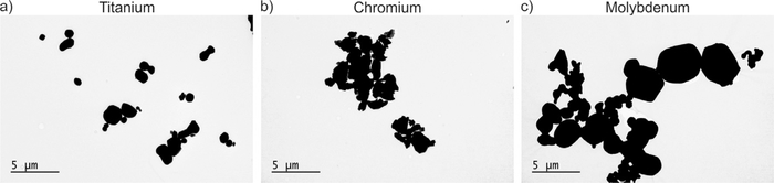
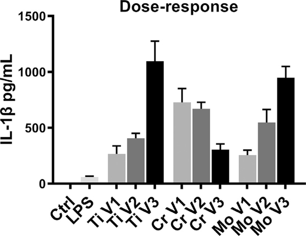
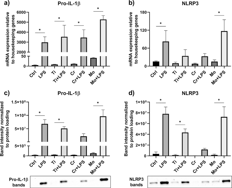
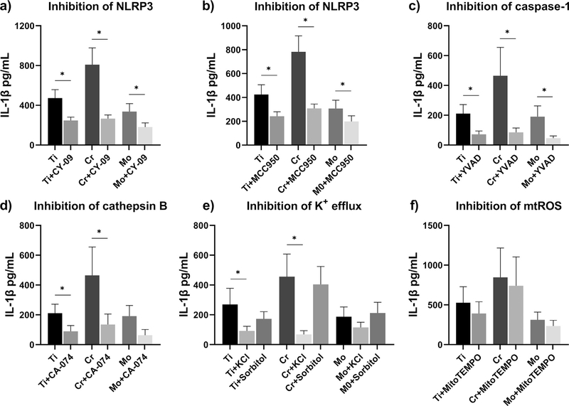
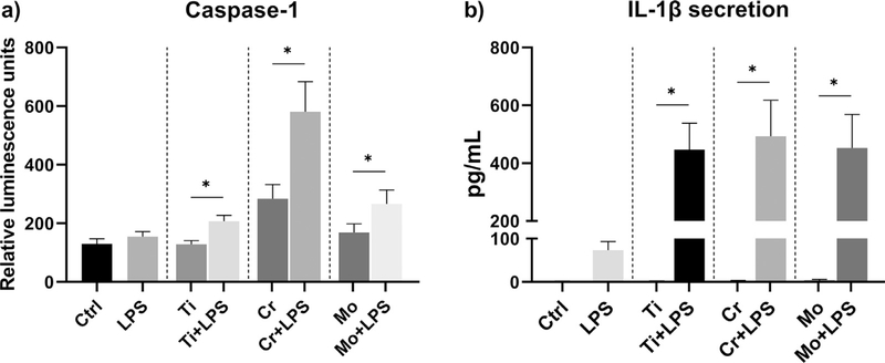
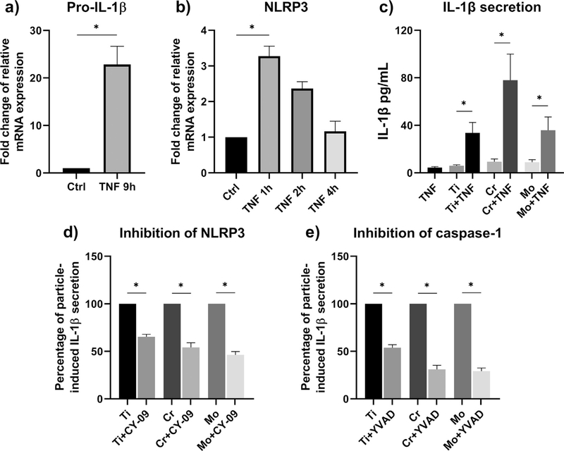
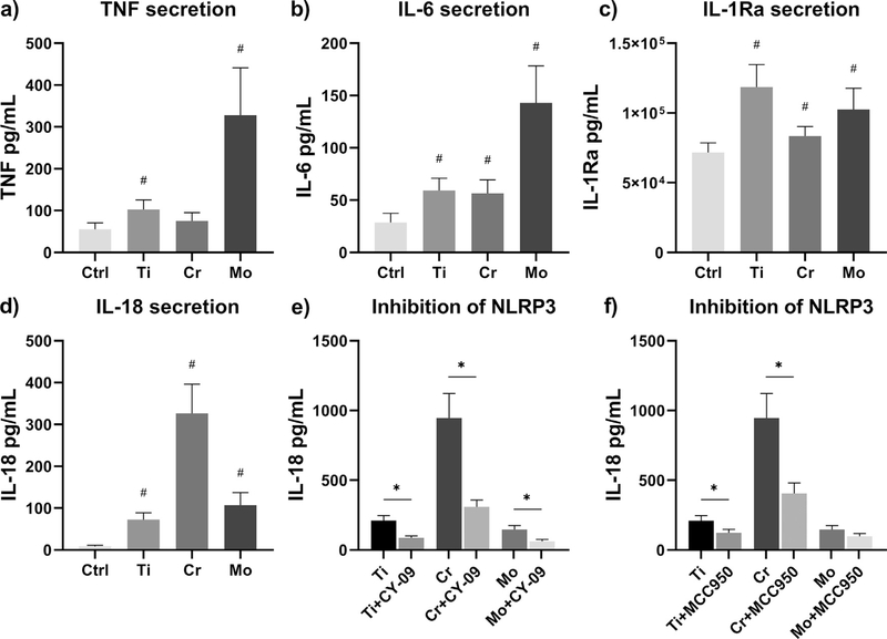
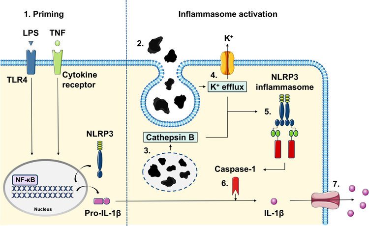
Aseptic loosening of total joint replacements is driven by a macrophage-mediated inflammatory reaction to implant-derived wear particles. Phagocytosis of implant debris has been suggested to activate the NLRP3 inflammasome leading to secretion of interleukin (IL)-1β. However, factors and molecular mechanisms driving the particle-induced inflammasome activation are yet to be fully elucidated. In this study, we investigated the inflammasome response of human primary macrophages to titanium, chromium, and molybdenum particles in vitro. We observed that particles alone were not sufficient to induce IL-1β secretion, but an additional priming signal-such as bacterial lipopolysaccharide (LPS)-was required to license the inflammasome activation. By using specific inhibitors against the inflammasome signaling pathway, we demonstrate that the particle-induced IL-1β secretion depended upon activation of the NLRP3 inflammasome. We further hypothesized that tumor necrosis factor (TNF) could substitute for LPS as a priming signal, and found that particle stimulation together with preceding TNF treatment resulted in inflammasome-dependent IL-1β production as well. Our results show that the NLRP3 inflammasome mediates wear particle responses in human primary macrophages, and its activation does not necessarily require the presence of bacterial components, but can be induced under aseptic conditions by TNF priming. STATEMENT OF SIGNIFICANCE: This study was conducted to elucidate the molecular mechanisms of metal particle-induced IL-1β secretion in human primary macrophages. Production of this pro-inflammatory mediator from wear particle-activated macrophages has been associated with increased bone loss around total joint replacements-a condition eventually requiring revision surgery. Our results confirm that together with a co-stimulatory priming signal, particles of common implant metals elicit macrophage-mediated IL-1β secretion through activation of the NLRP3 inflammasome pathway. We also present a concept of TNF priming in this context, demonstrating that the particle-related IL-1β secretion can take place in a truly sterile environment. Thus, inhibition of inflammasome signaling appears a means to prevent wear particle-induced inflammation and development of peri‑prosthetic osteolysis.

Keywords: IL-1β; Inflammasome; Macrophage; TNF; Wear particle.


Conflict of interest statement

Declaration of Competing Interest The authors declare that they have no known competing financial interests or personal relationships that could have appeared to influence the work reported in this paper.

Figures

Fig. 1.
Fig. 1.
Transmission electron microscopy (TEM) of metal particles. The appearance and size-distribution of (a) Ti, (b) Cr, and (c) Mo particles were assessed by TEM imaging. Scale bars 5 μm.
Fig. 2.
Fig. 2.
Dose-response of particle-challenged macrophages. Human primary macrophages (n = 6) were primed with LPS and stimulated for eight hours with increasing concentrations of Ti, Cr, and Mo particles. IL-1 β secretion induced by particle doses of (V1) 0.1, (V2) 0.25, and (V3) 0.5 mm3/well was determined from culture supernatants by ELISA.
Fig. 3.
Fig. 3.
The expression of pro-IL-1 β and NLRP3. Priming effects of LPS and particles alone on macrophages (n = 4) were analyzed by qRT-PCR and Western blotting. The relative mRNA expression of (a) pro-IL-1 β and (b) NLRP3, and the production of intracellular (c) pro-IL-1 β and (d) NLRP3 proteins were determined after an eight-hour stimulation with Ti, Cr, and Mo particles. Representative Western blot images are included with * illustrating statistically significant difference between indicated conditions (p < 0.05).
Fig. 4.
Fig. 4.
Inhibition of NLRP3 inflammasome activation. LPS-primed macrophages were treated with inhibitors of NLRP3 inflammasome pathway, and challenged with Ti, Cr, and Mo particles for eight hours. Inhibition of inflammasome components (a-b) NLRP3 and (c) caspase-1, and upstream inflammasome activating signals (d) cathepsin B, (e) K+ efflux, and (f) mtROS were assessed by studying the IL-1 β secretion from culture media with ELISA. A control group of sorbitol-treated macrophages was included to take into account the effect of increased osmolarity. In graphs (b) and (e) n = 5, whereas other experiments were performed with n = 4. * indicates statistical significance between the conditions (p < 0.05).
Fig. 5.
Fig. 5.
Activation of caspase-1. Human primary macrophages (n = 5) were primed with LPS and challenged with Ti, Cr, and Mo particles for eight hours. (a) The release of active caspase-1 was measured from culture supernatants using a luminescence assay, and (b) the corresponding IL-1 β secretion was determined by ELISA. * represents statistical significance between indicated conditions (p < 0.05).
Fig. 6.
Fig. 6.
The effect of TNF priming. Human primary macrophages were cultured in the presence of TNF up to nine hours, after which the relative mRNA expression of (a) pro-IL-1 β (n = 3) and (b) NLRP3 (n = 4) was assessed by qRT-PCR analysis. (c) TNF-primed macrophages (n = 5) were exposed to an eight-hour stimulation with Ti, Cr, and Mo particles, and the IL-1 β secretion was assessed by ELISA. (d-e) Particle-induced IL-1 β secretion from TNF-primed cells (n = 4) was further modified by inhibition of NLRP3 and caspase-1. Data in graphs (a) and (b) are presented as fold change compared to untreated control, whereas results from experiments (d) and (e) are shownas percentage of particle-induced IL-1 β secretion. * indicates statistically significant differences between groups (p < 0.05).
Fig. 7.
Fig. 7.
Particle-induced secretion of inflammatory cytokines. Unprimed human primary macrophages were challenged with Ti, Cr, and Mo particles for eight hours, and the secretion of (a) TNF, (b) IL-6, (c) IL-1Ra, and (d) IL-18 was analyzed from culture supernatants using ELISA assays. LPS-primed macrophages were treated with NLRP3 inhibitors and exposed to an eight-hour particle stimulation as well; the inhibitory effects of (e) CY-09 and (f) MCC950 on particle-induced IL-18 secretion were assessedby ELISA. In graphs (a-c) n = 8, whereas experiments illustrated in graphs (d-f) were performed with n = 4. # illustrates statistical significance for particle stimulation, and* represents significant difference between indicated conditions (p < 0.05).
Fig. 8.
Fig. 8.
Schematic view of the wear particle-induced inflammasome activation. 1. An NF-ĸB activating priming signal such as LPS or TNF is required to synthesize NLRP3 and pro-IL-1 β thus licensing macrophages for the NLRP3 inflammasome activation. 2. Recognition of foreign body particulate material results in particle phagocytosis and formation of phagolysosomes. Subsequent intracellular aberrations including 3. leakage of lysosomal protease cathepsin B and 4. outflow of potassium (K+) are sensed by the NLRP3 receptor. 5. Upon activation, NLRP3 triggers the assembly of the inflammasome complex by recruiting specific adaptor proteins. 6. NLRP3 inflammasome eventually generates active caspase-1, which cleaves precursor protein pro-IL-1 β into the secreted form. 7. Mature IL-1 β is released from the cell through membrane pores formed after the inflammasome activation.

References

    1. Karachalios T, Komnos G, Koutalos A, Total hip arthroplasty: survival and modes of failure, EFORT Open Rev. 3 (5) (2018) 232–239. - PMC - PubMed
    1. Abu-Amer Y, Darwech I, Clohisy JC, Aseptic loosening of total joint replace-ments: mechanisms underlying osteolysis and potential therapies, Arthritis Res. Therapy 9 (2007) S6 Suppl 1. - PMC - PubMed
    1. Nich C, Goodman SB, Role of macrophages in the biological reaction to wear debris from joint replacements, J. Long Term Eff. Med. Implants 24 (4) (2014) 259–265. - PMC - PubMed
    1. Nich C, Takakubo Y, Pajarinen J, Ainola M, Salem A, Sillat T, Rao AJ, Raska M, Tamaki Y, Takagi M, Konttinen YT, Goodman SB, Gallo J, Macrophages-Key cells in the response to wear debris from joint replacements, J. Biomed. Mater. Res. A 101 (10) (2013) 3033–3045. - PMC - PubMed
    1. Landgraeber S, Jager M, Jacobs JJ, Hallab NJ, The pathology of orthopedic implant failure is mediated by innate immune system cytokines, Mediat. In-flamm. 2014 (2014) 185150. - PMC - PubMed

Publication types

Substances