

Innovative Multiparametric Characterization of Carotid Plaque Vulnerability by Ultrasound
- PMID: 32194437
- PMCID: PMC7064056
- DOI: 10.3389/fphys.2020.00157
Innovative Multiparametric Characterization of Carotid Plaque Vulnerability by Ultrasound
Abstract
Objective: The degree of stenosis of a carotid plaque is a well-established risk factor for ischemic stroke. Nevertheless, the risk of ipsilateral stroke in asymptomatic carotid stenosis remains low and new imaging markers are needed to better target which patients would benefit most from endarterectomy or intensive medical therapy. Ultrafast ultrasound imaging offers parameters helping at characterizing the carotid plaque by shear wave elastography and Ultrafast Doppler (UFD). We aimed at using these techniques to characterize 3 different ultrasound biomarkers: plaque stiffness heterogeneity, wall shear stress (WSS) and intraplaque micro-flows and to correlate these biomarkers with findings on computed tomography angiography (CTA) and the pathological examination.
Methods: We present the case of a multimodal evaluation of a carotid plaque using ultrasound. Elastography has been coupled to the WSS assessment and the detection of intraplaque micro-flows by UFD. The data have been compared to CTA and to the pathology examination of the tissue after carotid endarterectomy.
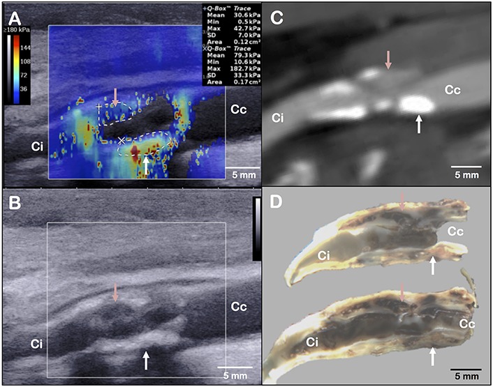
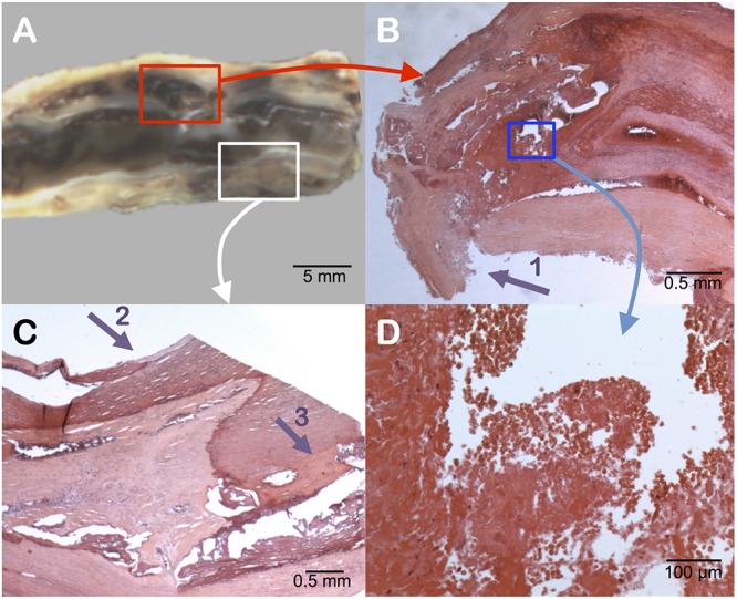
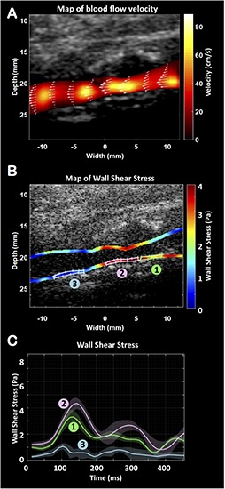
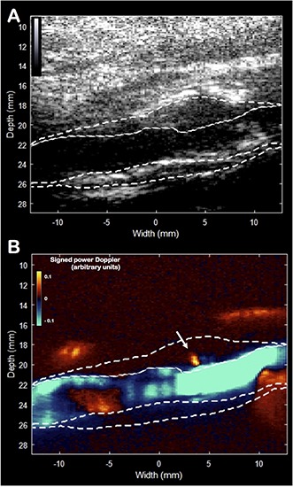
Results: Elastography allowed at identifying stiff areas corresponding to calcifications, as well as a soft area corresponding to an intraplaque hemorrhage. The flow evaluation with UFD showed an increase of the WSS along the plaque and identified the presence of a plaque rupture, confirmed by the pathologist.
Conclusion: Ultrafast ultrasound imaging is an innovative, easily accessible technique that provides imaging modalities on top of the conventional B-mode. Ultrafast ultrasound biomarkers such as plaque stiffness heterogeneity, WSS and intraplaque micro-flows could help to define the vulnerability of the carotid plaque in order to stratify patients that could benefit most from endarterectomy or intensive medical therapy.
Keywords: carotid plaque; elastography; plaque vulnerability; ultrafast ultrasound imaging; wall shear stress.
Copyright © 2020 Goudot, Khider, Pedreira, Poree, Julia, Alsac, Amemiya, Bruneval, Messas, Pernot and Mirault.
Figures
References
-
- Abbott A. L., Silvestrini M., Topakian R., Golledge J., Brunser A. M., de Borst G. J., et al. (2017). Optimizing the definitions of stroke, transient ischemic attack, and infarction for research and application in clinical practice. Front. Neurol. 8:537. 10.3389/fneur.2017.00537 - DOI - PMC - PubMed
-
- Aboyans V., Ricco J.-B., Bartelink M.-L. E. L., Björck M., Brodmann M., Cohnert T., et al. (2017). 2017 ESC guidelines on the diagnosis and treatment of peripheral arterial diseases, in collaboration with the european society for vascular surgery (ESVS). Eur. Heart J. 39 763–816. 10.1093/eurheartj/ehx095 - DOI - PubMed
LinkOut - more resources
Full Text Sources
Research Materials