. 2020 Mar 3:11:156.
doi: 10.3389/fmicb.2020.00156. eCollection 2020.

Study of the cwaRS-ldcA Operon Coding a Two-Component System and a Putative L,D-Carboxypeptidase in Lactobacillus paracasei

Affiliations

Study of the cwaRS-ldcA Operon Coding a Two-Component System and a Putative L,D-Carboxypeptidase in Lactobacillus paracasei

Hélène Scornec et al. Front Microbiol. .

Abstract

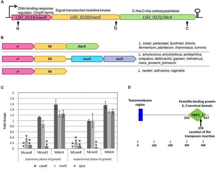
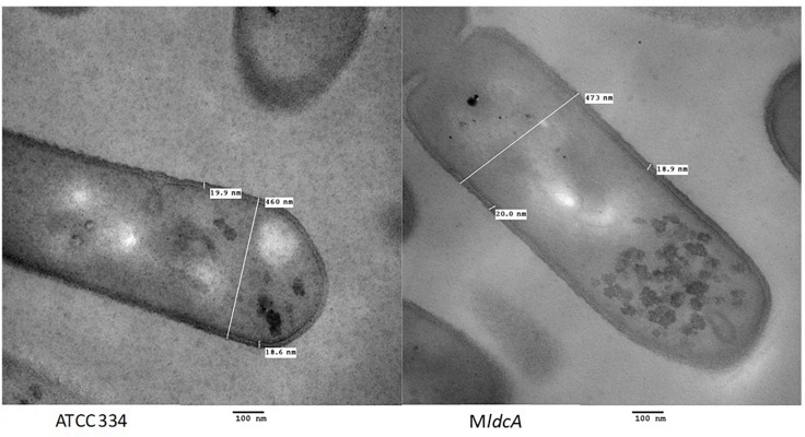
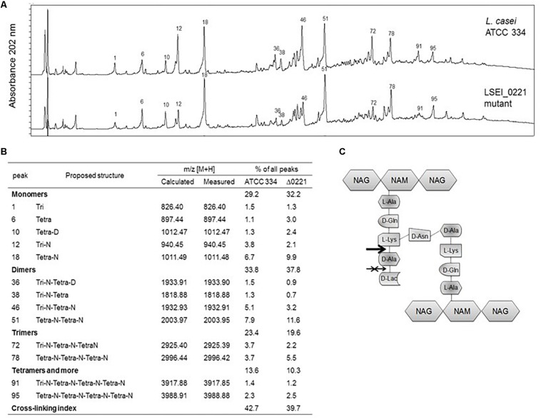
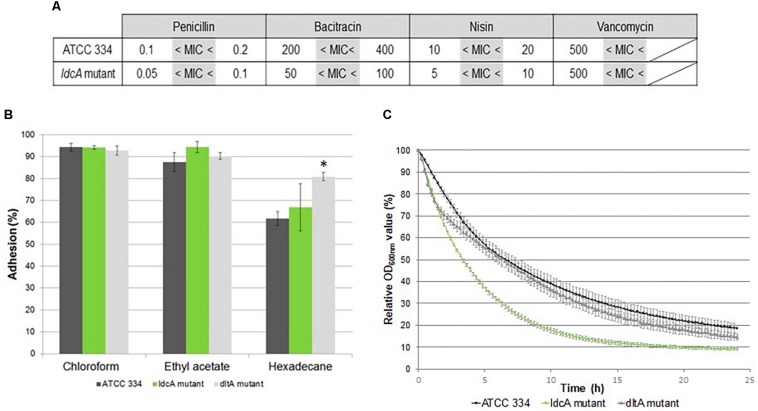
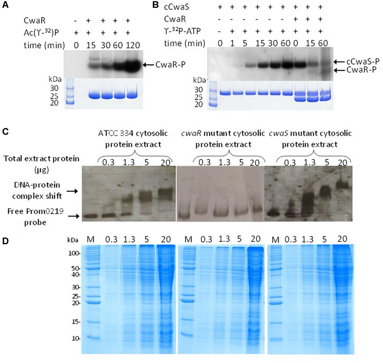
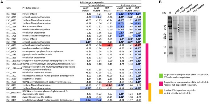
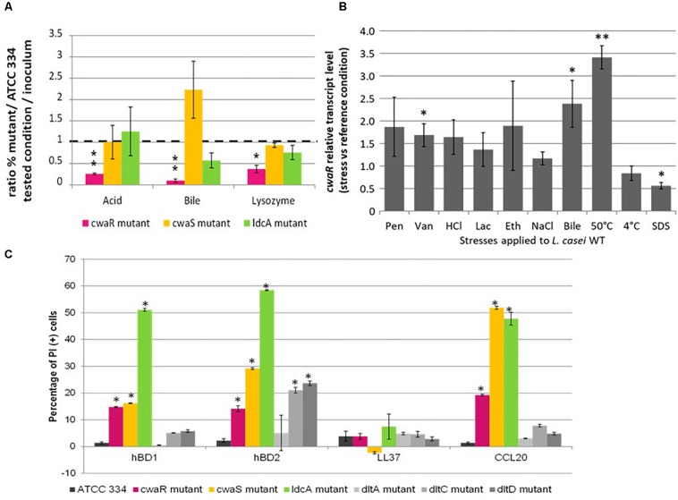
The cell surface is the primary recognition site between the bacterium and the host. An operon of three genes, LSEI_0219 (cwaR), LSEI_0220 (cwaS), and LSEI_0221 (ldcA), has been previously identified as required for the establishment of Lactobacillus paracasei in the gut. The genes cwaR and cwaS encode a predicted two-component system (TCS) and ldcA a predicted D-alanyl-D-alanine carboxypeptidase which is a peptidoglycan (PG) biosynthesis enzyme. We explored the functionality and the physiological role of these three genes, particularly their impact on the bacterial cell wall architecture and on the bacterial adaptation to environmental perturbations in the gut. The functionality of CwaS/R proteins as a TCS has been demonstrated by biochemical analysis. It is involved in the transcriptional regulation of several genes of the PG biosynthesis. Analysis of the muropeptides of PG in mutants allowed us to re-annotate LSEI_0221 as a putative L,D-carboxypeptidase (LdcA). The absence of this protein coincided with a decrease of two surface antigens: LSEI_0020, corresponding to p40 or msp2 whose implication in the host epithelial homeostasis has been recently studied, and LSEI_2029 which has never been functionally characterized. The inactivation of each of these three genes induces susceptibility to antimicrobial peptides (hBD1, hBD2, and CCL20), which could be the main cause of the gut establishment deficiency. Thus, this operon is necessary for the presence of two surface antigens and for a suitable cell wall architecture.

Keywords: antimicrobial peptides; carboxypeptidase; gene regulation; host–microbe interaction; lactic acid bacteria; peptidoglycan; two-component system.


Figures

FIGURE 1
FIGURE 1
Description of cwaRS-ldcA locus. (A) Locations of the different transposon insertions are shown with arrows a, b, and c for mutants McwaR, McwaS, and MldcA, respectively. (B) Comparison of the organization in different Lactobacillus species. rr, response regulator gene; hk, histidine kinase gene. (C) Relative transcript levels of L. casei mutants in stationary and exponential phase of growth in MRS broth. Transcript levels of each gene are expressed as the relative fold change, with L. paracasei ATCC 334 as the reference condition (fold change = 1). Three biological repeats were performed, and bars indicate standard deviations. Statistical analysis was performed using the unpaired Student t-test: P < 0.01; ∗∗P < 0.001. (D) Smart schema of the domains within LdcA protein and location of the transposon in mutant MldcA.
FIGURE 2
FIGURE 2
Transmission electron microscopy micrographs of L. paracasei ATCC 334 and LSEI_0221 mutant grown in MRS medium until the early stationary phase of growth.
FIGURE 3
FIGURE 3
Muropeptide profiles of L. paracasei ATCC 334 and lcdA mutant. (A) Reversed phase high-performance liquid chromatography (RP-HPLC) separation profiles of muropeptides of L. paracasei and lcdA mutant, peptidoglycans were digested by mutanolysin. Numbers correspond to the main muropeptides whose amounts changed between ATCC 334 and lcdA mutant. (B) Main muropeptide compositions, Tri, disaccharide tripeptide (L-Ala-D-iGln-L-Lys); Tetra, disaccharide tetrapeptide (L-Ala-D-iGln-L-Lys-D-Ala); Disaccharide, GlcNAc-MurNAc; iGln, isoglutamine; N, D-Asn; D, D-Asp. Percentage of each peak was calculated as the ratio of the peak area to all peak areas. Cross-linking index was calculated according to Glauner (1988) with the formula (1/2 Σ dimers + 2/3 Σ trimers + 3/4 Σ tetramers and more)/Σ all muropeptides. (C) Schematic structure of L. paracasei ATCC 334 peptidoglycan. The arrow indicates the cleavage site of LdcA enzyme, and the crossed arrow indicates the incorrect, predicted function.
FIGURE 4
FIGURE 4
Analysis of surface properties of ATCC 334 and ldcA mutant of L. paracasei. (A) Minimal inhibitory concentration (MIC) of ATCC 334 and the ldcA mutants in μg.ml–1. (B) Microbial Adhesion To Solvents (MATS) tests of ATCC 334 and the ldcA and dltA mutants. Error bars represent standard deviations between three biological repeats. Statistical analysis was performed for each mutant versus ATCC334 using the unpaired Student t test: P < 0.01. (C) Autolysis profiles of ATCC 334 and the ldcA and dltA mutants. Cultures were grown to the exponential phase of growth (OD of 0.8), then the cells were pelleted and suspended in phosphate-buffered saline (PBS) containing 0.05% Triton X-100 (v/v) and incubated at 30°C. Error bars represent standard deviations between three biological repeats.
FIGURE 5
FIGURE 5
In vitro characterizations of the two-component system (TCS). (A) Radiolabeling of RR0219 using [γ -32P] acetyl phosphate after different times of incubation. (B) Autophosphorylation of the cytosolic domain of HK0220 (cHK0220) when incubated with [γ -32P]ATP. After an incubation of 60 min, the RR0219 protein was added, and the phosphotransfer from radiolabeled cHK0220 (cHK0220-P) to RR0219 was observed after different times of co-incubation. (C) Electrophoretic mobility shift assay (EMSA) of cwaR promoter with cellular extract of L. paracasei ATCC 334 and the cwaR mutant. (D) The corresponding sodium dodecyl sulfate–polyacrylamide gel electrophoresis (SDS-PAGE) is 12.5%.
FIGURE 6
FIGURE 6
Consequences of cwaRS gene inactivation on genes encoding in peptidoglycan (PG) synthesis proteins and surface proteins. (A) Transcriptional profiles for genes implicated in PG synthesis of the McwaR, McwaS, and MldcA mutants relative to the parental strain ATCC 334 grown either exponential or stationary phase. Values are the mean RTL obtained for three biological repeats. In bold letters, significant values; in red and a, upregulation, p < 0.01; in light red and b, upregulation, p < 0.05; in blue and a, downregulation, p < 0.01; in light blue and b, downregulation, p < 0.05. Genes for which no significant value was obtained are not presented. The list of the tested genes and their corresponding annotations are presented in Supplementary Table S2. (B) Sodium dodecyl sulfate–polyacrylamide gel electrophoresis (SDS-PAGE) of surface protein extracts from L. paracasei ATCC 334 and McwaR, McwaS, and MldcA mutants in stationary phase of growth. (1) Surface antigen (LSEI_2029), (2) Surface antigen (LSEI_0020).
FIGURE 7
FIGURE 7
Role of cwaSR genes in adaptation to gut conditions and to reference stresses. (A) Competition in stressful conditions between L. paracasei mutants (McwaR, McwaS, and MldcA) and ATCC 334. The competitions were carried out in diluted MRS broth (1/20) in 16 h at 37°C; acid pH 3.5; bile 3 g.l–1; lysozyme 10 g.l–1. The percentage was determined by counting on Petri dishes. Three biological repeats were performed, and bars indicate standard deviations. Statistical analysis was performed using the unpaired Student t-test: P < 0.01; ∗∗P < 0.001. (B) Relative transcript levels of cwaR gene in L. paracasei ATCC 3334 under stressful conditions. L. paracasei was grown until OD reached 0.6. Then, a 15-min stress was applied. Transcript levels are expressed as the relative fold change, with L. paracasei cells incubated at 37°C as the reference condition (fold change = 1). Pen, penicillin 0.1 μg/ml; Van, vancomycin 0.5 mg/ml; HCl, HCl added to reach pH 3; Lac, lactic acid added to reach pH 3; Eth, ethanol 15% (vol/vol); NaCl, NaCl 1 M; Bile, bile 3 g/l; SDS, SDS 0.5 g/l. Four biological repeats were performed, and bars indicate standard deviations; p < 0.05; ∗∗p < 0.01. (C) Percentage of PI (+) bacteria (permeabilized cell) after treatment with hBD1, hBD2, LL37, and CCL20. Values were obtained by flow cytometry. Indicate significant differences (P < 0.05).

Similar articles

References

    1. Abo-Amer A. E., Munn J., Jackson K., Aktas M., Golby P., Kelly D. J., et al. (2004). DNA interaction and phosphotransfer of the C4-dicarboxylate-responsive DcuS-DcuR two-component regulatory system from Escherichia coli. J. Bacteriol. 186 1879–1889. 10.1128/jb.186.6.1879-1889.2004 - DOI - PMC - PubMed
    1. Alcantara C., Revilla-Guarinos A., Zuniga M. (2011). Influence of two-component signal transduction systems of Lactobacillus casei BL23 on tolerance to stress conditions. Appl. Environ. Microbiol. 77 1516–1519. 10.1128/aem.02176-10 - DOI - PMC - PubMed
    1. Arioli S., Koirala R., Taverniti V., Fiore W., Guglielmetti S. (2018). Quantitative recovery of viable Lactobacillus paracasei CNCM I-1572 (L. casei DG®) after gastrointestinal passage in healthy adults. Front. Microbiol. 9:1720 10.3389/fmicb.2018.01720 - DOI - PMC - PubMed
    1. Arumugam M., Raes J., Pelletier E., Le Paslier D., Yamada T., Mende D. R., et al. (2011). Enterotypes of the human gut microbiome. Nature 473 174–180. - PMC - PubMed
    1. Balandino G., Milazzo I., Fazio D. (2008). Antibiotic susceptibility of bacterial isolates from probiotic products available in Italy. Microbial. Ecol. Health Dis. 20 199–203. 10.1080/08910600802408111 - DOI - PubMed